What Causes Bladder Spasms? Causes, Risks, and Care 2026
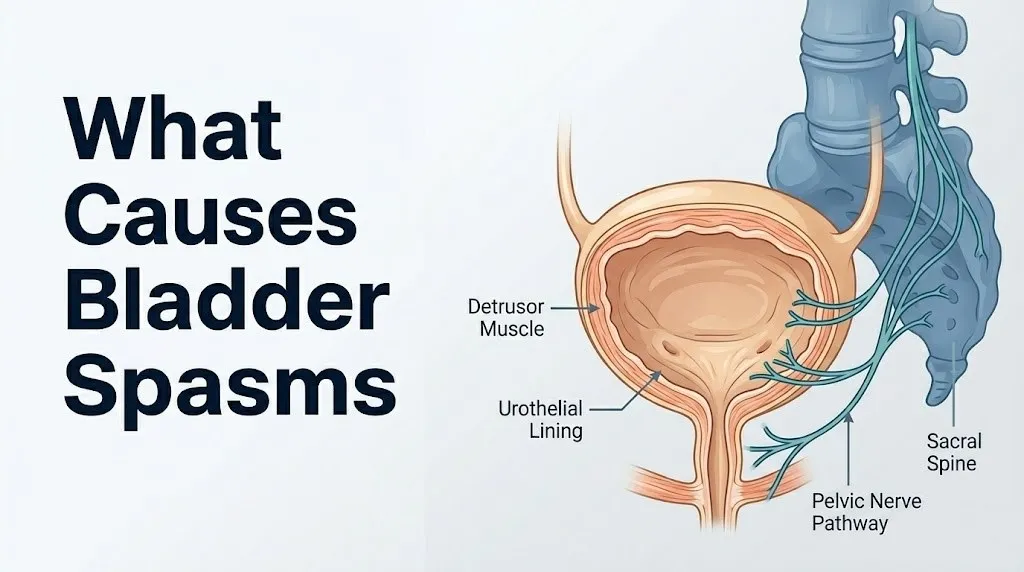
What causes bladder spasms comes down to one core event: the detrusor muscle, the smooth muscle wall of the bladder, contracts involuntarily before you are ready to void. The causes behind that involuntary contraction range from a straightforward urinary tract infection to complex neurological conditions, hormonal changes, and mechanical irritation, and correctly identifying which cause is driving your symptoms determines every decision that follows.
The National Institute of Diabetes and Digestive and Kidney Diseases (NIDDK) estimates that tens of millions of Americans experience urinary urgency and involuntary bladder contractions, with overactive bladder alone affecting approximately 33 million people in the United States. These numbers reflect a spectrum of underlying causes, not a single disease.
This article covers the specific physiological mechanism behind each recognized cause of bladder spasms, who is most at risk, which causes require urgent medical attention, and what a clinical evaluation actually involves. Every cause is explained at the level of the named nerve pathways, receptor subtypes, and urothelial signaling that produce the spasm.
What Causes Bladder Spasms?
Bladder spasms are caused by involuntary contractions of the detrusor muscle, the smooth muscle lining the bladder wall, triggered by disrupted neural control, physical irritation of the urothelium, infection, inflammation, or structural changes to the bladder or its nerve supply.
The underlying causes fall into several distinct categories. Some are infectious: bacteria in the bladder irritate the urothelial lining and trigger sensory nerve firing that produces urgency and spasm. Some are neurological: damage to or disease of the nerve pathways controlling the detrusor removes the brain’s inhibitory control, allowing the muscle to contract freely. Some are structural: bladder stones, tumors, or catheter tips physically irritate the trigone. Some are hormonal: estrogen deficiency in postmenopausal women reduces urothelial thickness, increasing sensory nerve sensitivity.
| Cause Category | Primary Mechanism | Who Is Most Affected | Resolves Without Treatment? |
|---|---|---|---|
| Urinary tract infection | Bacterial toxins and prostaglandins irritate urothelium | Women, elderly, catheter users | Yes, with antibiotics |
| Overactive bladder | Detrusor overactivity, excess urothelial ATP signaling | Older adults, postmenopausal women, men with BPH | No; requires management |
| Interstitial cystitis | Mast cell activation, urothelial barrier defect | Women aged 30-50 | No; chronic condition |
| Neurogenic bladder | Loss of supraspinal or spinal inhibition | People with MS, SCI, Parkinson’s | No; requires neurourological management |
| Catheter irritation | Mechanical trigone stimulation | Hospitalized patients, spinal cord injury | Yes, when catheter removed |
| Post-surgical | Surgical disruption of pelvic autonomic nerves | Post-hysterectomy, post-prostatectomy | Often improves over months |
| Hormonal | Estrogen deficiency causing urothelial thinning | Postmenopausal women | Partial improvement with hormone management |
| Dietary irritants | Direct mucosal irritation by acidic or caffeinated substances | Anyone with hypersensitive bladder | Yes, with elimination |
Most bladder spasms have an identifiable cause. The clinical priority is establishing which cause is present, because treatments target specific mechanisms and are not interchangeable.
People taking diuretics for blood pressure or heart failure are at increased risk of bladder spasms because rapid bladder filling from increased urine production places higher stretch demand on the detrusor wall in a shorter timeframe, activating mechanoreceptors earlier in the filling cycle.
Bladder Spasm Causes: The Detrusor Muscle Mechanism
Bladder spasms originate in the detrusor muscle, a three-layered smooth muscle structure forming the bladder wall, when its normal neurological inhibition is overcome or bypassed, producing an involuntary contraction that the person cannot consciously suppress.
Understanding the spasm mechanism requires understanding normal bladder control. During filling, the sympathetic nervous system keeps the detrusor relaxed via beta-3 adrenergic receptors on the detrusor smooth muscle cells. Simultaneously, the pontine micturition center in the brainstem maintains tonic inhibitory control over the sacral parasympathetic pathway, preventing premature detrusor contraction. When voiding is appropriate, the pontine micturition center (PMC), also called Barrington’s nucleus, releases its inhibition and activates the pelvic nerve (parasympathetic, S2-S4). Pelvic nerve terminals release acetylcholine, which binds to muscarinic M3 receptors on detrusor smooth muscle, causing the coordinated contraction of voiding.
Think of the detrusor muscle like a car engine that’s supposed to start only when you turn the key. The pelvic nerve is the ignition signal. The pontine micturition center is the key holder. Bladder spasms happen when the engine starts without anyone turning the key, because either the key holder has lost control, the ignition wires are crossed by inflammation, or something is pressing the starter button from inside the car.
The urothelium plays an active role that most health content ignores entirely. Research published in Neurourology and Urodynamics has documented that the urothelial cells release ATP (adenosine triphosphate) in response to bladder wall stretch during filling. This ATP activates P2X3 purinergic receptors on subepithelial sensory nerve fibers, generating the afferent signal that communicates bladder fullness to the spinal cord and brain. In pathological states such as infection, interstitial cystitis, and catheter irritation, urothelial ATP release is amplified. The sensory threshold drops, the urgency signal fires at lower bladder volumes, and the PMC loses its ability to suppress the detrusor contraction. The result is a spasm.
Muscarinic M2 receptors on the detrusor also contribute. While M3 receptors drive the primary contraction, M2 receptors prevent beta-3 adrenergic relaxation of the detrusor, effectively locking the muscle into a contracted state during overactive episodes. This is why drugs targeting M2 and M3 receptors are used in clinical management of detrusor overactivity.
Women experience detrusor overactivity at a higher rate than men, partly because female urethra length is shorter (approximately 4 cm compared to 20 cm in males), providing less anatomical resistance to urgency signals reaching the external sphincter. Children have higher rates of detrusor overactivity than adults because the inhibitory pathways from the prefrontal cortex to the PMC are not fully myelinated until age 5 to 7.
Urinary Tract Infection and Bladder Spasms
A urinary tract infection (UTI) causes bladder spasms by triggering an inflammatory cascade in the urothelial lining that directly sensitizes the sensory nerve endings in the bladder wall, lowering the contraction threshold and producing urgent, painful, involuntary detrusor contractions.
The mechanism begins when gram-negative bacteria, most commonly Escherichia coli (responsible for approximately 80-85% of uncomplicated UTIs per the NIDDK), adhere to urothelial cells via type 1 fimbriae binding to uroplakin surface proteins. Once adhered, the bacteria produce toxins including lipopolysaccharide (LPS) and hemolysin, which trigger urothelial inflammatory mediator release. The urothelium responds by producing prostaglandins E2 and I2, which directly sensitize the C-fiber afferent nerve endings in the subepithelial space. These C-fibers, normally silent at rest, begin firing at lower bladder volumes. The result is the classic UTI symptom triad: urgency, frequency, and dysuria (burning with urination), with bladder spasms superimposed.
UTI-related bladder spasms are typically acute in onset. They appear over hours to one to two days and resolve as the infection is treated. This acute onset pattern distinguishes UTI-related spasms from the chronic progressive pattern of interstitial cystitis or overactive bladder.
Women have a lifetime risk of at least one UTI of approximately 50-60% according to the NIDDK, compared to approximately 12% in men over a lifetime. The anatomical reason is the female urethra’s shorter length and its proximity to the vaginal and anal orifices, facilitating bacterial ascent to the bladder. This makes UTI the single most common identifiable cause of acute bladder spasms in premenopausal women.
Important red flags to recognize alongside UTI-related spasms:
- Fever above 38°C (100.4°F) with spasms: suggests the infection has ascended to the kidneys (pyelonephritis), requiring prompt evaluation by a primary care physician or emergency medicine physician the same day
- Flank pain with spasms: also suggests pyelonephritis or ureteral calculus, both requiring same-day evaluation
- Blood in urine (hematuria) that is gross (visible) and not clearing: warrants urological evaluation to rule out bladder malignancy, which can mimic UTI presentation
Overactive Bladder as a Cause of Bladder Spasms
Overactive bladder (OAB) causes bladder spasms through persistent detrusor overactivity in the absence of active infection, defined by the American Urological Association (AUA) as urinary urgency, often with frequency and nocturia, with or without urge urinary incontinence, not caused by metabolic or local pathological factors.
The pathophysiology of OAB involves both myogenic and neurogenic mechanisms. The myogenic component involves autonomous detrusor smooth muscle micro-contractions that develop independently of nerve input. Research published in the Journal of Urology has documented that detrusor smooth muscle cells in OAB patients show increased spontaneous electrical activity and heightened coupling between adjacent cells, allowing small focal contractions to propagate across the entire bladder wall as a spasm. The neurogenic component involves increased urothelial ATP signaling and altered C-fiber afferent sensitivity, as described above.
OAB affects an estimated 33 million Americans, with the NIDDK reporting that prevalence increases with age in both sexes. In men, OAB frequently co-occurs with benign prostatic hyperplasia (BPH), where bladder outlet obstruction from the enlarged prostate causes secondary detrusor overactivity. The detrusor hypertrophies and becomes unstable in response to the increased voiding effort required against the obstructed outlet.
Quick Tip:
- OAB-related bladder spasms are typically characterized by strong, sudden urgency that occurs even when the bladder is not full, distinguishing them from the gradually increasing urgency of a normally filling bladder
- Keeping a bladder diary for three to five days before a urology appointment (recording voiding times, volumes, urgency scores, and fluid intake) provides the urologist with the most useful clinical information for distinguishing OAB from other causes
- Men over age 50 with bladder spasms should specifically mention any hesitancy, weak stream, or double voiding to their primary care physician or urologist, because these symptoms suggest BPH-related secondary detrusor overactivity
Key Takeaway: Overactive bladder causes spasms through both autonomous smooth muscle micro-contractions and amplified urothelial ATP signaling, making it a fundamentally different cause from UTI despite producing similar urgency symptoms.
Interstitial Cystitis and Bladder Spasm Causes
Interstitial cystitis (IC), also called painful bladder syndrome (PBS), causes bladder spasms through a combination of urothelial barrier defect, mast cell activation, and chronic C-fiber nerve sensitization, producing some of the most severe and persistent bladder pain and spasm of any condition affecting the lower urinary tract.
The urothelium normally maintains a protective glycosaminoglycan (GAG) layer that prevents urine solutes from penetrating the urothelial surface and irritating underlying sensory nerves. In IC/PBS, this GAG layer is deficient or disrupted. Urine components, particularly potassium ions, penetrate the urothelium and directly depolarize subepithelial C-fiber afferent nerve endings. Research published in the Journal of Urology has demonstrated that intravesical potassium chloride instillation reproduces urgency and pain in IC patients at concentrations that do not affect healthy controls, supporting the GAG barrier deficiency model.
Mast cells are present in elevated numbers in the bladder wall in IC/PBS, particularly in cases with Hunner’s lesions (discrete inflammatory ulcers on the bladder mucosa visible on cystoscopy). These mast cells degranulate in response to urothelial irritation, releasing histamine, tryptase, and prostaglandins, which sensitize C-fiber afferents and trigger smooth muscle irritability. The result is spasm superimposed on chronic pain.
IC/PBS predominantly affects women, with the NIDDK reporting that approximately 3-8 million women in the United States may be affected. The condition is underdiagnosed because its symptoms overlap with recurrent UTI, but a key distinguishing feature is that IC/PBS does not produce a positive urine culture, while a UTI does. People with IC/PBS who repeatedly test negative on urine culture but continue to have urgency, frequency, and pelvic pain should be evaluated by a urologist or urogynecologist, specifically requesting cystoscopy under anesthesia and bladder biopsy to assess for Hunner’s lesions and mast cell infiltration.
Catheter-Related Bladder Spasms
Catheter-related bladder spasms are caused by the physical presence of an indwelling urethral catheter, particularly the balloon of the catheter, mechanically irritating the trigone of the bladder, the most sensory-dense region of the lower urinary tract.
The trigone is a triangular area at the base of the bladder, bounded by the two ureteral orifices superiorly and the internal urethral orifice inferiorly. It has a significantly higher density of sensory nerve endings than the rest of the bladder wall, because it serves as the primary detector of bladder fullness and coordinates the initiation of voiding. When a Foley catheter balloon is inflated in the bladder, typically with 10-30 mL of sterile water, the balloon sits at or near the trigone and continuously stimulates these sensory receptors. The brain interprets this as a signal that the bladder is full and needs to be voided, triggering detrusor contraction even though the catheter is draining the bladder continuously.
People with spinal cord injuries, neurological conditions, or post-surgical indwelling catheters are particularly susceptible to severe catheter-related spasms because their baseline bladder sensitivity is already altered. In a person without a neurological condition, catheter spasms are uncomfortable but manageable. In a person with a complete spinal cord injury above T6, catheter-related detrusor contractions can trigger autonomic dysreflexia, a dangerous episode of uncontrolled sympathetic nervous system activation producing severe hypertension, severe headache, and profuse sweating above the level of injury.
To minimize catheter-related bladder spasms in a home setting:
- Ensure the catheter drainage bag is always positioned below the level of the bladder to prevent urine from refluxing back into the bladder, which amplifies trigone irritation
- Check that the catheter is not kinked, obstructed, or pulling tightly, because tension on the catheter transmits mechanical irritation directly to the trigone
- Maintain fluid intake of at least 1.5-2 liters per day to dilute urine and reduce chemical irritation of the urothelium
- Report any spasms that are severe, worsening, or accompanied by fever, foul-smelling urine, or visible blood to the prescribing physician or urologist promptly
Older adults with indwelling catheters are at higher risk of catheter-associated urinary tract infections (CAUTIs), which compound catheter-related mechanical spasms with infectious urothelial inflammation, producing a combined spasm trigger that is more severe than either alone.
Neurogenic Bladder Spasm Causes
Neurogenic bladder refers to bladder dysfunction caused by disruption of the neural pathways that normally control detrusor muscle activity, and it is one of the most mechanistically distinct causes of bladder spasms because the source of the problem is in the nervous system, not the bladder itself.
Normal bladder control requires an intact circuit connecting the sacral micturition center (S2-S4 spinal cord), the pontine micturition center (brainstem), and the prefrontal cortex (voluntary control of voiding). Disruption at any level of this circuit produces a form of neurogenic bladder with distinct characteristics depending on the site of the lesion.
Lesions above the pontine micturition center (e.g., stroke, Parkinson’s disease, frontal lobe tumors) typically produce detrusor overactivity with coordinated sphincter relaxation: the bladder contracts urgently and frequently because the cortical and basal ganglia inhibitory input to the PMC is lost, but the voiding reflex itself remains coordinated because the PMC is intact. This produces urgency and frequency without significant obstruction or retention.
Lesions between the pons and the sacral cord (e.g., cervical or thoracic spinal cord injury, multiple sclerosis lesions in the spinal cord) produce detrusor-sphincter dyssynergia (DSD): the detrusor contracts but the external sphincter contracts simultaneously instead of relaxing, because the coordination center in the pons is disconnected from the sphincter relaxation pathway. This produces incomplete bladder emptying, high intravesical pressures, and spasms.
The European Association of Urology (EAU) guidelines on neurogenic lower urinary tract dysfunction classify neurogenic bladder by the site of neurological lesion and by its functional consequences, specifically whether it produces overactivity or underactivity of the detrusor and sphincter. This classification directly guides treatment selection.
People with neurological conditions causing bladder spasms should be evaluated by a neurourologist or a physiatrist with expertise in neurogenic bladder management, because standard overactive bladder treatments may be insufficient or may worsen incomplete bladder emptying, which is a risk for renal damage in this population.
Key Takeaway: Neurogenic bladder spasms originate above the bladder itself, in disrupted nerve pathways from the spinal cord to the brainstem, and their treatment must address the neurological lesion level, not just the bladder, to be effective and safe.
Multiple Sclerosis and Spinal Cord Injury Bladder Spasms
Multiple sclerosis (MS) and spinal cord injury (SCI) are the two most common neurological causes of severe, medically complex bladder spasms, each producing spasms through distinct mechanisms that require specialized urological and neurological management.
In MS, demyelinating plaques in the spinal cord white matter disrupt the long descending tracts that carry the PMC’s inhibitory signals to the sacral micturition center. When these pathways are demyelinated, nerve conduction slows or fails, removing the cortical and pontine inhibitory brake on the sacral parasympathetic output. The sacral micturition center becomes hyperexcitable, producing uninhibited detrusor contractions. Research published in Neurourology and Urodynamics has found that approximately 75-80% of people with MS develop lower urinary tract symptoms during the course of their disease, with detrusor overactivity being the most common urodynamic finding.
The location and severity of MS lesions determine the bladder symptom pattern. Lesions in the cervical cord are more likely to produce DSD (detrusor-sphincter dyssynergia), while thoracic lesions may produce either overactivity alone or DSD. People with MS who develop new or worsening bladder spasms should have a urodynamic study performed to characterize the specific dysfunction, because empirical antimuscarinic treatment without ruling out significant post-void residual urine carries a risk of worsening urinary retention.
In spinal cord injury, the severity of bladder dysfunction depends critically on the level and completeness of the injury. Injuries above T6 produce a specific and dangerous complication: autonomic dysreflexia (AD). In AD, a noxious stimulus below the level of injury, most commonly a full or spasming bladder, triggers massive uncontrolled sympathetic discharge below the injury level, producing severe hypertension, pounding headache, profuse sweating above the injury level, and facial flushing. If untreated, AD can cause hypertensive stroke or cardiac arrest.
People with SCI above T6 and their caregivers must be trained to recognize autonomic dysreflexia as a medical emergency. Call 911 immediately if a person with a spinal cord injury above T6 develops sudden severe headache, profuse sweating, and high blood pressure in the context of a perceived full bladder or known catheter obstruction.
Bladder Spasms After Surgery
Bladder spasms after surgery arise from several simultaneous mechanisms: surgical disruption of pelvic autonomic nerve fibers, direct surgical trauma to the bladder wall or trigone, the presence of a postoperative indwelling catheter, and residual irritation from surgical instruments.
Pelvic surgeries with the highest rates of postoperative bladder spasms include hysterectomy, prostatectomy, bladder tumor resection, and colorectal surgery involving the posterior pelvic dissection. These procedures pass through the same anatomical territory as the pelvic nerve plexus (the network of autonomic nerve fibers that supplies the bladder, arising from the inferior hypogastric plexus at the level of S2-S4). Even when surgeons use nerve-sparing techniques, temporary neurapraxia (conduction block without axon disruption) is common, producing a period of detrusor overactivity as the nerve recovers.
Transurethral resection of bladder tumors (TURBT) directly cauterizes and removes bladder wall tissue, including urothelium, and leaves a raw mucosal surface that is highly sensitive to urine contact. The inflammatory response to surgical trauma produces prostaglandins E2 and I2 at the surgical site, sensitizing the remaining afferent nerve endings and producing spasms for days to weeks postoperatively.
Post-surgical bladder spasms in women who have undergone hysterectomy require particular attention. The American College of Obstetricians and Gynecologists (ACOG) acknowledges that radical hysterectomy carries a higher risk of bladder denervation than simple hysterectomy, potentially producing a long-term neurogenic component to post-surgical bladder dysfunction. Women with persistent bladder spasms beyond six weeks after hysterectomy should be evaluated by a urogynecologist, specifically asking for urodynamic testing to characterize whether the dysfunction is primarily sensory (overactive afferent signaling) or motor (detrusor overactivity without adequate sphincter coordination).
Dietary and Lifestyle Causes of Bladder Spasms
Dietary and lifestyle factors cause bladder spasms by directly irritating the urothelium, altering urine pH, or stimulating the autonomic nervous system in ways that lower the detrusor contraction threshold.
The bladder’s urothelial lining is directly exposed to urine, which means every substance excreted in urine has the potential to act as a chemical irritant to the mucosal surface. The most clinically consistent dietary bladder irritants are caffeine, alcohol, and acidic foods. Caffeine acts as a direct bladder stimulant through two mechanisms: it is a phosphodiesterase inhibitor, increasing intracellular cyclic AMP in detrusor smooth muscle cells and amplifying contractile signals, and it is a diuretic, increasing urine production and hastening bladder filling. Research published in BJU International found that caffeine intake above 100 mg per day was associated with increased detrusor overactivity in urodynamic studies.
Alcohol increases urine production through inhibition of antidiuretic hormone (ADH) secretion from the posterior pituitary gland, causing rapid bladder filling and frequent urgency. Acidic foods and beverages, including citrus fruits, tomatoes, spicy foods, and carbonated drinks, may alter urine pH and directly irritate the GAG layer of the urothelium in people with baseline bladder hypersensitivity.
Dehydration is a paradoxical contributing factor. Reducing fluid intake concentrates urine, increasing the concentration of irritating solutes that contact the urothelium with each fill cycle, often worsening spasm frequency despite the reduced urine volume.
Dietary triggers vary substantially between individuals. People with IC/PBS or chronic OAB report a broader range of dietary sensitivities than people with UTI-related spasms. Men with BPH-related bladder spasms may find that dietary triggers have less influence on their symptoms compared to the dominant role of outlet obstruction.
Key Takeaway: Caffeine above 100 mg per day has documented urodynamic evidence linking it to increased detrusor overactivity, making caffeine reduction one of the few lifestyle modifications with published clinical support for reducing bladder spasms.
Bladder Spasms in Females and Postmenopausal Causes
Bladder spasms in females are driven by a combination of anatomical, hormonal, and reproductive factors that differ fundamentally from the causes in males, and postmenopausal women face a specific urothelial mechanism not present in premenopausal women or in men.
Premenopausal women have a higher baseline risk of UTI-related bladder spasms due to the anatomical proximity of the urethra to the vaginal introitus and rectum, facilitating bacterial colonization of the periurethral area. Sexual activity is an established risk factor for UTI in premenopausal women because it can mechanically introduce periurethral bacteria into the urethra. Hormonal fluctuations across the menstrual cycle also affect bladder sensory thresholds: progesterone has a mild relaxing effect on smooth muscle including the detrusor, while estrogen supports urothelial thickness. In the late luteal phase, when both hormones decline before menstruation, some women experience increased bladder sensitivity and spasm frequency.
After menopause, the loss of estrogen profoundly affects the lower urinary tract through a mechanism called urogenital atrophy (now more precisely termed genitourinary syndrome of menopause, or GSM). Estrogen receptors are present in the urothelium, urethral epithelium, periurethral connective tissue, and pelvic floor muscles. As estrogen levels fall, the urothelium thins, the GAG protective layer becomes less robust, and the density of sensory nerve endings relative to protective epithelial cells increases. The lower urinary tract becomes more sensitive to normal urine constituents, producing urgency, frequency, dysuria, and bladder spasms without infection.
The North American Menopause Society recognizes genitourinary syndrome of menopause as a clinically distinct condition affecting up to 50-60% of postmenopausal women. Postmenopausal women with new or worsening bladder spasms in the absence of a positive urine culture should be evaluated by a urogynecologist or gynecologist, who may assess for urogenital atrophy using a pelvic examination and discuss the evidence base for localized vaginal estrogen therapy as a management option.
Bladder Spasms in Elderly Adults
Bladder spasms in elderly adults occur at higher rates and with greater clinical complexity than in younger populations because aging produces multiple simultaneous changes in the bladder, its nerve supply, and the central nervous system pathways that regulate voiding.
The NIDDK reports that overactive bladder affects approximately 30-40% of adults over age 65, compared to approximately 10-15% in adults under age 45. This dramatic increase with age reflects a combination of structural and functional changes. The detrusor muscle undergoes fibrotic changes with aging, developing increased collagen deposition between smooth muscle bundles. This fibrosis reduces the muscle’s compliance (its ability to accommodate increasing volumes without a pressure rise) and creates areas of electrical instability that can generate autonomous micro-contractions, much like scar tissue in a cardiac muscle can generate arrhythmia.
Aging also produces a reduction in the myelination and conduction efficiency of the afferent nerve pathways from the bladder to the spinal cord, paradoxically increasing the firing rate and urgency signal rather than reducing it. The inhibitory input from the prefrontal cortex to the PMC is reduced in elderly adults with white matter changes, which are present in approximately 60-70% of people over age 65 per neuroimaging studies. This cortical-PMC disconnection removes the higher-order suppression of urgency, producing the clinical picture of “can’t make it to the bathroom in time.”
Elderly adults with bladder spasms also face a specific diagnostic challenge: bladder malignancy, which peaks in incidence in the seventh and eighth decades of life, can present with urgency and bladder spasm indistinguishable from OAB or UTI. New onset bladder spasms with hematuria in any person over age 60 require urine cytology and urological evaluation, including cystoscopy, to rule out bladder carcinoma before attributing symptoms to age-related OAB. A primary care physician managing an elderly patient with new bladder spasms should order a urinalysis, urine culture, and urine cytology as the minimum initial evaluation.
Bladder Spasms During Pregnancy
Bladder spasms during pregnancy arise from the combined mechanical and hormonal effects of pregnancy on the lower urinary tract, which simultaneously increase susceptibility to urinary tract infection and alter the autonomic control of the detrusor.
Mechanical factors begin in the first trimester, when the expanding uterus rises out of the pelvis and starts compressing the bladder anteriorly. By the third trimester, the uterus exerts continuous pressure on the bladder, reducing its functional capacity and triggering detrusor contractions at lower fill volumes. The fetal head in vertex position in late pregnancy can intermittently compress the urethra and bladder base during fetal movement, producing sudden sharp spasm-like sensations.
Progesterone, elevated throughout pregnancy, has a relaxing effect on smooth muscle including the ureteral smooth muscle, which is why pregnant women are at higher risk of hydronephrosis (ureteral dilation). However, this same smooth muscle relaxation can alter detrusor contractility patterns, and the combination of reduced bladder capacity and increased susceptibility to bacterial colonization of the urinary tract significantly elevates UTI risk during pregnancy.
The American College of Obstetricians and Gynecologists (ACOG) recommends routine urine culture screening in pregnant women at the first prenatal visit because asymptomatic bacteriuria (bacteria in the urine without symptoms) occurs in approximately 2-7% of pregnant women and can progress to symptomatic UTI and pyelonephritis with spasm symptoms if untreated. Pregnant women with bladder spasms should have a urine culture performed promptly, as untreated UTI in pregnancy carries risks for preterm labor and low birth weight per ACOG clinical guidelines.
Bladder spasms in pregnancy accompanied by fever, flank pain, or contractions in a pregnant woman before 37 weeks of gestation require evaluation by an obstetrician the same day.
Medications That Cause Bladder Spasms
Certain medications cause bladder spasms by directly stimulating the detrusor, reducing bladder outlet resistance, increasing urine production, or altering the neural signals controlling detrusor activity.
Diuretics (thiazides, loop diuretics such as furosemide, potassium-sparing agents) increase urine production, causing rapid bladder filling that overwhelms the detrusor’s accommodation capacity and triggers contractions at higher frequency. People on diuretics frequently report urgency and frequency as side effects, and those with underlying OAB may find that diuretic use significantly worsens their spasm frequency.
Cholinergic medications, including bethanechol (used to treat urinary retention) and certain Alzheimer’s disease medications such as donepezil and rivastigmine (acetylcholinesterase inhibitors), increase parasympathetic activity at muscarinic receptors throughout the body, including the M3 receptors of the detrusor. This can produce de novo bladder spasms in people without a prior history of bladder symptoms, or worsen existing spasms significantly.
Medications that cause or worsen bladder spasms:
- Diuretics: furosemide, hydrochlorothiazide, spironolactone (increase urine production, accelerate bladder filling)
- Acetylcholinesterase inhibitors: donepezil, rivastigmine, galantamine (increase acetylcholine availability at muscarinic receptors)
- Bethanechol: direct cholinergic agonist (prescribed for urinary retention but can trigger spasms in a hypersensitive bladder)
- Alpha-blockers: tamsulosin, doxazosin (reduce internal urethral sphincter resistance; may unmask underlying OAB by reducing outlet resistance)
- Opioid analgesics (paradoxically): long-term use is associated with detrusor overactivity in some patients through poorly characterized central mechanisms
People starting a new medication who develop new bladder spasms within days to weeks of initiation should report this to the prescribing physician, specifically noting the timing of onset relative to the new medication. A pharmacist conducting a medication review can identify cholinergic or diuretic contributors to spasm symptoms in people on multiple medications.
Emergency Symptoms: When Bladder Spasms Require Immediate Care
Certain symptoms associated with bladder spasms require immediate emergency evaluation. Do not wait to see if these resolve on their own.
Call 911 or go to the nearest emergency room immediately if bladder spasms occur alongside:
- Sudden severe headache, profuse sweating above the injury level, and facial flushing in a person with a spinal cord injury above T6, because this pattern indicates autonomic dysreflexia, a potentially life-threatening hypertensive emergency requiring immediate intervention
- High fever above 38.5°C (101.3°F), rigors (uncontrollable shaking chills), and flank pain, because this constellation indicates pyelonephritis or early urosepsis requiring emergency evaluation and intravenous antibiotic treatment
- Gross hematuria (blood clearly visible in the urine as pink, red, or brown discoloration) that is new, painless, and not attributable to a known cause, because painless gross hematuria is the most common presenting symptom of bladder carcinoma and requires urgent urological evaluation within one to two weeks per AUA guidelines, or same-day emergency evaluation if accompanied by clot retention (inability to urinate due to blood clots obstructing the urethra)
- Inability to urinate despite bladder fullness and spasms, which indicates acute urinary retention, requiring catheterization to relieve the obstruction and prevent renal damage from back-pressure
- Bladder spasms in a pregnant woman accompanied by uterine contractions before 37 weeks of gestation and fever, because this combination raises concern for both ascending UTI and preterm labor
These presentations can indicate autonomic dysreflexia, urosepsis, bladder malignancy, acute urinary retention, or preterm labor, and require emergency medical assessment or urgent specialist evaluation, not watchful waiting.
Diagnosing the Cause of Bladder Spasms
Diagnosing the cause of bladder spasms requires a systematic clinical approach that begins with the most common and treatable causes and advances to specialized testing when initial evaluation is unrevealing.
The first step in any evaluation is a urinalysis and urine culture. Urinalysis detects pyuria (white blood cells indicating infection or inflammation), bacteriuria (bacteria suggesting UTI), hematuria (blood, warranting further evaluation), and nitrites (produced by gram-negative bacteria like E. coli). A positive urine culture with identified bacteria and sensitivities confirms UTI as the cause. A negative culture in a symptomatic patient directs the workup toward non-infectious causes.
A postvoid residual (PVR) measurement, performed by bladder ultrasound immediately after urination, quantifies how much urine remains in the bladder after voiding. A PVR above 100 mL suggests impaired bladder emptying, pointing toward neurogenic causes, outlet obstruction (BPH in men), or detrusor underactivity. This single measurement significantly changes the diagnostic and management pathway.
Diagnostic sequence for evaluating bladder spasm causes:
- Urinalysis and urine culture: rule out UTI and assess for hematuria or pyuria without bacteria (suggesting IC or malignancy)
- Urine cytology: for patients over age 50, or any age with hematuria, to screen for bladder carcinoma cells in the urine
- Postvoid residual by ultrasound: rule out incomplete emptying and outlet obstruction
- Bladder diary (3-5 days): quantify voiding frequency, volumes, urgency episodes, and fluid intake to characterize the functional pattern
- Urodynamic testing (cystometry and uroflowmetry): measure bladder pressure during filling, identify involuntary detrusor contractions, quantify flow rates; the gold standard for diagnosing OAB, neurogenic bladder, and DSD
- Cystoscopy: direct visualization of the bladder interior; identifies Hunner’s lesions in IC, bladder tumors, bladder stones, and catheter-related mucosal changes
- MRI of the lumbar and thoracic spine: for any patient with bladder spasms and neurological symptoms, to identify spinal cord or nerve root compression contributing to neurogenic bladder
Specialized testing such as urodynamics and cystoscopy is managed by a urologist, urogynecologist, or neurourologist. Primary care physicians typically complete steps 1 through 4 and refer based on findings.
Frequently Asked Questions About What Causes Bladder Spasms
What causes bladder spasms at night?
Bladder spasms at night are most commonly caused by reduced antidiuretic hormone (ADH) secretion during sleep in older adults, producing higher-than-normal urine volumes at night (nocturnal polyuria) that fill the bladder more rapidly and trigger detrusor contractions.
Other causes of nocturnal spasms include OAB with nocturia (defined by the AUA as waking one or more times per night to void with urgency), UTI, and catheter-related irritation in people with indwelling catheters.
Adults over age 65 who wake more than twice per night with bladder spasms should mention this to their primary care physician, because nocturnal polyuria in this group can also reflect uncontrolled heart failure or sleep apnea causing nocturnal fluid mobilization.
Can stress cause bladder spasms?
Psychological stress can trigger or worsen bladder spasms through the autonomic nervous system’s response to stress, which includes increased sympathetic and parasympathetic activity that can alter detrusor muscle tone and lower the bladder’s contraction threshold.
Research published in Neurourology and Urodynamics has found associations between anxiety disorders and increased urinary urgency and frequency, with proposed mechanisms including corticotropin-releasing hormone (CRH) acting on bladder mast cells and stress-related alterations in spinal cord pain modulation pathways.
Stress-related bladder spasms are a diagnosis of exclusion, meaning infection, structural, and neurological causes must be ruled out before attributing spasms to stress alone.
What causes bladder spasms without a UTI?
Bladder spasms without a UTI can be caused by overactive bladder, interstitial cystitis, neurogenic bladder from multiple sclerosis or Parkinson’s disease, catheter irritation, dietary irritants, hormonal changes, or bladder stones.
The absence of bacteria on urine culture rules out UTI but does not rule out other inflammatory causes like IC/PBS, where white blood cells may be present in the urine without bacteria.
A person with recurrent bladder spasms consistently testing negative on urine culture should be evaluated by a urologist, who may perform a bladder diary analysis, postvoid residual measurement, urodynamic testing, and cystoscopy to identify the specific underlying cause.
Are bladder spasms a sign of something serious?
Bladder spasms are sometimes a sign of a serious condition, particularly when they are accompanied by gross hematuria, new onset in a person over age 60, severe headache in a person with spinal cord injury, or high fever and flank pain suggesting pyelonephritis or urosepsis.
Most bladder spasms in otherwise healthy adults are caused by UTI, OAB, or dietary irritants and resolve with appropriate management, but any new or worsening pattern of bladder spasms warrants a minimum evaluation of urinalysis, urine culture, and urine cytology in adults over age 50.
The most important red flag is painless gross hematuria with urgency in any adult, which the American Urological Association recommends be evaluated with cystoscopy to rule out bladder carcinoma.
What do bladder spasms feel like?
Bladder spasms typically feel like sudden, intense pressure or cramping in the lower abdomen or pelvic area, often accompanied by a desperate urge to urinate that is difficult or impossible to delay.
The sensation is produced by the involuntary contraction of the detrusor muscle against a closed or partially closed urethra, generating intravesical pressure that the person perceives as urgency and pelvic discomfort.
In conditions like interstitial cystitis, spasms may be accompanied by burning or sharp pelvic pain; in neurogenic bladder from spinal cord injury, the person may not feel the spasm as pain but may observe autonomic responses like sweating or erection as indirect indicators.
How do doctors find the cause of bladder spasms?
Doctors begin diagnosing the cause of bladder spasms with a urinalysis and urine culture to rule out infection, followed by a postvoid residual measurement to assess bladder emptying.
A bladder diary documenting voiding frequency, volumes, and urgency episodes over three to five days provides key functional information, and urodynamic testing (cystometry) can directly record involuntary detrusor contractions and measure bladder pressures during filling.
Cystoscopy by a urologist or urogynecologist allows direct visualization of the bladder interior to identify structural causes including Hunner’s lesions from IC/PBS, bladder tumors, and bladder stones.
Closing
Bladder spasms have a specific physiological cause in every person who experiences them. The cause determines the treatment, and the treatment does not work when the cause is wrong. A UTI causing spasms responds to antibiotics. Neurogenic spasms from MS require urodynamic characterization before any treatment decision. Postmenopausal urothelial atrophy causing spasms has a different clinical pathway from OAB-related autonomous detrusor contractions.
The clearest first step is a urinalysis, urine culture, and a direct conversation with your primary care physician that includes your full medication list, your voiding pattern, and whether your spasms are new, recurrent, or worsening. If initial evaluation is unrevealing, request referral to a urologist or urogynecologist for urodynamic testing.
You deserve a specific answer about why your bladder is doing this, and the clinical tools to find that answer exist and are accessible. Start with the basics, ask for the next step when those don’t explain it, and do not accept “it’s probably OAB” without the urodynamic evidence to support it.