What Causes Lie Bumps on the Tongue in 2026: Full Guide
What causes lie bumps is a question that sounds simple but actually involves a specific piece of tongue anatomy, a local inflammatory cascade, and at least three clinically distinct triggering patterns, each with its own set of likely causes and a different prognosis. The short answer is that lie bumps form when one or more fungiform papillae on the tongue become acutely inflamed, producing the painful, raised, red or white bump that resolves, in most cases, within a few days without any treatment.
Understanding why lie bumps happen at this specific location on the tongue, and why some people get one isolated bump while others get dozens simultaneously with a fever, requires understanding the unique anatomy of the fungiform papilla and the different inflammatory pathways each variant of this condition likely involves. According to a clinical review published in the Journal of Oral Pathology and Medicine, transient lingual papillitis (the formal name for lie bumps) remains incompletely understood, with its exact triggering mechanisms not yet confirmed by controlled research, but the clinical patterns are well-documented enough to guide practical decisions.
This article covers the specific tongue anatomy involved, the primary triggers (food, trauma, stress, infection), the three distinct clinical types with their likely causes, the conditions that can mimic a lie bump, and the specific signs that tell you when a bump on your tongue warrants evaluation by a physician or dentist rather than watchful waiting.
What Are Lie Bumps and Why Do They Form on the Tongue
Lie bumps are the colloquial name for transient lingual papillitis (TLP), a condition in which one or more fungiform papillae on the tongue become acutely inflamed, swollen, and painful, appearing as small white or red elevated bumps most commonly at the tip and sides of the tongue.
The name “lie bump” comes from a folk belief that they appear when someone tells a lie, a charming origin story with no biological basis whatsoever. The actual reason these bumps appear relates entirely to the unique anatomy of the fungiform papilla and its relative vulnerability to the inflammatory triggers discussed throughout this article.
Fungiform papillae are normally present and functional but largely inconspicuous. They sit among the much more numerous filiform papillae (the fine, thread-like projections that give the tongue its rough texture) and are most easily seen at the front and sides of the tongue as tiny, rounded, slightly redder protrusions. When one becomes inflamed, it swells from its baseline diameter of approximately 1 to 2 millimeters to 3 to 5 millimeters and turns visibly red or develops a white surface coating, making it suddenly noticeable against the surrounding tongue surface.
The inflammation typically resolves on its own within one to three days in the classic form, which is why the word “transient” appears in the formal name. No scarring occurs. The papilla returns to its normal appearance and function after the inflammatory episode resolves.
The American Academy of Oral Medicine notes that transient lingual papillitis is likely underreported because most people experience it as a minor nuisance that resolves without medical attention, making true population prevalence difficult to establish from published data.
Children, adolescents, and adults of all ages can develop lie bumps, though the pattern of involvement differs by age group and clinical variant, as discussed in subsequent sections.
What Causes Lie Bumps: The Primary Triggers
The primary triggers of lie bumps include local chemical irritation from acidic or spicy foods, mechanical trauma to the tongue surface, psychological stress, gastrointestinal disturbances, and hormonal fluctuation, though the exact mechanism connecting each of these triggers to the inflammatory response within the fungiform papilla is not fully established by controlled clinical research.
What is clear from clinical observation and case reports documented in the oral medicine literature is that lie bumps are reactive: something triggers the local inflammatory response in the fungiform papilla, and the bump is the visible result. The trigger list reflects the diversity of inputs that can activate the mast cells, local nerve terminals, and vascular components of the papilla’s immune response.
The most frequently reported triggers in clinical practice, ranked from most to least consistently cited:
- Acidic or spicy foods: consumption of highly acidic foods (citrus fruits, vinegar-based foods, tomatoes) or capsaicin-containing spicy foods directly before or coinciding with lie bump onset
- Mechanical trauma: biting the tongue accidentally, eating sharp or hard foods (chips, crackers, hard candy), dental appliance pressure (orthodontic braces, retainers, dentures)
- Psychological stress: periods of heightened stress or anxiety temporally associated with lie bump episodes in patient-reported histories
- Gastrointestinal disturbances: constipation, gastrointestinal upset, and symptoms associated with Helicobacter pylori infection have been reported by patients as temporally linked to lie bump onset, though no confirmed causal mechanism has been established
- Hormonal fluctuation: some women report lie bumps more frequently during specific phases of the menstrual cycle or during pregnancy
It bears stating clearly: none of these associations has been confirmed as a direct causal mechanism by controlled human clinical trials. The evidence base for all proposed causes of classic TLP is clinical observation and patient-reported correlation, which is the appropriate evidence quality level to apply when discussing this topic.
Fungiform Papillae Anatomy: Why These Specific Tongue Structures Get Inflamed
Fungiform papillae are mushroom-shaped protrusions on the anterior two-thirds of the tongue’s dorsal surface that are specifically targeted by transient lingual papillitis because of their thin overlying epithelium, rich vascular and nerve supply, and direct exposure to oral contents, making them uniquely vulnerable to local inflammatory responses.
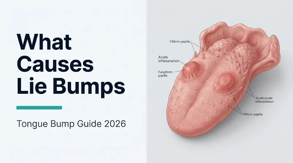
The human tongue dorsum is covered by four types of papillae, each with a distinct morphology and function. Filiform papillae are the most numerous, covering most of the tongue surface with keratinized, hair-like projections that provide mechanical friction and texture. Circumvallate (vallate) papillae are the large, rounded structures arranged in a row at the back of the tongue, forming the apex of a “V” shape visible at the junction of the anterior two-thirds and posterior one-third. Foliate papillae are the parallel ridges on the lateral edges of the posterior tongue. None of these three types is affected in transient lingual papillitis.
Fungiform papillae sit among the filiform papillae across the anterior tongue surface, most densely distributed at the tip. They are easily recognized by their slightly redder color (from the dense capillary network just below the thin epithelium) and their mushroom shape. Their superior surfaces contain taste buds, specialized epithelial structures housing taste receptor cells with microvilli projecting into taste pores on the surface. These taste buds receive innervation from the chorda tympani nerve, a branch of the facial nerve (cranial nerve VII), which carries taste information to the brain.
Think of the fungiform papilla like a tiny, richly decorated island surrounded by the simpler, thicker-walled filiform papillae. It has a thinner “roof,” a denser blood supply, and a richer nerve supply than its neighbors. When any irritant reaches the island, the inflammatory response is faster, the blood vessels dilate more visibly, and the resulting pain is amplified by the dense sensory innervation. That is precisely why a tiny inflamed fungiform papilla produces pain that seems disproportionate to its size.
According to data summarized in Oral Surgery, Oral Medicine, Oral Pathology, and Oral Radiology, fungiform papillae contain the highest concentration of taste buds in the anterior tongue, averaging approximately 3 to 5 taste buds per papilla in adults.
Key Takeaway: Lie bumps form specifically on fungiform papillae because these structures have thinner surface epithelium and denser nerve and blood vessel networks than any other tongue structure, making them the most reactive site on the tongue surface to any chemical, mechanical, or inflammatory trigger.
Food and Dietary Causes of Lie Bumps
Food-related lie bumps are caused by chemical irritation from acids, capsaicin, or other bioactive food compounds directly contacting the fungiform papillae’s thin epithelial surface, triggering local mast cell degranulation and the release of histamine and prostaglandins that produce the characteristic swelling and redness.
Highly acidic foods are one of the most consistently reported food-related triggers. Citric acid (in citrus fruits, lemonade, and many carbonated drinks), acetic acid (in vinegars and pickled foods), and the organic acids in tomatoes and tomato products all lower the local pH at the tongue surface when held in contact. The fungiform papillae’s thin epithelium is more vulnerable to acid-induced barrier disruption than the thicker keratinized filiform papillae surrounding them. When the epithelial barrier is compromised, even transiently, local immune cells are activated.
Capsaicin, the active compound in hot chili peppers and spicy condiments, binds TRPV1 receptors (transient receptor potential vanilloid 1 channels) on sensory nerve terminals in the tongue mucosa, including those of the chorda tympani and lingual nerves. TRPV1 activation at high intensity triggers the release of substance P and calcitonin gene-related peptide (CGRP) from sensory nerve terminals, which in turn stimulate mast cell degranulation and local vasodilation in a process called neurogenic inflammation. This explains why very spicy meals can produce lie bumps within hours in susceptible individuals.
Some individuals also report lie bumps associated with highly sweetened foods, artificial sweeteners, or alcohol-containing mouthwashes. The proposed mechanism involves the same local epithelial irritation pathway, though these associations are based on patient-reported experiences and have not been studied in controlled research.
People with gastroesophageal reflux disease (GERD) may experience food-related lie bumps from a secondary mechanism: gastric acid reaching the oral cavity during reflux episodes provides additional chemical irritation to the tongue surface beyond what the food itself contributed. Dental erosion patterns consistent with acid exposure are sometimes observed alongside recurrent TLP in individuals with significant untreated reflux.
Mechanical Trauma as a Cause of Lie Bumps
Mechanical trauma causes lie bumps by directly disrupting the epithelial surface of one or more fungiform papillae, initiating the same local inflammatory cascade that chemical irritants trigger through a physical rather than chemical mechanism.
The most common traumatic cause is accidentally biting the tongue. During eating, speaking, or during sleep in individuals who brux (grind their teeth), the tongue can be caught between the teeth. A bite sufficient to produce mild ecchymosis (bruising) or to break the surface epithelium of a fungiform papilla can trigger an acute inflammatory response that produces the characteristic swollen, painful bump within hours. The bump appears at the precise location of the bite, which sometimes allows a person to connect the event directly to the onset.
Sharp, hard, or rough foods can abrade the tongue surface during eating. Potato chips, corn tortilla chips, hard candies, and dry crackers are frequently cited in patient accounts as mechanical triggers. The edges of these foods scrape across the tongue dorsum with each chewing motion, and if a chip or edge makes repeated contact with the same fungiform papilla during a meal, cumulative mechanical stress can initiate the inflammatory response.
Dental appliances are an underrecognized cause of chronic or recurrent lie bumps in specific populations. Orthodontic bracket wires that project toward the tongue, removable retainers with lingual clasps, lower complete dentures that contact the tongue, and tongue piercings that impinge on specific papillae can all produce persistent low-grade mechanical irritation that repeatedly inflames the same papilla or papillae. In these cases, the lie bumps are recurrent and appear at the same location with each episode, which is a clinically useful pattern for identifying the cause.
According to clinical descriptions in Oral Surgery, Oral Medicine, Oral Pathology, and Oral Radiology, mechanically induced TLP typically involves a single papilla at the site of contact and resolves within the standard one-to-three-day timeframe once contact is avoided.
Stress and Hormonal Changes as Lie Bump Causes
Psychological stress and hormonal fluctuation are reported by a subset of individuals as triggers for lie bumps, and while the precise biological mechanism linking these systemic states to focal fungiform papillary inflammation is not established by controlled research, plausible pathways exist through the immune-modulating effects of cortisol and reproductive hormones on oral mucosal tissue.
Chronic stress activates the hypothalamic-pituitary-adrenal (HPA) axis, driving elevated cortisol production from the adrenal cortex. Cortisol has complex effects on mucosal immunity: at acutely elevated levels, it initially suppresses inflammation, but at chronically elevated levels, it impairs mucosal healing and alters the function of local immune cells, including mast cells and macrophages, in ways that may increase sensitivity to minor irritants. Clinicians generally observe that patients with recurrent TLP report higher episode frequency during high-stress periods, which is consistent with this proposed mechanism but does not confirm it.
Hormonal fluctuation, particularly during the luteal phase of the menstrual cycle (days 14 to 28, when progesterone rises and estrogen falls from its mid-cycle peak) and during pregnancy, has been reported by women as a period of increased lie bump frequency. Estrogen and progesterone both influence oral mucosal vascularity, immune cell activity, and epithelial turnover rates. The increased mucosal vascular permeability associated with elevated progesterone may lower the threshold for visible papillary swelling in response to routine minor irritants that would not produce a visible bump in other hormonal phases.
Women who track their menstrual cycles and notice a consistent correlation between their lie bump episodes and the week before menstruation have an identifiable hormonal pattern worth mentioning to their gynecologist or primary care physician, particularly if the episodes are severe or recurrent enough to impair eating.
Pregnant women who develop lie bumps should not be alarmed, as the combination of increased mucosal vascularity and potential dietary changes during pregnancy can facilitate papillary inflammation through the same mechanisms, but any new or persistent oral lesion during pregnancy warrants mention at the next prenatal visit with an obstetrician-gynecologist.
Key Takeaway: Stress and hormonal fluctuation are the two most frequently self-reported lie bump triggers that have the weakest direct evidence supporting them as independent causes, but the biological pathways through cortisol and sex hormone effects on mucosal immunity are mechanistically plausible and worth tracking if your episodes follow a consistent pattern.
Gastrointestinal Factors and Lie Bumps
Gastrointestinal disturbances, particularly constipation, upper gastrointestinal upset, and possible associations with Helicobacter pylori infection, are reported in the clinical TLP literature as associated conditions, though the mechanism connecting gastrointestinal state to focal tongue papillary inflammation remains hypothetical.
Patient-reported associations between lie bumps and constipation or bowel irregularity are mentioned in older dermatological and oral medicine literature, possibly reflecting a broader pattern of oral mucosal reactivity during systemic GI stress. The proposed mechanism involves systemic changes in immune mediator levels or gut microbiome-derived metabolites that alter mucosal barrier function, though this is speculation at this point in the research evidence.
A more mechanistically specific association involves Helicobacter pylori (H. pylori), a gram-negative bacterium that colonizes the gastric mucosa and is associated with peptic ulcer disease, chronic gastritis, and, in some published case reports, oral mucosal changes. H. pylori has been detected in dental plaque and oral tissue samples, raising the possibility that it can directly influence oral mucosal inflammatory responses. A case series published in the Journal of Oral Pathology and Medicine described patients with recurrent TLP whose episodes resolved after successful H. pylori eradication therapy, though this represents case-level evidence and cannot establish causation.
GERD is the gastrointestinal condition with the most mechanistically direct connection to lie bumps. When gastric acid reaches the oral cavity through reflux, it acts as a direct chemical irritant to the fungiform papillae through the same mechanism as dietary acidic foods discussed earlier. Individuals with frequent nighttime acid reflux may wake with tongue irritation and lie bumps without having consumed any obvious dietary trigger.
Individuals with recurrent lie bumps who also have unexplained upper gastrointestinal symptoms (heartburn, indigestion, bloating, nausea) should mention both to their primary care physician, as treating an underlying gastrointestinal condition may incidentally reduce TLP episode frequency.
Viral and Infectious Causes of Transient Lingual Papillitis
A viral or infectious cause has been proposed for certain patterns of transient lingual papillitis, specifically the household-clustering and febrile variant, based on epidemiological evidence that this form spreads between individuals in close contact, though no specific causative virus has been definitively identified and confirmed in peer-reviewed research.
The epidemiological evidence supporting a viral etiology for the eruptive variant of TLP is notable. Multiple published reports in the oral medicine literature describe household clusters where one family member (often a child) develops eruptive TLP with dozens of simultaneous papillary eruptions and mild systemic symptoms (fever, malaise, lymph node tenderness), and within days, other household members develop similar presentations. This pattern of household clustering is characteristic of viral transmission rather than shared environmental exposures.
Several candidate viruses have been proposed, including members of the herpesvirus family and various enteroviruses, but none has been consistently identified across independent studies. A 2004 case series published in Oral Surgery, Oral Medicine, Oral Pathology, and Oral Radiology described multiple affected household members and proposed viral transmission as the most probable explanation, a finding replicated in subsequent smaller reports. The current scientific consensus, as expressed by the American Academy of Oral Medicine, is that the eruptive febrile variant likely has an infectious trigger but the specific pathogen has not been confirmed.
Immunocompromised individuals (those on chemotherapy, organ transplant recipients, people living with HIV, and individuals on long-term corticosteroid therapy) may experience atypical or more severe presentations of any oral viral infection, including conditions that mimic or overlap with TLP. For this group, any new or unusual oral lesion warrants prompt evaluation by their primary care physician or infectious disease specialist rather than watchful waiting.
The Three Types of Transient Lingual Papillitis and Their Distinct Causes
Transient lingual papillitis is not a single uniform condition but a clinical label applied to three distinct presentations with likely different underlying causes, different affected populations, different clinical courses, and different implications for whether professional evaluation is warranted.
The three recognized clinical forms, as described in oral medicine literature:
| TLP Variant | Typical Presentation | Likely Cause | Primary Population | Duration |
|---|---|---|---|---|
| Classic/localized | One to two papillae affected at tongue tip or sides, intensely painful, no systemic symptoms | Local mechanical trauma or chemical irritation to individual fungiform papilla | Adults, all ages | One to three days; fully self-resolving |
| Eruptive/febrile | Dozens of papillae affected simultaneously across the tongue dorsum, may include fever, malaise, lymph node tenderness, household clustering | Suspected viral or infectious etiology; specific pathogen unconfirmed | Children aged 2 to 14 most commonly, household contacts secondarily | Seven to ten days; self-resolving but warranting evaluation in children with fever |
| Papulokeratotic | Chronic or recurrent, keratinized white papillary lesions with a firmer texture, less acutely painful than classic form | Proposed chronic irritation or distinct immunological mechanism; poorly understood | Adults, often associated with tobacco use or chronic irritants | Weeks; may persist and recur; warrants evaluation |
The classic/localized form is what most adults mean when they say they have a “lie bump.” One or two papillae, maximum pain, gone in two to three days. No systemic symptoms. No evaluation needed if there is an identifiable recent trigger and it resolves completely within the expected timeframe.
The eruptive/febrile form is a different clinical entity that requires a different response, particularly when it occurs in a child with fever. This form is discussed in detail in the following section.
The papulokeratotic form is the least common and least understood. It presents as raised, white, keratinized bumps on the tongue that persist longer than the classic form and tend to recur. Because white persistent tongue lesions require evaluation to exclude premalignant or malignant changes, this variant warrants evaluation by a board-certified dentist or oral medicine specialist if it does not resolve within two weeks.
Key Takeaway: If your “lie bump” involves dozens of swollen papillae at once, is accompanied by any fever, or has spread to other family members, it is likely the eruptive febrile variant of TLP and a pediatrician should be contacted, particularly if the affected person is a child.
What Causes Eruptive Transient Lingual Papillitis in Children
Eruptive transient lingual papillitis in children is the specific variant of lie bumps most likely to have a viral or infectious cause, characterized by the simultaneous eruption of multiple inflamed fungiform papillae across the tongue surface, often accompanied by mild systemic symptoms including low-grade fever, excessive salivation, and refusal to eat, in children most commonly between ages 2 and 14.
The presentation is dramatically different from the adult classic form. Instead of one or two isolated bumps at the tongue tip, the child’s entire visible tongue surface may be studded with swollen, pale or white-tipped papillae. The tongue may appear bumpy and irregular across its full dorsal surface. The child is often distressed and reluctant to eat because the diffuse papillary inflammation makes any oral contact painful. Parents who bring their child to a pediatrician with this presentation may be met with unfamiliarity, as the condition is not universally known by all primary care providers.
According to case series published in the Journal of Oral Pathology and Medicine, affected children recover fully within seven to ten days in the reported cases. However, the household clustering pattern means that parents and siblings of an affected child should be alert for their own onset of papillary symptoms in the days following the child’s presentation.
The practical approach for a parent whose child develops multiple simultaneous tongue bumps with fever: contact the child’s board-certified pediatrician within 24 to 48 hours of symptom onset, particularly if the child has a fever above 38°C (100.4°F) or is refusing all food and fluids. The pediatrician may perform a clinical examination to exclude herpes simplex stomatitis, hand foot and mouth disease (caused by coxsackievirus), and other viral oral conditions that require specific management or isolation precautions.
Infants under 12 months with any oral lesion and fever should be seen by a pediatrician the same day rather than monitored at home, because the differential diagnosis in this age group is broader and the consequences of missed infections (herpes simplex neonatal) are more serious.
What Causes Recurring Lie Bumps
Recurring lie bumps are caused by a persistent or repeated trigger that continues to activate the fungiform papillary inflammatory response despite individual episodes resolving, with the most common recurring triggers being an unidentified dietary sensitivity, a dental appliance or structural issue causing repeated mechanical contact, an unresolved gastrointestinal condition, or an underlying tendency toward oral mucosal reactivity that may be hormonally driven.
A single lie bump episode that resolves in two days and never recurs requires no investigation. Recurrent episodes, defined by clinical observation as four or more episodes per year, warrant a structured approach to identifying the trigger.
To systematically identify what is causing your recurring lie bumps:
- Keep a symptom diary for four to six weeks, recording the date each episode starts, its location on the tongue, what you ate in the 24 hours before onset, your stress level, and (for women) where you are in your menstrual cycle.
- Note whether the bumps consistently appear at the same location on the tongue, which suggests a mechanical trigger (dental appliance, sharp tooth edge, habit of tongue chewing at that location).
- Identify whether episodes cluster around specific dietary habits, such as regular citrus consumption, daily vinegar use, or weekly spicy food occasions.
- Consider whether you use a toothpaste containing sodium lauryl sulfate (SLS), a foaming detergent that disrupts the oral mucosal barrier and is associated with recurrent aphthous stomatitis; switching to an SLS-free toothpaste for four to six weeks may reduce episode frequency if SLS is a contributing factor.
- If no pattern emerges after six weeks of tracking, schedule an appointment with a board-certified dentist and bring your symptom diary; the dentist can assess for dental appliance fit issues, sharp tooth surfaces, and other structural causes during a clinical oral examination.
Individuals who smoke or use tobacco products have increased rates of all oral mucosal conditions and should discuss recurrent tongue lesions with both their primary care physician and dentist, as tobacco use increases the probability that a persistent or recurrent lesion reflects a more serious process requiring biopsy evaluation.
Lie Bumps vs. Canker Sores: How to Tell the Difference
Lie bumps and canker sores (aphthous stomatitis) are frequently confused because both produce painful oral lesions that resolve without treatment in most cases, but they differ in location, appearance, the tissue structures they affect, and their underlying causes.
The single most reliable distinguishing feature is location. Lie bumps (TLP) appear specifically on the fungiform papillae of the dorsal tongue surface, presenting as raised, swollen protrusions. Canker sores (aphthous ulcers, ICD-10 K12.0) are shallow ulcerations that develop in the non-keratinized oral mucosa: the inner surface of the cheeks, the inner lips, the floor of the mouth, the soft palate, and the lateral and ventral (underside) tongue. Canker sores can appear on the tongue but develop in the smooth mucosal areas, not on the papillae.
The appearance also differs. A lie bump is a raised, swollen protrusion of the papilla, which may be red or have a white surface coating from the superficial epithelial layer, but it does not ulcerate or develop a crater. A canker sore is a true ulcer: a crater with a loss of surface epithelium, typically 2 to 10 millimeters in diameter, with a characteristic grey-white fibrinous base surrounded by a red halo.
| Feature | Lie Bump (TLP) | Canker Sore (Aphthous Ulcer) |
|---|---|---|
| Location | Dorsal tongue, on fungiform papillae | Non-keratinized oral mucosa: inner cheek, inner lip, floor of mouth, soft palate |
| Appearance | Raised, swollen protrusion; no crater | Crater with grey-white fibrinous base and red halo; true ulcer |
| Duration | One to three days typically | Seven to fourteen days for minor aphthae |
| Cause | Local papillary inflammation from irritation or infection | Multifactorial: immune-mediated, SLS toothpaste, nutritional deficiency, stress |
| Contagious risk | Eruptive variant may be transmissible | Not contagious |
| Recurrence | Variable | Recurrent aphthous stomatitis in 20% of adults (per NIDCR data) |
According to the National Institute of Dental and Craniofacial Research (NIDCR), recurrent aphthous stomatitis affects approximately 20% of the general population at some point in their lives, making it one of the most common oral mucosal conditions. Individuals with frequent, large, or slow-healing aphthous ulcers (major aphthae) should be evaluated by a board-certified dentist or oral medicine specialist to assess for Behçet’s disease, inflammatory bowel disease, or nutritional deficiency as contributing causes.
Lie Bumps vs. Herpes Simplex Oral Lesions
A lie bump and an oral herpes simplex virus type 1 (HSV-1) lesion can initially look similar in their early stage, but they differ in important ways that affect whether the presentation is contagious, requires antiviral treatment, and warrants clinical evaluation.
HSV-1 primary oral infection (primary herpetic gingivostomatitis, ICD-10 B00.2) typically presents with multiple small vesicles (fluid-filled blisters) on the tongue, gingiva (gums), palate, and inner lips, accompanied by fever, lymph node tenderness, and malaise. The vesicles rupture within hours, leaving shallow ulcers that are extremely painful. This presentation is more dramatic than a typical lie bump and occurs most commonly in children and young adults experiencing their first HSV-1 exposure.
Recurrent oral HSV-1 (cold sores, fever blisters) typically appears on the outer lip border (herpes labialis) rather than inside the mouth, because the virus lives latently in the trigeminal nerve ganglion and recurrences tend to follow the same dermatome. However, intraoral recurrent HSV can produce small clusters of painful erosions inside the mouth in some individuals, usually on keratinized mucosa (the palate or gingiva) rather than on fungiform papillae specifically.
The key clinical distinctions are:
- HSV lesions begin as vesicles (blisters) that rupture to leave ulcers; TLP begins as a swollen protrusion without a blister stage
- HSV lesions in primary infection are multiple, widespread, and accompanied by systemic symptoms (fever, lymphadenopathy); classic TLP involves one to two isolated papillae with no systemic symptoms
- HSV is contagious through direct oral contact, saliva, and respiratory droplets; classic TLP is not considered contagious
- HSV recurs in a predictable pattern related to stress, UV exposure, or febrile illness; TLP recurrence is less predictable
Any person who experiences recurrent intraoral painful lesions with a blister stage, accompanied by tingling or burning prodrome before the lesion appears, should be evaluated by a primary care physician or dermatologist, as antiviral therapy initiated during the prodrome can reduce the severity and duration of HSV recurrences.
Key Takeaway: The single fastest way to distinguish a lie bump from a canker sore or herpes lesion is location: lie bumps are always on a raised fungiform papilla on the tongue top, canker sores are always in a crater in the smooth mouth lining, and herpes lesions begin as blisters that rupture.
What Causes White Bumps on the Tongue That Are Not Lie Bumps
Several conditions produce white bumps or white patches on the tongue that can be mistaken for lie bumps but have entirely different causes, mechanisms, and clinical implications, making the distinction practically important for determining whether evaluation is needed.
Oral candidiasis (thrush, ICD-10 B37.0) caused by the fungus Candida albicans is the most commonly mistaken condition. Oral candidiasis presents as white, creamy plaques on the tongue and inner cheeks that wipe off when rubbed with a gauze pad or tongue depressor, leaving a reddened, sometimes bleeding surface beneath. Lie bumps do not wipe off. The white appearance of a lie bump is caused by surface epithelial changes on a raised protrusion; the white of candidiasis is a fungal pseudomembrane overlying the mucosa. Oral candidiasis is associated with antibiotic use (which disrupts the oral bacterial microbiome and allows Candida overgrowth), corticosteroid inhaler use without mouth rinsing after each puff, dry mouth (xerostomia) from medications or Sjögren’s syndrome, and immunosuppression.
Geographic tongue (benign migratory glossitis, ICD-10 K14.1) produces irregular, map-like patches of denuded (smooth, filiform-papilla-free) tongue surface surrounded by slightly elevated white borders, which migrate to different locations on the tongue over days to weeks. It is not a bump condition but can produce white border areas that are noticed as visible tongue changes. Geographic tongue is generally considered benign, though it is associated with psoriasis in some individuals.
Oral leukoplakia is a white or grey plaque on the oral mucosa or tongue that cannot be wiped off and is not attributable to an identifiable benign cause. Unlike lie bumps, leukoplakia does not resolve in days, does not appear on fungiform papillae specifically, and requires biopsy evaluation because a proportion of leukoplakic lesions harbor dysplastic (precancerous) cells.
Any white lesion on the tongue that persists beyond two weeks, grows over time, is firm or hard to the touch, bleeds without provocation, or is associated with pain that worsens rather than resolves, requires evaluation by a board-certified dentist or board-certified oral medicine specialist to exclude leukoplakia, oral squamous cell carcinoma, or other conditions requiring biopsy.
When to See a Doctor for Tongue Bumps
Most lie bumps resolve within one to three days without any intervention and do not require a physician or dentist visit, but specific clinical features distinguish the straightforward self-resolving case from the presentation that warrants professional evaluation.
Circumstances that warrant seeing a board-certified dentist or primary care physician:
- Any tongue bump that does not resolve within two weeks: the two-week threshold is the standard clinical guideline for oral lesion evaluation, because conditions ranging from persistent TLP variants to oral cancer can present as persistent tongue lesions
- A bump that grows over the observation period rather than shrinking: growth is inconsistent with the expected TLP resolution pattern and warrants evaluation
- A tongue bump with an indurated (hard, firm) base when pressed: hardness indicates tissue changes beyond simple edema, which can reflect squamous cell carcinoma or other conditions requiring biopsy
- Unprovoked bleeding from a tongue lesion: normal lie bumps do not bleed spontaneously
- Multiple simultaneous eruptions with fever in a child: the eruptive febrile TLP variant warrants pediatrician evaluation within 24 to 48 hours
- Recurrent episodes four or more times per year: recurrent oral lesions warrant evaluation to assess for recurrent aphthous stomatitis, nutritional deficiency, or systemic disease contribution
- Any oral lesion in an immunocompromised individual: people on chemotherapy, immunosuppressants, or living with HIV should contact their primary care physician for any new oral lesion rather than observing at home
For tongue swelling that extends beyond a single papilla and involves the broader tongue body, or that causes difficulty swallowing, difficulty breathing, or throat tightness, Call 911 immediately or go to the nearest emergency room. Rapidly spreading tongue swelling can indicate angioedema (a potentially airway-compromising allergic or immune reaction) which is a medical emergency unrelated to lie bumps but may be confused with them initially by a worried patient.
Quick Tip:
- A mirror and a flashlight are all you need to distinguish a raised papillary bump (consistent with TLP) from an ulcer with a crater (consistent with aphthous stomatitis) or a white plaque that wipes off (consistent with candidiasis).
- Photograph the lesion on day one, day three, and day seven. The photo record is useful if you later see a physician, as it documents whether the lesion was resolving, stable, or growing.
- Immunocompromised individuals, pregnant women, and tobacco users should have a lower threshold for evaluation than otherwise healthy adults with a single self-resolving bump.
Frequently Asked Questions About Lie Bump Causes
What causes lie bumps on your tongue?
Lie bumps are caused by the acute inflammation of one or more fungiform papillae on the tongue’s surface, triggered most often by local chemical irritation from acidic or spicy foods, mechanical trauma from biting or sharp food contact, psychological stress, or gastrointestinal disturbances.
The specific mechanism involves local mast cell degranulation and release of histamine and prostaglandins within the papilla, causing swelling, redness, and pain disproportionate to the bump’s small size.
In children who develop multiple simultaneous bumps with fever, a suspected viral or infectious cause is more likely, based on the household-clustering pattern documented in clinical case series.
How long do lie bumps last?
Classic lie bumps (one or two isolated inflamed fungiform papillae) typically resolve completely within one to three days without any treatment.
The eruptive febrile variant in children, which involves dozens of papillae simultaneously, typically resolves within seven to ten days, according to case series published in the Journal of Oral Pathology and Medicine.
Any tongue bump that has not resolved within two weeks warrants evaluation by a board-certified dentist to exclude other conditions.
Are lie bumps contagious?
The classic, localized lie bump (one or two papillae affected in an adult with an identifiable food or trauma trigger) is not considered contagious.
The eruptive febrile variant, which produces multiple simultaneous bumps with mild systemic symptoms and clusters in households, is suspected to be contagious through an unidentified viral mechanism, based on epidemiological patterns documented in published clinical reports.
Until a causative pathogen is confirmed by controlled research, the American Academy of Oral Medicine has not issued specific isolation guidelines for the eruptive variant, but household members should be aware of the possible transmission pattern.
Can stress cause lie bumps on your tongue?
Stress is one of the patient-reported triggers most consistently associated with lie bumps in clinical observations, but a direct causal mechanism has not been confirmed by controlled clinical research.
The proposed biological pathway involves cortisol’s immune-modulating effects on oral mucosal mast cells and epithelial barrier function through the hypothalamic-pituitary-adrenal axis, potentially lowering the threshold for papillary inflammation in response to minor routine irritants.
Individuals who notice a consistent pattern of lie bump onset during periods of high stress may benefit from tracking this correlation and discussing it with their primary care physician, particularly if episodes are frequent or severe.
How do I tell the difference between lie bumps and canker sores?
Lie bumps are raised, swollen protrusions on the fungiform papillae of the top of the tongue; canker sores are shallow craters with a grey-white fibrinous base surrounded by a red halo, located in the smooth mucosal areas of the inner cheek, inner lip, floor of mouth, or soft palate.
Lie bumps do not form a crater or ulcerate; canker sores are ulcers by definition, with visible loss of surface epithelium.
If the painful sore is on the top surface of the tongue and raised like a tiny swollen bump, it is likely a lie bump; if it is inside the cheek or on the inner lip and has a visible crater, it is more likely a canker sore.
When should I see a doctor about a bump on my tongue?
See a board-certified dentist or primary care physician if a tongue bump has not resolved within two weeks, is growing over the observation period, has a hard or firm base, bleeds without being touched, or is associated with difficulty swallowing.
Children with multiple simultaneous tongue bumps accompanied by fever should be seen by a board-certified pediatrician within 24 to 48 hours rather than observed at home, because the eruptive febrile variant of TLP requires clinical differentiation from herpes simplex stomatitis and hand foot and mouth disease.
Call 911 immediately if tongue swelling extends beyond a single papilla and causes difficulty breathing or swallowing, as this indicates possible angioedema, which is an airway emergency.
Closing
The practical rule with lie bumps is straightforward: one or two painful bumps on the tongue tip that resolve in two to three days after you ate a spicy or acidic meal, or bit your tongue, are almost certainly classic transient lingual papillitis and require nothing more than avoidance of additional irritants and gentle oral hygiene while they heal.
If the bumps are multiple, accompanied by fever, spreading to household contacts, or simply not resolving within the expected window, those are the signals to make a call. A board-certified dentist can examine the tongue, assess for dental causes, and refer to an oral medicine specialist if needed. A pediatrician should evaluate a child with febrile eruptive TLP.
The two-week rule applies to any oral lesion: if something is still there in two weeks without resolving, it needs professional eyes on it, because a lesion that does not follow the TLP pattern may not be a TLP.