What Causes Your Heel to Hurt? 15 Causes Explained 2026
Your heel hurts most commonly because of plantar fasciitis, a condition in which the thick band of connective tissue spanning the bottom of your foot develops microtears at its attachment to the heel bone, triggering pain that can range from a dull morning ache to a sharp, stabbing sensation with the first steps of the day. Understanding what causes your heel to hurt requires knowing which structure is failing and exactly why, because the treatment and urgency differ substantially depending on the cause.
According to the National Institute of Arthritis and Musculoskeletal and Skin Diseases (NIAMS), plantar fasciitis alone affects an estimated 2 million Americans annually, making it one of the most frequently treated foot conditions in clinical practice. Heel pain accounts for approximately 15 percent of all foot complaints presenting to primary care physicians, according to the American Academy of Orthopaedic Surgeons (AAOS), and a meaningful proportion of those cases involve causes other than plantar fasciitis that require different management.
This article explains fifteen specific causes of heel pain with the anatomical and physiological mechanism behind each one, identifies which populations face distinct risks, distinguishes causes that respond to home care from those that need medical attention, and provides clear guidance on the symptoms that require prompt evaluation.
What Causes Your Heel to Hurt?
Your heel hurts when one or more of the structures in and around the calcaneus (heel bone) experience mechanical overload, inflammatory damage, nerve compression, or structural degeneration that activates the pain-sensing nerve fibers concentrated in that region of the foot.
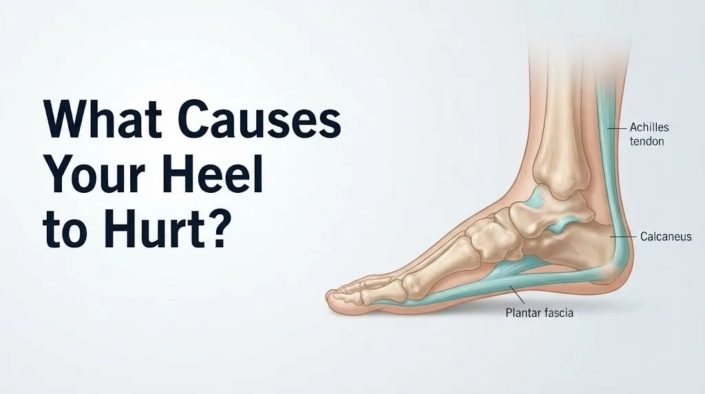
The heel is not a single structure. It is a complex anatomical junction where the plantar fascia, the Achilles tendon, two bursae, the heel fat pad, and multiple nerve branches all converge within a compact space. Any one of these structures can become the source of pain, and each produces a distinct pain pattern depending on its location and the mechanism of injury.
Heel pain broadly divides into three anatomical zones. Pain on the bottom of the heel (plantar surface) most commonly originates from the plantar fascia or the subcalcaneal fat pad. Pain at the back of the heel (posterior) most commonly originates from the Achilles tendon insertion or the retrocalcaneal bursa. Pain on the inside of the heel (medial) most commonly originates from the tibial nerve or its branches within the tarsal tunnel, or from the medial plantar fascia attachment.
The most frequent causes, in order of clinical prevalence, are plantar fasciitis, Achilles tendinitis, heel bursitis, and calcaneal stress fracture. Less frequent but clinically important causes include tarsal tunnel syndrome, Haglund deformity, heel fat pad atrophy, and referred pain from lumbar nerve root compression at the L5-S1 intervertebral level.
Common causes of heel pain ranked by frequency:
| Cause | Location of Pain | Frequency |
|---|---|---|
| Plantar fasciitis | Bottom of heel | Most common (approx. 80% of plantar heel pain cases per AAOS) |
| Achilles tendinitis/tendinopathy | Back of heel | Common in runners and active adults |
| Heel bursitis (retrocalcaneal) | Back/inside of heel | Moderately common |
| Calcaneal stress fracture | Diffuse or pinpoint heel | Less common; higher risk in women with osteoporosis |
| Tarsal tunnel syndrome | Inside of heel, arch | Less common but frequently misdiagnosed |
| Haglund deformity | Back of heel, prominence | Moderately common, often with footwear friction |
| Fat pad atrophy | Bottom of heel | Common in adults over 60 |
| Peripheral neuropathy | Diffuse, burning | Common in diabetics, often underrecognized |
The Anatomy of Heel Pain: Why the Heel Is Uniquely Vulnerable
The heel is uniquely vulnerable to pain because it is the first point of ground contact during normal walking, absorbing a force equal to approximately 1.25 times body weight with each step, according to biomechanical research published in Foot and Ankle International.
The calcaneus is the largest bone in the foot. Its plantar surface bears the insertion of the plantar fascia at the medial calcaneal tubercle, a bony prominence on the undersurface of the heel that you can roughly locate by pressing on the inside edge of the very bottom of your heel. Its posterior surface bears the insertion of the Achilles tendon, the thickest and strongest tendon in the human body, transmitting the force of the gastrocnemius and soleus muscles of the calf.
Think of the calcaneus as a keystone under constant tension from two opposing directions. The Achilles tendon pulls upward and backward from above. The plantar fascia pulls forward from below. Every step you take cycles this tension repeatedly. Over thousands of steps per day, even small imbalances in loading, foot mechanics, or tissue flexibility can accumulate into pathology at the weakest point of attachment.
Between the Achilles tendon and the calcaneal tuberosity sits the retrocalcaneal bursa, a fluid-filled sac designed to reduce friction during ankle movement. On the plantar surface, just beneath the skin and the plantar fascia origin, sits the subcalcaneal fat pad, a specialized shock-absorbing structure with fibrous septa that compartmentalize fat globules to distribute ground reaction force. Both structures can become sources of pain independently of the plantar fascia or Achilles tendon.
Running alongside these mechanical structures, the tibial nerve passes through the tarsal tunnel on the inside of the ankle, dividing into the medial plantar nerve, lateral plantar nerve, and medial calcaneal nerve branch as it enters the foot. Compression of this nerve at the tarsal tunnel produces a distinct burning, tingling pain pattern in the heel and arch that is frequently mistaken for plantar fasciitis.
Plantar Fasciitis: The Most Common Cause of Heel Pain
Plantar fasciitis is the most common cause of heel pain, occurring when repetitive tensile overload at the medial calcaneal tubercle produces collagen fiber microtears in the proximal plantar fascia, triggering an inflammatory response that generates pain, stiffness, and tenderness on the bottom of the heel.
The plantar fascia functions as a passive tension cable, maintaining the longitudinal arch of the foot during weight-bearing. During each step, as the toes extend (dorsiflex) during push-off, the Windlass mechanism tightens the plantar fascia like a bowstring, transmitting force to the calcaneal attachment. In people with tight calf muscles, flat feet (pes planus), high arches (pes cavus), high body weight, or occupations requiring prolonged standing, the cumulative tensile load on the calcaneal attachment exceeds the tissue’s capacity to repair itself between loading cycles.
The resulting pathology in early-stage plantar fasciitis is genuinely inflammatory: prostaglandin E2 (PGE2), interleukin-1 beta, and tumor necrosis factor-alpha (TNF-alpha) are elevated at the enthesis, producing local swelling, warmth, and sensitization of nearby nociceptors through substance P and calcitonin gene-related peptide (CGRP) release. However, in cases persisting beyond three to six months, the histological picture shifts. Research published in Foot and Ankle International has found that chronic plantar heel pain specimens show angiofibroblastic hyperplasia (disorganized collagen replacement) rather than inflammatory cell infiltration, a condition more accurately termed plantar fasciosis. This distinction matters: anti-inflammatory treatments that work in the acute phase become less effective in the degenerative chronic phase.
The American College of Foot and Ankle Surgeons (ACFAS) notes that plantar fasciitis affects both sexes but is diagnosed more frequently in women, and that people with a body mass index (BMI) above 30 face substantially elevated risk due to increased mechanical load per unit area of the plantar fascia.
Risk factors for plantar fasciitis:
- BMI above 30 (increased tensile load per step)
- Tight gastrocnemius and soleus muscles (reduced ankle dorsiflexion, increasing plantar fascia strain during push-off)
- Pes planus (flat feet, causing medial column overpronation)
- Pes cavus (high arch, reducing shock absorption)
- Prolonged standing on hard surfaces (occupational)
- Sudden increase in running mileage or activity level
- Age 40 to 60 (peak incidence period per AAOS)
- Inappropriate footwear (inadequate arch support or heel cushioning)
Achilles Tendinitis and Posterior Heel Pain Causes
Achilles tendinitis causes posterior heel pain when repetitive mechanical loading produces microtrauma and subsequent inflammation or degeneration in the Achilles tendon, either at its insertion on the calcaneal tuberosity (insertional tendinopathy) or approximately 2 to 6 centimeters above the insertion in the tendon’s midsubstance (non-insertional tendinopathy).
The Achilles tendon transmits the contractile force of the triceps surae muscle group (the gastrocnemius and soleus muscles) to the heel during push-off. These muscles generate the highest muscular forces in the lower extremity during walking and running. The tendon itself tolerates loads up to 6 to 8 times body weight during running, according to biomechanical studies cited in the British Journal of Sports Medicine. When cumulative loading exceeds the tendon’s capacity for repair, collagen fiber disruption occurs, followed by a failed healing response characterized by disorganized neovascularization and fibroblast proliferation.
Insertional Achilles tendinopathy (at the calcaneal attachment) and non-insertional tendinopathy (midsubstance) present differently. Insertional pain is located directly at the back of the heel, is worsened by footwear with a rigid heel counter, and may be accompanied by bony calcification within the tendon at the attachment site (visible on lateral heel radiograph). Non-insertional pain occurs slightly higher up the back of the leg, approximately at the height of the top of most shoe heel counters.
People with tight gastrocnemius muscles who suddenly increase their running distance or begin running on hills face elevated risk. Older male runners aged 40 to 60 represent the highest-risk demographic for non-insertional Achilles tendinopathy per data from the British Journal of Sports Medicine, while insertional tendinopathy is more equally distributed between sexes and more common in recreational than competitive athletes.
Quick Tip:
- Posterior heel pain that is worst with the first steps after waking suggests insertional Achilles tendinopathy. Plantar heel pain worst in the morning suggests plantar fasciitis. These are different structures and require different stretching approaches.
- If posterior heel pain is accompanied by a palpable gap in the Achilles tendon or an inability to rise onto the tiptoe of that foot, seek evaluation from a primary care physician or orthopedic surgeon the same day. A complete Achilles tendon rupture requires prompt clinical assessment.
- Fluoroquinolone antibiotics (such as ciprofloxacin) carry an FDA black box warning for increased Achilles tendon rupture risk. People currently taking or recently completing a fluoroquinolone course should tell their physician before starting any exercise program that loads the Achilles tendon.
Key Takeaway: Posterior heel pain located at the very back of the heel that worsens with shoe friction is more likely Achilles tendinopathy or bursitis than plantar fasciitis, and they require different management approaches.
Heel Bursitis: Causes of Pain at the Back and Inside of the Heel
Heel bursitis causes pain at the back or inside of the heel when the retrocalcaneal bursa (located between the Achilles tendon and the calcaneal tuberosity) or the subcalcaneal bursa (located between the plantar fascia origin and the skin) becomes inflamed, producing fluid accumulation, pressure, and pain.
Bursae are thin-walled, fluid-filled sacs whose function is to reduce friction between adjacent moving structures. The retrocalcaneal bursa reduces friction between the Achilles tendon and the posterior calcaneal surface during ankle flexion and extension. When the ankle is repeatedly loaded in positions that compress this bursa (particularly plantarflexion combined with direct pressure from a rigid shoe heel counter), the bursal lining (synovium) responds with synovial fluid overproduction and inflammatory cell infiltration, producing pain and swelling directly behind the heel.
Retrocalcaneal bursitis is frequently associated with Haglund deformity (discussed separately), in which an enlargement of the posterior superior calcaneal surface mechanically compresses the bursa. It can also occur in isolation from footwear friction, particularly in women who wear rigid-backed dress shoes or certain athletic shoes with high, stiff heel counters.
Subcalcaneal bursitis produces pain on the bottom of the heel that closely mimics plantar fasciitis in location but differs in character. Subcalcaneal bursitis pain is typically more diffuse and less sharply localized to the medial calcaneal tubercle, and it may produce visible localized swelling at the bottom of the heel in more severe cases.
An important safety note: when heel bursitis is accompanied by fever above 38.5°C (101.3°F), rapidly spreading redness, warmth, and swelling of the surrounding soft tissue, septic bursitis (bacterial infection of the bursa) must be excluded immediately. Septic bursitis requires urgent evaluation at an emergency room or urgent care facility that day, as it can progress to cellulitis and systemic infection without prompt antibiotic treatment. People with type 2 diabetes or who are immunocompromised face particularly high risk of rapid progression.
Calcaneal Stress Fracture: Severe Heel Pain Without an Obvious Injury
A calcaneal stress fracture causes severe heel pain when repetitive mechanical loading produces microscopic fatigue fractures in the trabecular bone of the calcaneus, leading to progressive bone failure that can cause intense pain with weight-bearing even in the absence of a single traumatic event.
Unlike the acute traumatic fracture that occurs from falling from a height, a stress fracture develops gradually from accumulated loading that outpaces the bone’s remodeling capacity. The calcaneus is the most commonly stress-fractured tarsal bone in military recruits and distance runners, according to research published in the Journal of Bone and Joint Surgery. Pain is typically diffuse across the heel, worsens progressively with activity over days to weeks, and may persist even at rest in more advanced cases.
The squeeze test is a useful clinical screening tool: squeezing the sides of the calcaneus simultaneously (medial and lateral walls) with both hands produces sharp, localized pain in the presence of a stress fracture, at a sensitivity reported between 80 and 95 percent in studies reviewed by the AAOS. Standard weight-bearing radiographs may appear normal in early stress fractures because the fracture line is not yet visible on X-ray. MRI or bone scan is required for definitive early diagnosis.
Postmenopausal women with reduced bone mineral density (osteoporosis or osteopenia) face the highest risk of calcaneal stress fracture from relatively low-impact activities, including normal walking on hard surfaces. The National Institute of Arthritis and Musculoskeletal and Skin Diseases reports that women over 65 with osteoporosis can sustain stress fractures from loading forces that would be entirely tolerated by younger adults with normal bone density. A postmenopausal woman who develops progressively worsening heel pain without trauma and without the characteristic morning pattern of plantar fasciitis should have a bone density assessment discussed with her primary care physician alongside heel imaging.
Tarsal Tunnel Syndrome and Inside Heel Pain Causes
Tarsal tunnel syndrome causes medial heel pain and burning when the tibial nerve is compressed within the tarsal tunnel, a fibro-osseous channel located on the inside of the ankle, bounded by the medial malleolus (the bony prominence on the inner ankle) above and the flexor retinaculum (a tough fibrous band) below.
The tibial nerve carries sensory and motor fibers to the sole of the foot. When compressed within the tarsal tunnel (from space-occupying lesions, swelling, varicose veins within the tunnel, or mechanical narrowing from flat feet), the nerve becomes ischemic: blood flow to the nerve fibers is restricted, disrupting normal electrical conduction. This produces pain, burning, tingling, and numbness along the distribution of the medial calcaneal nerve branch (inside heel) and the plantar surface of the foot.
Think of tarsal tunnel syndrome as the foot’s equivalent of carpal tunnel syndrome in the wrist. Just as the median nerve is compressed at the carpal tunnel producing hand numbness and tingling, the tibial nerve is compressed at the tarsal tunnel producing heel and arch symptoms. The comparison is more than metaphorical: both conditions result from nerve ischemia within a fixed anatomical tunnel, and both share the clinical feature of symptoms that worsen with prolonged standing or activity and may disturb sleep.
Tarsal tunnel syndrome is frequently misdiagnosed as plantar fasciitis, because both conditions produce medial heel pain. The distinguishing features are the quality of pain (burning or electric in tarsal tunnel syndrome versus sharp or aching in plantar fasciitis) and the presence of tingling or numbness extending into the arch or toes (present in tarsal tunnel syndrome, absent in plantar fasciitis). Confirmation requires a nerve conduction velocity (NCV) study and electromyography (EMG), ordered by a primary care physician or neurologist.
People with pes planus (flat feet) face elevated tarsal tunnel syndrome risk because overpronation during walking places a repetitive traction stress on the tibial nerve as it passes through the tunnel’s inferior aspect. Women during pregnancy are also at elevated risk due to foot edema and arch collapse from relaxin-mediated ligament laxity.
Key Takeaway: Inside heel pain with burning or tingling that extends into the arch is a specific red flag for tarsal tunnel syndrome rather than plantar fasciitis, and it requires nerve conduction testing for accurate diagnosis.
Haglund Deformity as a Cause of Posterior Heel Pain
A Haglund deformity causes posterior heel pain when an abnormal bony enlargement of the posterior superior corner of the calcaneus mechanically compresses the retrocalcaneal bursa and irritates the adjacent Achilles tendon insertion, producing a painful prominence at the back of the heel sometimes called a “pump bump.”
The pathological sequence begins with the bony prominence. The posterosuperior corner of the calcaneus is enlarged relative to normal, either from hereditary bone morphology or from remodeling stimulated by chronic friction. When the heel is loaded in a shoe with a rigid posterior heel counter (classically in rigid dress shoes or certain pumps with a firm cup at the back), the heel counter impinges directly on this prominence, compressing the retrocalcaneal bursa between the bone and the counter. The bursal compression triggers inflammation, and if the bony prominence also contacts the Achilles tendon, the tendon itself develops insertional tendinopathy at its lower fibers.
Haglund deformity is diagnosed on lateral weight-bearing X-ray, where the posterior calcaneal angle can be measured (Fowler-Philip angle greater than 75 degrees suggests Haglund morphology, though clinical correlation is required). MRI is more informative for assessing the degree of associated bursitis and Achilles tendon involvement.
Women who regularly wear rigid-backed shoes face disproportionately higher risk, which is why the condition earned the colloquial name “pump bump.” However, the bony morphology is also present in people who have never worn fashion footwear: runners in rigid training shoes and men in dress shoes can develop identical pathology. The initial management focus is footwear modification (open-backed shoes, heel pads that offload the prominence, heel cups that cushion without compressing). Persistent cases that do not respond to conservative management over three to six months warrant evaluation by a podiatrist or board-certified orthopedic surgeon with foot and ankle subspecialty training for discussion of corticosteroid injection into the bursa or, in refractory cases, surgical calcaneal osteotomy.
Heel Fat Pad Atrophy and Heel Pain in Older Adults
Heel fat pad atrophy causes diffuse, burning pain on the bottom of the heel when the specialized adipose tissue of the subcalcaneal fat pad loses its structural integrity with age, reducing the heel’s ability to absorb and distribute ground reaction force and exposing the calcaneal periosteum to direct mechanical stress.
The subcalcaneal fat pad is not ordinary adipose tissue. Its fat lobules are compartmentalized within a lattice of fibrous septa attached to the calcaneal periosteum and the plantar skin. This architecture allows it to function like a multi-cell air cushion: each septum-bounded chamber deforms under load and then recoils. As the body ages, particularly after 60, the collagen within the fibrous septa weakens, the fat cells lose volume, and the septa begin to flatten under repeated loading. The result is a significantly thinner, less shock-absorptive heel pad.
According to research reviewed in Foot and Ankle International, heel fat pad thickness decreases measurably after age 40 and can thin by 20 to 30 percent by age 70. Pain from fat pad atrophy is diffuse rather than localized to the medial calcaneal tubercle (the specific trigger point of plantar fasciitis), affects both heels more symmetrically than plantar fasciitis typically does, and is characteristically worsened by hard surfaces and barefoot walking rather than being worst immediately after rest.
People who have received multiple corticosteroid injections into the heel for plantar fasciitis face accelerated fat pad atrophy, because corticosteroids cause adipocyte apoptosis (fat cell death) and collagen degradation in soft tissue. The American College of Foot and Ankle Surgeons recommends limiting corticosteroid injections into the plantar heel to no more than two to three lifetime injections for this specific reason. Older adults with bilateral heel pain that is worst on hard floors and in thin-soled shoes, particularly those who have had previous heel injections, should discuss fat pad atrophy specifically with a podiatrist rather than assuming plantar fasciitis recurrence.
What Causes Heel Pain Without Injury
Heel pain without injury occurs when cumulative mechanical overload, systemic inflammatory disease, nerve compression, or metabolic bone pathology gradually damages heel structures without any single traumatic event to explain the onset.
Most people who develop heel pain cannot identify a specific moment of injury because the majority of heel pain causes are degenerative or repetitive in nature rather than traumatic. Plantar fasciitis, Achilles tendinopathy, fat pad atrophy, and tarsal tunnel syndrome all develop progressively. The absence of injury does not mean the pain is less real or less anatomically grounded.
One category of non-traumatic heel pain that receives insufficient attention in general health content is enthesitis from systemic inflammatory disease. Enthesitis is inflammation specifically at the point where a tendon or ligament attaches to bone (the enthesis). In seronegative spondyloarthropathies including ankylosing spondylitis, psoriatic arthritis, and reactive arthritis, the calcaneal entheses (both the plantar fascia origin and the Achilles tendon insertion) are among the most commonly affected sites. A 2021 review published in Annals of Internal Medicine noted that enthesitis-associated heel pain in spondyloarthropathy is distinguishable from plantar fasciitis by bilateral distribution, association with morning stiffness lasting more than 30 minutes that improves with movement (rather than worsening), young age of onset (typically under 40), and concurrent symptoms including inflammatory back pain, uveitis, or skin lesions.
Causes of heel pain without injury, by mechanism:
- Repetitive microtrauma accumulation (plantar fasciitis, Achilles tendinopathy, fat pad atrophy)
- Systemic inflammatory enthesitis (ankylosing spondylitis, psoriatic arthritis, reactive arthritis)
- Metabolic bone disease (osteoporosis causing stress fracture from normal walking)
- Nerve compression (tarsal tunnel syndrome from progressive flat foot deformity)
- Peripheral neuropathy (diabetic, autoimmune, or idiopathic, producing diffuse burning heel pain)
- Gout (monosodium urate crystal deposition in periarticular tissue around the calcaneus)
- Referred pain from lumbar spine pathology at L5-S1 (a nerve root cause that produces no foot injury at all)
Key Takeaway: Bilateral heel pain with morning stiffness lasting over 30 minutes that improves with movement in a person under 45 warrants a rheumatology evaluation to rule out seronegative spondyloarthropathy before attributing the pain to mechanical plantar fasciitis.
What Causes Heel Pain in One Foot Only
Heel pain in one foot only most commonly indicates a localized mechanical cause, such as plantar fasciitis or Achilles tendinopathy affecting one side preferentially due to asymmetric foot mechanics, footwear, or activity patterns, rather than a systemic disease, which typically causes bilateral symptoms.
Several specific reasons explain unilateral heel pain. Leg length discrepancy, even when less than one centimeter, can cause the shorter leg’s foot to pronate more aggressively during walking to compensate, placing asymmetrically increased tensile load on the plantar fascia of that foot. A previous ankle sprain on one side alters biomechanics and can shift loading patterns in a way that overloads the plantar fascia or Achilles tendon months or years after the original injury heals. Asymmetric gastrocnemius tightness (often from habitual posture, previous lower leg injury, or a prior period of immobilization) creates unequal Achilles tendon load between sides.
Unilateral heel pain that starts suddenly, is accompanied by swelling and redness of the heel and surrounding skin, and occurs in someone with a history of type 2 diabetes or peripheral vascular disease warrants same-day evaluation. Infection (septic bursitis, osteomyelitis of the calcaneus) can produce sudden unilateral heel pain with soft tissue changes, and delayed diagnosis in diabetic patients carries a risk of serious tissue complications.
A less commonly recognized cause of unilateral heel pain is referred pain from a lumbar disc herniation at L5-S1, where the S1 nerve root is compressed. The S1 nerve root carries sensory fibers to the lateral and plantar heel, the lateral foot, and the small toe. Compression of this root can produce burning, aching, or electric heel pain with no local foot pathology whatsoever. The distinguishing feature is the presence of associated low back pain, buttock pain, or pain that radiates down the back of the leg in a sciatic pattern. If unilateral heel pain coexists with any of these features, a primary care physician should evaluate the lumbar spine as part of the diagnostic workup.
Heel Pain Causes by Population: Pregnancy, Diabetes, Children, Athletes
Heel pain causes differ by population in ways that standard general guidance consistently fails to address, because the underlying anatomy, physiology, and risk mechanisms are genuinely distinct in each group.
Pregnancy: Pregnant women experience a distinct pattern of heel pain driven by two simultaneous physiological changes. First, relaxin, a peptide hormone produced by the corpus luteum and later the placenta, causes systemic ligament laxity beginning in the first trimester. In the foot, this relaxin-mediated laxity reduces the structural support of the plantar ligaments, allowing the medial longitudinal arch to flatten and increasing tensile load on the plantar fascia at the calcaneal attachment. Second, progressive weight gain across the pregnancy increases absolute ground reaction force on the heel with every step. Pregnant women who develop heel pain in the second or third trimester should discuss supportive footwear with arch support and cushioned insoles with their obstetrician-gynecologist, and can be referred to a podiatrist for custom orthotics if symptoms are disabling.
Type 2 diabetes: People with type 2 diabetes and diabetic peripheral neuropathy can experience heel pain that does not follow the classic morning-pain pattern of plantar fasciitis, because sensory nerve damage reduces the intensity and sharpness of pain perception. They may instead report vague burning, pressure, or numbness rather than sharp heel tenderness. The risk is that painlessness can mask serious pathology: calcaneal osteomyelitis (bone infection) can develop from a heel ulcer or skin breakdown without the severe pain that would alert a non-diabetic person to seek care. Any heel discoloration, skin breakdown, warmth, or swelling in a person with type 2 diabetes requires evaluation by a primary care physician or endocrinologist the same day.
Children (Sever’s disease): Children aged 8 to 15 who participate in running sports frequently develop Sever’s disease (calcaneal apophysitis), an overuse injury of the calcaneal apophysis (the secondary ossification center of the heel bone that fuses to the main calcaneus between ages 14 and 16). The Achilles tendon and plantar fascia attach to this growth plate, and repetitive traction during sporting activity causes inflammation at the apophysis. Pain is located at the back and bottom of the heel, worsens with activity, and is reproduced by squeezing the sides of the calcaneus. Sever’s disease is self-limiting and resolves once the apophysis fuses, but activity modification, heel lifts, and calf stretching support comfort during the symptomatic period.
Distance runners: Runners face elevated risk of plantar fasciitis, Achilles tendinopathy, and calcaneal stress fracture compared to non-runners. A sudden increase in weekly mileage (increasing by more than 10 percent per week is a recognized risk threshold, cited in the British Journal of Sports Medicine) or transition to minimalist footwear without an appropriate adaptation period are the most common precipitating factors in runners who develop heel pain.
What Causes Heel Pain in the Morning and After Rest
Heel pain that is worst in the morning or after a period of rest, and improves within the first 10 to 20 minutes of walking, is the hallmark presentation of plantar fasciitis caused by the behavior of the plantar fascia during periods of non-weight-bearing.
During sleep or prolonged sitting, the foot naturally assumes a plantarflexed position (toes pointing slightly downward). In this position, the plantar fascia is at its shortest resting length. At the site of micro-tears at the medial calcaneal tubercle, early phases of repair and scar tissue formation occur overnight in this shortened position. When you stand up and take your first steps, the foot immediately dorsiflexes (toes come up as the heel strikes the ground), and the plantar fascia is suddenly stretched from its shortened resting state. This abrupt lengthening re-tears the fragile new tissue forming at the attachment site, producing the characteristic sharp “first step” pain.
As walking continues, the plantar fascia warms up, local circulation improves, the pain-sensitizing chemicals (PGE2, substance P) disperse, and pain decreases. This is not recovery: the tissue is still damaged. The morning pain pattern is a daily cycle of overnight partial repair followed by re-tearing on first loading.
The morning pain pattern is so specific to plantar fasciitis that its absence should prompt consideration of alternative diagnoses. Achilles tendinopathy at the insertional site also produces morning stiffness, but it is felt at the back of the heel rather than the bottom. Fat pad atrophy produces diffuse bottom-of-heel pain that does not specifically peak with first steps. Tarsal tunnel syndrome produces burning and tingling that may worsen with prolonged standing or walking but does not specifically peak in the morning after rest. Systemic inflammatory enthesitis (spondyloarthropathy) produces morning stiffness that is worst after extended rest and improves with movement, similar to plantar fasciitis in timing but distinct in the pattern of improvement: spondyloarthropathy stiffness improves with movement and warmth rather than simply with time and stretching.
Quick Tip:
- Before your first step in the morning, perform a seated plantar fascia stretch: sitting on the edge of the bed, cross one foot over the other knee, pull the toes back toward the shin until you feel a stretch along the arch, and hold for 10 seconds, repeating 10 times. Research in the Journal of Bone and Joint Surgery found this specific stretch reduces first-step plantar fascia pain more effectively than Achilles tendon stretching alone in people with plantar fasciitis.
- If morning heel pain has not improved meaningfully after 6 weeks of consistent stretching and footwear modification, a podiatrist or primary care physician should assess the foot specifically for the pattern and location of tenderness.
Red Flags: Heel Pain That Needs Urgent or Emergency Evaluation
Certain heel pain presentations indicate conditions that require urgent or emergency medical evaluation and should not be managed with home care while waiting for a routine appointment.
Most heel pain is benign and responds to conservative management over weeks to months. However, the following presentations are not appropriate for watchful waiting.
Seek same-day urgent care or emergency evaluation if your heel pain is accompanied by:
- Fever above 38.5°C (101.3°F) combined with redness, warmth, and progressive swelling of the heel or surrounding tissue. This combination suggests septic bursitis or cellulitis, which requires immediate antibiotic treatment and, in some cases, surgical drainage. Do not wait for a routine appointment.
- Sudden severe heel pain following a fall from a height, a motor vehicle accident, or a direct blow to the heel, particularly if you cannot bear weight. This pattern suggests a calcaneal fracture requiring emergency room evaluation and imaging.
- Complete inability to rise on the tiptoe of the affected foot following sudden posterior heel pain during activity, particularly if accompanied by a snap or pop. This pattern suggests Achilles tendon rupture, which requires emergency orthopedic evaluation. Delay in diagnosis worsens surgical and functional outcomes.
- Numbness, tingling, or weakness in the foot or lower leg combined with heel pain and low back pain. This pattern may indicate cauda equina syndrome from a large central lumbar disc herniation, a neurological emergency requiring immediate emergency room evaluation. Do not drive yourself.
- Heel pain with skin discoloration, an open wound, or visible tissue breakdown in a person with type 2 diabetes or peripheral vascular disease. These findings suggest possible osteomyelitis or ischemic tissue injury and require same-day evaluation by a primary care physician or emergency physician.
Go to the nearest emergency room immediately if heel pain is accompanied by fever, inability to bear weight after trauma, signs of an open wound with redness spreading up the leg, or any neurological symptoms including leg weakness or loss of bladder or bowel control.
These presentations require emergency medical assessment, not home treatment, urgent care phone advice, or a routine appointment scheduled for the following week.
How Heel Pain Is Diagnosed and When to See a Doctor
Heel pain is diagnosed through a clinical examination that combines the location and character of the pain, specific physical examination findings, and targeted imaging or nerve testing depending on the suspected cause.
A primary care physician or podiatrist begins by identifying the exact location of maximal tenderness. Point tenderness at the medial calcaneal tubercle (inner bottom of the heel) strongly suggests plantar fasciitis. Tenderness at the posterior calcaneal tuberosity (back of the heel at the Achilles insertion) suggests insertional Achilles tendinopathy or retrocalcaneal bursitis. Tenderness along the medial ankle behind the malleolus, particularly with a positive Tinel’s sign (tapping the tibial nerve at the tarsal tunnel produces tingling into the heel), suggests tarsal tunnel syndrome. Diffuse calcaneal tenderness with positive squeeze test suggests stress fracture.
The following diagnostic tests are ordered based on clinical suspicion:
To describe your symptoms accurately to your clinician:
- Note the exact location on the heel where pain is most intense (bottom, back, or inside).
- Describe the character of the pain (sharp, burning, aching, or electric).
- Record the timing pattern (worst in the morning, after sitting, during activity, or at rest).
- Note whether both heels or only one is affected, and whether the pain has been present for less than 6 weeks or longer.
- Report any associated symptoms: numbness, tingling, back pain, morning stiffness in other joints, fever, or skin changes.
- Bring a list of all medications, including any fluoroquinolone antibiotics taken in the past 12 months, as well as any corticosteroid injections received previously.
| Diagnostic Test | What It Shows | When It Is Ordered |
|---|---|---|
| Weight-bearing lateral foot X-ray | Bone morphology, heel spurs, Haglund deformity, calcaneal fracture (late-stage) | First-line for most heel pain evaluations |
| Diagnostic ultrasound of plantar fascia | Plantar fascia thickness, tears, bursitis | When plantar fasciitis or bursitis is suspected clinically |
| MRI of foot and ankle | Detailed soft tissue imaging; confirms stress fracture, tendon tears, nerve compression | When X-ray is normal but symptoms are severe or progressive |
| Bone scintigraphy (bone scan) | Detects stress fracture before X-ray changes appear | When stress fracture is suspected and MRI is unavailable |
| Nerve conduction velocity (NCV) and EMG | Confirms tibial nerve compression at tarsal tunnel | When burning/tingling in heel suggests tarsal tunnel syndrome |
| Blood tests (ESR, CRP, uric acid, HLA-B27) | Detects systemic inflammation, gout, or spondyloarthropathy | When bilateral heel pain, joint involvement, or inflammatory pattern is present |
If heel pain has been present for more than 6 weeks without improvement from stretching, ice, rest, and appropriate footwear, schedule an appointment with a primary care physician or podiatrist. If the pain is severe, rapidly worsening, or accompanied by any of the red flags described in the previous section, seek evaluation urgently rather than waiting.
Key Takeaway: A primary care physician or podiatrist can distinguish plantar fasciitis from a stress fracture, nerve entrapment, or systemic inflammatory cause with a focused clinical examination and targeted imaging, none of which require waiting months for a specialist referral in most cases.
Frequently Asked Questions About Heel Pain Causes
What is the most common cause of heel pain?
Plantar fasciitis is the most common cause of heel pain, accounting for approximately 80 percent of plantar heel pain presentations according to the American Academy of Orthopaedic Surgeons.
It occurs when repetitive tensile overload at the medial calcaneal tubercle produces collagen fiber microtears in the proximal plantar fascia, triggering pain that is characteristically worst with the first steps of the morning.
A primary care physician or podiatrist can confirm this diagnosis through physical examination and ultrasound imaging when the clinical picture is unclear.
Can heel pain go away on its own without treatment?
Mild plantar fasciitis in otherwise healthy adults can improve over several months with activity modification, appropriate footwear, and calf stretching, without formal medical treatment.
However, the American College of Foot and Ankle Surgeons reports that untreated or inadequately managed plantar fasciitis becomes chronic in approximately 10 percent of cases, persisting beyond 12 months and requiring more intensive intervention.
Heel pain that does not show measurable improvement after 6 weeks of consistent home management warrants evaluation by a primary care physician or podiatrist to rule out alternative causes.
What does it mean when only one heel hurts?
Unilateral heel pain most often indicates a localized mechanical cause, such as asymmetric foot pronation, leg length discrepancy, or footwear-related friction affecting one side preferentially.
It can also indicate a referred pain pattern from lumbar nerve root compression at L5-S1, which produces heel and lateral foot pain in the distribution of the S1 dermatome without any local foot injury.
Sudden unilateral heel pain with redness and swelling, particularly in people with type 2 diabetes or vascular disease, warrants same-day medical evaluation to exclude infection.
Why does my heel hurt so much in the morning?
Morning heel pain that is worst with the first steps after waking and improves within 10 to 20 minutes of walking is the hallmark presentation of plantar fasciitis.
During sleep, the plantar fascia rests in a shortened position, and early scar tissue forms at the site of microtears at the calcaneal attachment overnight.
The sharp first-step pain occurs as the fascia is abruptly lengthened on weight-bearing, re-tearing the fragile repair tissue that formed during the night.
What causes heel pain without any injury?
Heel pain without injury occurs from cumulative repetitive microtrauma (plantar fasciitis, Achilles tendinopathy), systemic inflammatory enthesitis (ankylosing spondylitis, psoriatic arthritis), metabolic bone disease (stress fracture from osteoporosis), nerve compression (tarsal tunnel syndrome), or referred pain from lumbar disc pathology.
The absence of a single traumatic event does not indicate that the pain is psychosomatic or non-anatomical: all of these causes have clear structural mechanisms that produce genuine tissue damage.
Bilateral heel pain with morning stiffness lasting over 30 minutes that improves with movement in a person under 45 specifically warrants a rheumatology evaluation.
When should I see a doctor for heel pain?
Schedule an appointment with a primary care physician or podiatrist when heel pain has not improved after 6 weeks of consistent home management including stretching, ice, rest, and appropriate supportive footwear.
Seek same-day urgent or emergency evaluation for heel pain accompanied by fever, inability to bear weight, rapidly spreading redness and swelling, or any neurological symptoms including leg weakness or loss of bowel and bladder control.
People with type 2 diabetes, peripheral vascular disease, or osteoporosis should have a lower threshold for early evaluation because their heel pain presentations can mask serious underlying pathology.
Closing
Heel pain is almost never “just” something to push through. The structure that’s failing, whether it’s the plantar fascia, the Achilles tendon, a bursa, the fat pad, or a nerve, determines everything about how it should be managed and how long recovery realistically takes.
The most actionable step right now is identifying your pain pattern precisely: where on the heel the pain is located, when it is worst, whether it burns or aches or stabs, and whether it affects one foot or both. That pattern points toward a specific anatomical structure, and that structure points toward the right professional and the right diagnostic test.
If your heel pain has been present for more than six weeks, schedule an appointment with a primary care physician or podiatrist and bring notes on your pain location, timing, and any previous treatments. You deserve a specific anatomical answer, not a generic “rest and ice” instruction.