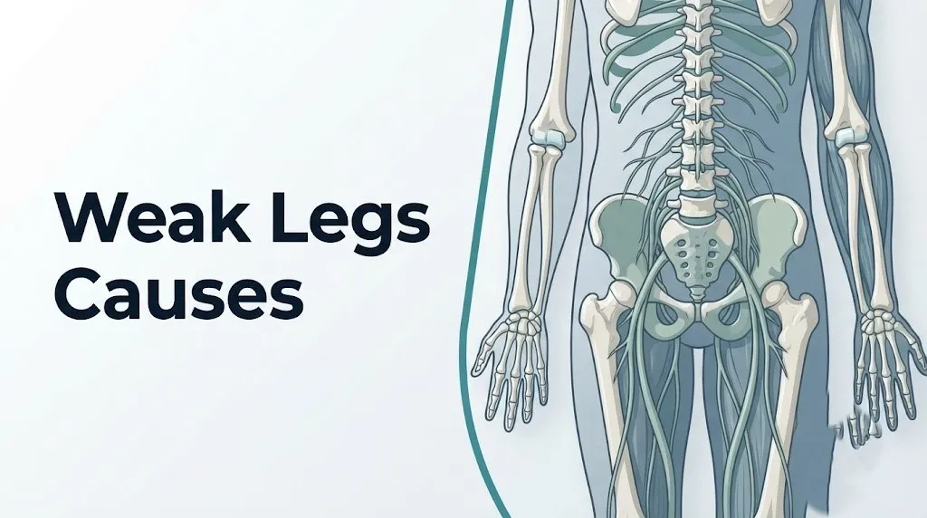
Weak Legs Causes: What Is Making Your Legs Give Out
Weak legs causes range from compressed spinal nerve roots to vascular insufficiency, electrolyte imbalances, and neurological conditions that affect the motor pathway from the brain down through the peripheral nerves to the muscle fibers themselves. Understanding the specific mechanism behind your leg weakness is what separates a situation you can manage with a scheduled appointment from one that requires you to call 911 right now.
Leg weakness is not a single condition. It is a symptom that can originate at any point along a complex chain of anatomy: the motor cortex in the brain, the spinal cord, a nerve root, a peripheral nerve, the neuromuscular junction, or the muscle fiber itself. According to the National Institute of Neurological Disorders and Stroke (NINDS), conditions affecting the nervous system account for the majority of clinically significant leg weakness presentations, but vascular, metabolic, and medication-related causes are frequently underestimated.
This article explains each major cause of leg weakness with its specific physiological mechanism, identifies which patterns are most common versus rare, distinguishes the chronic and progressive presentations from the acute emergencies, and tells you exactly what type of provider you need and under what circumstances.
Weak Legs Causes Overview: Why Your Legs Lose Strength
The causes of weak legs are classified by where in the motor pathway the disruption occurs: the central nervous system, the peripheral nervous system, the neuromuscular junction, the muscles themselves, or the vascular supply delivering oxygen to those structures.
Your legs move because of a precise chain of command. The primary motor cortex in the frontal lobe of the brain generates a motor signal. That signal travels down the corticospinal tract (also called the pyramidal tract), a bundle of approximately one million nerve fibers that descend through the brainstem, cross sides in the pyramidal decussation at the medulla oblongata, and continue down the lateral columns of the spinal cord. At the appropriate spinal level, these upper motor neurons synapse with anterior horn cells (lower motor neurons). Those lower motor neurons exit through spinal nerve roots, form the lumbosacral plexus, and ultimately reach the leg muscles through named peripheral nerves including the sciatic nerve (L4-S1) and the femoral nerve (L2-L4).
Disruption anywhere along this chain produces weakness. The character of the weakness tells you which part of the chain is affected.
| Location of Disruption | Type of Weakness | Key Associated Features |
|---|---|---|
| Brain (motor cortex, internal capsule) | Spastic, unilateral, upper motor neuron | Increased reflexes, Babinski sign, facial weakness |
| Spinal cord | Bilateral below level of lesion | Sensory level, possible bowel/bladder change |
| Nerve root (e.g., L4-L5 disc) | Specific muscle group, dermatomal sensory loss | Pain radiating into leg, worsened by Valsalva |
| Peripheral nerve | Distal, stocking-glove pattern | Numbness, tingling, burning, reduced reflexes |
| Neuromuscular junction | Fatigable weakness, worse with activity | Eyelid drooping, diplopia in myasthenia gravis |
| Muscle (myopathy) | Proximal limb weakness | Difficulty rising from chair, symmetrical |
The frequency of each category matters enormously. Lumbar disc-related nerve root compression is by far the most common cause of new-onset unilateral leg weakness in adults under 60. Peripheral neuropathy is the most common cause of bilateral progressive leg weakness in adults with type 2 diabetes. Stroke is the most time-sensitive cause of sudden unilateral leg weakness in adults over 50.
What Leg Weakness Actually Means Clinically
Clinically, leg weakness refers to a measurable reduction in the force-generating capacity of one or more muscle groups in the lower extremity, distinct from fatigue, pain-limited movement, or the sensation of heaviness without actual strength deficit.
This distinction matters practically. When a person says their legs feel weak after a long walk, they may be describing muscle fatigue from metabolic substrate depletion in the quadriceps or gastrocnemius muscle fibers, not true neurological weakness. True weakness means the muscle cannot generate its expected force even when the person is rested and trying maximally. A clinician tests this with formal muscle strength grading on the Medical Research Council (MRC) scale, rating each muscle group from 0 (no contraction) to 5 (normal strength against full resistance).
Many people describe weakness when they actually mean pain-limited movement. If hip pain stops you from fully extending your leg during the swing phase of walking, that looks like weakness but originates from pain guarding, not from a failure of motor signal delivery. Pain-limited movement and true weakness require different workups and reflect different underlying problems.
There is also the important distinction between objective weakness (which a clinician can confirm on examination) and the subjective experience of heaviness, fatigue, or unreliability in the legs. Both deserve evaluation, but they travel different diagnostic pathways.
Key distinctions to communicate to your clinician:
- Whether the weakness is constant or comes and goes
- Whether it is in one leg or both
- Whether it is worse at a specific time of day or after a specific activity
- Whether it is accompanied by pain, numbness, tingling, or changes in bladder or bowel function
- Whether it came on suddenly over minutes to hours or gradually over weeks to months
These details are not peripheral background information. They are the clinical data points that allow a physician or neurologist to narrow the differential diagnosis from dozens of possible causes to a manageable short list before any test is ordered.
Most Common Causes of Weakness in Legs
The most common causes of weakness in the legs in the general adult population are lumbar disc herniation with nerve root compression, peripheral neuropathy (most often from type 2 diabetes), lumbar spinal stenosis in older adults, and stroke in adults over 60 with vascular risk factors.
Understanding frequency matters when you are trying to evaluate your own situation. Rare conditions like amyotrophic lateral sclerosis (ALS) and spinal tumors generate significant search traffic and significant anxiety, but they account for a small fraction of leg weakness presentations in primary care and neurology clinics.
According to the American Academy of Orthopaedic Surgeons (AAOS), lumbar disc herniation affects approximately 2% of adults per year, with L4-L5 and L5-S1 disc levels accounting for the vast majority of clinically relevant herniations. Among adults with type 2 diabetes, the Centers for Disease Control and Prevention (CDC) reports that approximately 50% will develop peripheral neuropathy over the course of their illness, making diabetic peripheral neuropathy one of the single most common causes of progressive leg weakness and sensory loss globally.
| Cause | Common Population | Onset Pattern | Most Common Symptom Pattern |
|---|---|---|---|
| Lumbar disc herniation / sciatica | Adults 30 to 55, physically active | Days to weeks | Unilateral leg weakness + pain, dermatomal distribution |
| Peripheral neuropathy (diabetic) | Adults with type 2 diabetes | Months to years | Bilateral distal weakness, burning, numbness |
| Lumbar spinal stenosis | Adults over 60 | Months to years | Bilateral leg heaviness worse with walking, relieved by sitting |
| Stroke / TIA | Adults over 50 with vascular risk factors | Sudden (minutes) | Sudden unilateral leg weakness, possible facial or arm involvement |
| Multiple sclerosis | Adults 20 to 40, predominantly women | Weeks, relapsing-remitting | Unilateral or bilateral, often with sensory symptoms |
| Electrolyte imbalance | Any age, often after illness or medication change | Hours to days | Diffuse weakness, cramps, fatigue |
Quick Tip:
- If weakness developed over seconds to minutes, treat it as a potential stroke until proven otherwise.
- If weakness developed over days to two weeks with ascending pattern from feet upward, consider Guillain-Barré syndrome and seek same-day evaluation.
- If weakness is bilateral, slowly progressive over months, and occurs in an adult over 60, lumbar stenosis and peripheral neuropathy are the leading possibilities.
Key Takeaway: The most practically useful thing you can know about weak legs causes is this: the speed of onset is your first and most important clinical clue. Sudden weakness in minutes points to a vascular or neurological emergency; weakness developing over months points to a structural or metabolic cause that warrants a scheduled evaluation with a primary care physician.
Neurological Causes of Leg Weakness
Neurological causes account for the largest and most clinically diverse category of leg weakness, spanning problems that originate anywhere from the motor cortex in the brain to the individual peripheral nerve fibers innervating the lower limb muscles.
The neurological category breaks cleanly into central nervous system (CNS) causes and peripheral nervous system (PNS) causes. CNS causes involve damage to or disruption of the motor cortex, internal capsule, brainstem, or spinal cord. They typically produce what clinicians call upper motor neuron signs: spasticity (increased muscle tone), hyperreflexia (exaggerated deep tendon reflexes), and a positive Babinski sign (upward toe extension when the sole is stroked). PNS causes involve damage at the level of the spinal nerve root, the plexus, or the peripheral nerve itself, producing lower motor neuron signs: flaccid weakness (reduced or absent muscle tone), reduced or absent reflexes, and muscle wasting over time.
This distinction is not academic. A clinician who identifies upper motor neuron signs on examination knows to look toward the brain and spinal cord with a brain MRI or spinal cord MRI. Lower motor neuron signs direct the workup toward an electromyography (EMG) and nerve conduction velocity (NCV) study to evaluate the peripheral nerves and nerve roots.
Major neurological causes of leg weakness by anatomical level:
- Brain (stroke, TIA, brain tumor, multiple sclerosis lesion in motor cortex or internal capsule): typically unilateral, often with upper limb or facial involvement
- Spinal cord (myelopathy from cervical or thoracic disc disease, MS lesion, spinal cord infarction, tumor): typically bilateral below the lesion level, often with sensory changes
- Nerve root (lumbar disc herniation, spinal stenosis, cauda equina syndrome): dermatomal pattern, associated with back or buttock pain radiating into the leg
- Peripheral nerve (diabetic neuropathy, Guillain-Barré syndrome, compression neuropathy): distal-predominant, stocking-glove sensory loss pattern, reduced reflexes
- Neuromuscular junction (myasthenia gravis): fatigable weakness that worsens with repeated use and improves after rest
Adults with autoimmune conditions, particularly women between ages 20 and 50, face elevated risk of multiple sclerosis and myasthenia gravis as neurological causes. Men over 60 with chronic cigarette smoking history carry a significantly higher risk of stroke as the precipitating neurological event.
Lumbar Disc Herniation and Sciatica as Causes of Weak Legs
Lumbar disc herniation causes leg weakness when the displaced nucleus pulposus material compresses a spinal nerve root in the lumbar region, interrupting motor signal transmission to the specific muscle groups that nerve root supplies.
Think of the intervertebral disc as a shock absorber with a gel center (the nucleus pulposus) surrounded by a tough fibrous outer ring (the annulus fibrosus). When the annulus develops a tear, the nucleus pulposus can push through and press against an adjacent nerve root. At the L4-L5 level, this compresses the L5 nerve root, impairing the tibialis anterior muscle and producing foot drop: the inability to lift the front of the foot when walking. At the L5-S1 level, compression of the S1 nerve root weakens the gastrocnemius and soleus complex, reducing push-off strength and causing the person to shuffle rather than stride.
This is why sciatica is not simply pain. The sciatic nerve is formed from the L4, L5, S1, S2, and S3 nerve roots. Compression of any of these roots produces a specific pattern of motor weakness and sensory loss that maps to the anatomy of the root compressed.
According to the American Academy of Orthopaedic Surgeons (AAOS), the L4-L5 and L5-S1 disc levels account for over 90% of clinically significant lumbar disc herniations because these are the levels that bear the greatest mechanical loading during bending, lifting, and rotation.
Weakness from lumbar disc herniation is almost always unilateral (affecting one leg). It is typically accompanied by pain that radiates from the lower back or buttock into the leg, worsens with sitting, coughing, sneezing, or straining (Valsalva maneuver), and may be relieved by walking or lying flat. Purely motor symptoms without pain are less common but do occur, particularly with large central disc herniations.
Individuals with occupational heavy lifting, sedentary desk work, obesity (which increases lumbar disc loading), and a history of prior disc disease are at meaningfully elevated risk. Evaluation by a primary care physician with lumbar MRI is the appropriate first step for any leg weakness pattern consistent with radiculopathy that has persisted beyond four to six weeks or that includes a foot drop component, which warrants more urgent evaluation.
Cauda Equina Syndrome and Spinal Cord Compression
Cauda equina syndrome is a surgical emergency caused by compression of the bundle of nerve roots that descend below the spinal cord (the cauda equina) at the L1 level and below, producing a specific and recognizable pattern of bilateral leg weakness, saddle anesthesia, and loss of bladder or bowel control.
Below the level of L1, the spinal cord ends (at what is called the conus medullaris) and continues as a loose bundle of nerve roots resembling a horse’s tail (the Latin term “cauda equina” literally means horse tail). When a large central disc herniation, a spinal tumor, an epidural abscess, or severe spinal stenosis compresses this bundle, the result is not simply pain or single-root weakness. It is a multi-root emergency affecting bladder function, bowel function, sexual function, and bilateral leg strength simultaneously.
The clinical hallmark is saddle anesthesia: numbness or tingling in the perineum, inner thighs, and genitalia, the areas that would contact a saddle when riding a horse. This symptom, combined with bilateral leg weakness and sudden urinary retention or incontinence, constitutes a surgical emergency. The American Academy of Orthopaedic Surgeons and published guidelines in the journal Spine are clear: cauda equina syndrome requires emergency surgical decompression within 24 to 48 hours of symptom onset to prevent permanent bowel, bladder, and motor deficits.
This is not a “schedule an appointment” situation. If saddle anesthesia, bilateral leg weakness, and urinary changes appear together, go to the nearest emergency room immediately.
Spinal cord compression from a different mechanism (myelopathy from cervical or thoracic disc disease, a spinal tumor, or an epidural hematoma) produces a different but equally serious pattern: bilateral leg weakness with spasticity, a sensory level (below which sensation is impaired), and possible bowel or bladder changes. This also requires urgent evaluation, though the mechanism differs from cauda equina syndrome.
Any patient who has received a cancer diagnosis and develops new back pain with leg weakness deserves same-day evaluation to exclude spinal metastasis with cord or cauda equina compression.
Stroke and Sudden Leg Weakness
Stroke causes sudden leg weakness when an interruption of blood flow to the motor cortex or the descending corticospinal tract deprives those structures of the oxygen they require to maintain motor signal transmission, typically within minutes of the vascular event.
The American Heart Association (AHA) estimates that stroke affects approximately 795,000 people in the United States each year, making it one of the leading causes of sudden-onset leg weakness in adults. Stroke-related leg weakness is almost always unilateral, reflecting the anatomy: the corticospinal tract controls motor function contralateral to the lesion (the right motor cortex controls the left side of the body). Weakness that suddenly affects only the right or only the left leg, particularly in an adult with hypertension, atrial fibrillation, high cholesterol, or smoking history, should be treated as a stroke until definitively proven otherwise.
Think of the corticospinal tract as a high-speed fiber optic cable running from the brain’s command center to the leg’s muscles. A stroke is a power outage that cuts the cable mid-run. The signal stops. The muscle loses its command input. Weakness appears immediately, without a gradual build-up.
Stroke-related leg weakness almost never appears in isolation. The AHA’s BE-FAST criteria (Balance, Eyes, Face, Arms, Speech, Time) highlight the accompanying signs that, together with leg weakness, confirm the stroke pattern:
- Sudden loss of balance or coordination
- Sudden vision change in one or both eyes
- Facial drooping on one side
- Arm weakness accompanying the leg weakness
- Speech difficulty (slurred speech, inability to find words)
- Time: sudden onset within minutes
A transient ischemic attack (TIA) produces identical symptoms that resolve within 24 hours (often within minutes), but this does not reduce the urgency. A TIA is a neurological emergency because approximately 10 to 15% of people who experience a TIA have a completed stroke within 90 days, with the highest risk in the first 48 hours, according to research published in the New England Journal of Medicine.
Women experiencing stroke are more likely than men to report non-traditional symptoms including generalized weakness, confusion, and headache alongside or instead of classic unilateral motor deficits, which can delay recognition. Any sudden, unexplained change in neurological function in a woman warrants immediate emergency evaluation.
Key Takeaway: Sudden leg weakness that appeared within minutes, particularly on one side of the body and particularly in an adult with cardiovascular risk factors, is a stroke emergency. Call 911 immediately. Every minute of delay in a stroke allows approximately 1.9 million neurons to die, according to research published in Stroke journal.
Multiple Sclerosis and Progressive Leg Weakness
Multiple sclerosis causes leg weakness when autoimmune-mediated demyelination destroys the myelin sheaths surrounding nerve fibers in the central nervous system, slowing or completely blocking motor signal conduction through the corticospinal tract or its connections within the spinal cord.
Multiple sclerosis (MS) is a chronic inflammatory and demyelinating disease of the CNS. The myelin sheath functions like the plastic insulation around an electrical wire. When the immune system mistakenly attacks and strips this insulation from the nerve fibers (axons), the electrical signal either slows dramatically or fails to propagate at all. The result, depending on which fibers are affected, is weakness, spasticity, fatigue, sensory abnormalities, or coordination problems.
Leg weakness is one of the most common presenting and ongoing complaints in MS. The National Institute of Neurological Disorders and Stroke (NINDS) states that MS affects approximately one million people in the United States, with women being approximately three times more likely to be diagnosed than men. The disease typically presents between ages 20 and 40, though diagnosis can occur at any age.
In relapsing-remitting MS (RRMS), the most common form, leg weakness tends to worsen during relapses and partially or fully recover during remission. The weakness is often asymmetric (worse on one side), associated with spasticity (stiffness and increased tone), exacerbated by heat (Uhthoff’s phenomenon, in which elevated body temperature further impairs conduction through already-demyelinated fibers), and accompanied by sensory symptoms including numbness, tingling, or the Lhermitte sign (an electric shock-like sensation running down the spine and into the limbs with neck flexion).
Diagnosis of MS requires meeting the McDonald criteria, which incorporate clinical history, MRI findings of white matter lesions demonstrating dissemination in space and time, and in some cases cerebrospinal fluid analysis showing oligoclonal bands. Evaluation by a board-certified neurologist with experience in demyelinating disease is essential for anyone with progressive or relapsing leg weakness accompanied by sensory symptoms, particularly in a woman between ages 20 and 45.
Peripheral Artery Disease and Vascular Causes of Weak Legs
Peripheral artery disease (PAD) causes leg weakness when atherosclerotic narrowing of the iliac, femoral, or popliteal arteries reduces blood flow to the lower extremity muscles during exercise, creating an oxygen deficit that impairs muscle fiber contraction and produces the characteristic cramping weakness called intermittent claudication.
PAD is an atherosclerotic disease. Plaques of cholesterol, calcium, and inflammatory cells progressively narrow the inner diameter of the peripheral arteries supplying the legs. At rest, the reduced flow may be adequate. During walking or climbing stairs, the oxygen demand of active leg muscles outpaces the reduced supply. The result is a cramping, aching, or heavy weakness that begins in the calf, thigh, or buttock during walking and resolves predictably after one to five minutes of rest. This cramping-with-walking, relief-with-rest pattern is called intermittent claudication.
The American Heart Association estimates that PAD affects approximately 8 to 12 million people in the United States, with prevalence increasing sharply after age 60 and among people with diabetes, hypertension, hyperlipidemia, and tobacco smoking history. Among people with diabetes, PAD can produce significantly greater tissue ischemia at any given degree of arterial narrowing because diabetic microvascular disease compounds the macrovascular obstruction.
One important and underrecognized fact: women with PAD are substantially more likely than men to present with atypical or absent claudication symptoms, instead reporting leg fatigue, heaviness, or functional decline rather than the classic calf cramping pattern. This means PAD is frequently undiagnosed and untreated in women for longer periods than in men, according to data published in the Journal of the American Heart Association.
Diagnostic evaluation of suspected PAD begins with the ankle-brachial index (ABI), a non-invasive measurement comparing blood pressure at the ankle to blood pressure at the arm. An ABI below 0.90 indicates significant arterial obstruction. A primary care physician can order this test; a vascular surgeon or vascular medicine specialist manages confirmed PAD.
Peripheral Neuropathy and Electrolyte Imbalances Causing Weak Legs
Peripheral neuropathy causes progressive bilateral leg weakness when damage to the axons or myelin sheaths of peripheral motor nerves impairs signal transmission from the lower motor neurons to the leg muscles, most commonly as a complication of type 2 diabetes, chronic alcohol use, or autoimmune disease.
The peripheral nerves are vulnerable to metabolic insult in a way that central nerve fibers are not. In diabetic peripheral neuropathy, persistently elevated blood glucose promotes the formation of advanced glycation end-products (AGEs), which accumulate in Schwann cells and nerve axons. This disrupts axonal transport, impairs myelin synthesis, and eventually produces axonal degeneration. The longest nerve fibers are damaged first, which is why diabetic neuropathy classically begins in the toes and feet and progresses proximally: the “length-dependent” or “stocking-glove” pattern.
The CDC reports that diabetic peripheral neuropathy affects approximately 50% of people with type 2 diabetes over the course of the illness, making it one of the leading causes of progressive bilateral leg weakness and foot weakness globally.
Electrolyte imbalances represent a distinct and often rapidly reversible cause of leg weakness. The ability of motor neuron membranes to generate and propagate an action potential depends on precise ionic gradients across the cell membrane. Potassium (maintained at approximately 3.5 to 5.0 mEq/L in serum) is essential for repolarization. Hypokalemia (serum potassium below 3.5 mEq/L) reduces the potassium gradient, making motor neurons and muscle fibers harder to depolarize. The result is weakness that can range from mild leg heaviness to, in severe cases, complete flaccid paralysis.
Hypokalemia causes include:
- Diuretic medications (thiazides, loop diuretics) that promote renal potassium excretion
- Vomiting or diarrhea causing gastrointestinal potassium loss
- Inadequate dietary potassium intake in the context of poor nutrition or eating disorders
- Hyperaldosteronism (excess aldosterone causing renal potassium wasting)
- Certain laxatives and enemas used chronically
Low magnesium (hypomagnesemia), low calcium (hypocalcemia), and severe hyponatremia can each also produce clinically meaningful leg weakness, and electrolyte panels are among the first tests ordered when weakness has a metabolic character (bilateral, generalized, without focal neurological signs).
Key Takeaway: If leg weakness appeared over hours to days, especially alongside vomiting, diarrhea, or recent diuretic medication changes, an electrolyte imbalance is a reversible cause worth checking immediately with a blood test ordered by a primary care physician before extensive neurological workup begins.
Medication Side Effects and Systemic Causes of Weak Legs
Several widely prescribed medication classes can cause leg weakness directly by damaging skeletal muscle fibers (myopathy), impairing peripheral nerve function (drug-induced neuropathy), or altering neuromuscular junction transmission.
Statins, the cholesterol-lowering medications taken by tens of millions of adults globally, are the most commonly reported drug class associated with muscle-related side effects. Statin-induced myopathy occurs when statins inhibit the mevalonate pathway in skeletal muscle cells, reducing coenzyme Q10 synthesis and impairing mitochondrial energy production. The spectrum ranges from mild myalgia (muscle pain without weakness) to myositis (muscle inflammation with weakness and creatine kinase elevation) to, rarely, rhabdomyolysis (severe muscle destruction with markedly elevated creatine kinase, myoglobinuria, and risk of acute kidney injury). According to a systematic review published in the Journal of the American Medical Association, clinically meaningful muscle symptoms affect approximately 5 to 10% of statin users, with weakness accompanying pain in a smaller proportion.
Fluoroquinolone antibiotics (ciprofloxacin, levofloxacin) are associated with peripheral neuropathy that can include leg weakness and sensory changes. The U.S. Food and Drug Administration (FDA) added a black box warning to fluoroquinolone labels specifically addressing peripheral neuropathy risk because the deficit can be persistent in some patients.
Other medications with documented leg weakness potential include:
- Corticosteroids (prednisone, dexamethasone) taken long-term: cause steroid myopathy affecting proximal muscle groups including hip flexors and quadriceps
- Amiodarone (antiarrhythmic): can cause both peripheral neuropathy and myopathy
- Colchicine (gout medication): causes myopathy particularly in the setting of renal impairment
- Vincristine and other chemotherapy agents: cause peripheral neuropathy as a known toxicity
- Checkpoint inhibitor immunotherapy (pembrolizumab, nivolumab): can trigger inflammatory myopathy or neuromuscular junction dysfunction as immune-related adverse events
Systemic conditions beyond medication effects that produce leg weakness include hypothyroidism (reduced thyroid hormone slows sarcoplasmic ATPase function, impairing muscle contraction velocity and producing proximal limb weakness), inflammatory myopathies such as polymyositis and dermatomyositis (autoimmune conditions attacking skeletal muscle, producing symmetric proximal weakness with markedly elevated creatine kinase), and severe vitamin B12 deficiency (which causes subacute combined degeneration of the spinal cord, producing progressive weakness with spasticity and sensory changes).
Older adults on multiple medications and adults who have recently started or changed a statin, corticosteroid, or antibiotic regimen and notice new leg weakness should inform their prescribing physician promptly. The workup begins with a creatine kinase (CK) blood test and a review of the medication list.
What Causes Your Legs to Suddenly Give Out
Legs that suddenly give out (buckle or collapse without warning) typically result from one of four mechanisms: acute nerve root or spinal cord compression that abruptly disrupts motor signal delivery, a vascular event such as stroke or TIA, severe acute electrolyte disturbance, or a neuromuscular junction failure as seen in severe myasthenic crisis.
Sudden leg giving out is genuinely alarming and deserves to be taken seriously as a clinical sign. The distinction between the mechanisms matters because the urgency and the required response differ substantially.
Acute nerve root compression (from a disc herniation that suddenly worsens or an acute foraminal fragment) can cause a leg to give out due to sudden loss of L4 or L5 motor innervation. The L5 nerve root supplies the tibialis anterior, the muscle that dorsiflexes the foot during the swing phase of walking. Sudden L5 root compression can produce acute foot drop, where the foot slaps against the ground and the person trips or falls without any pain warning.
Transient ischemic attacks affecting the motor cortex or corticospinal tract can cause legs to give out for seconds to several minutes, then recover. This presentation is called a limb-shaking TIA when it involves involuntary shaking with weakness, and it carries high risk of early stroke.
Falls in older adults caused by “legs giving out” are frequently attributed to musculoskeletal causes but often reflect a neurological basis. A study published in JAMA Internal Medicine found that neurological conditions, including unrecognized peripheral neuropathy and occult spinal stenosis, accounted for a substantially higher proportion of falls in adults over 70 than standard falls prevention protocols recognized.
When legs give out with any accompanying neurological symptoms (facial asymmetry, speech change, arm weakness, visual change, confusion, or saddle region numbness), treat the event as an emergency.
If legs give out repeatedly in a pattern related only to position (worsening with standing, relieved by sitting or bending forward), lumbar spinal stenosis is the leading structural explanation, and a lumbar MRI ordered by a primary care physician or orthopedic spine specialist is the appropriate first diagnostic step.
Bilateral vs. Unilateral Leg Weakness and What Each Pattern Means
Unilateral leg weakness (affecting one leg) most commonly points to a problem on one side of the nervous system, typically at the level of the brain, spinal cord, or a single nerve root or peripheral nerve, while bilateral leg weakness (affecting both legs) suggests a lesion at or below the level of the spinal cord, or a systemic cause affecting all peripheral nerves simultaneously.
This anatomical logic is not just theoretical. It is the first clinical question a neurologist asks when evaluating weakness, because the answer immediately narrows the differential diagnosis and determines the direction of the diagnostic workup.
Unilateral leg weakness with upper motor neuron signs (increased reflexes, spasticity, Babinski sign) points to a stroke, brain tumor, or MS lesion in the contralateral motor cortex or descending corticospinal tract. Unilateral leg weakness with lower motor neuron signs (reduced reflexes, muscle wasting, fasciculations) points to a single nerve root or peripheral nerve problem, most commonly lumbar disc herniation.
| Pattern | Most Likely Causes | Urgency Level |
|---|---|---|
| Sudden unilateral + facial or arm involvement | Stroke, TIA | 911 emergency |
| Subacute unilateral + back pain radiating to leg | Lumbar disc herniation, sciatica | Urgent (days to week) |
| Bilateral + saddle anesthesia + bladder change | Cauda equina syndrome | 911 emergency or immediate ER |
| Bilateral progressive over weeks, ascending from feet | Guillain-Barré syndrome | Urgent same-day evaluation |
| Bilateral progressive over months, worse with walking | Lumbar spinal stenosis, PAD | Scheduled evaluation |
| Bilateral distal, stocking-glove pattern | Peripheral neuropathy | Scheduled evaluation |
| Bilateral proximal, symmetrical | Myopathy, inflammatory myositis | Scheduled evaluation with urgency based on rate of progression |
Bilateral leg weakness that develops over days to two weeks and progresses from the feet upward, accompanied by reduced or absent reflexes, should raise immediate concern for Guillain-Barré syndrome (GBS), an acute immune-mediated demyelinating polyneuropathy in which antibodies attack peripheral nerve myelin. GBS can progress to respiratory muscle paralysis within days. Any suspected GBS warrants same-day evaluation by emergency medicine and admission to a facility capable of respiratory monitoring.
Key Takeaway: The single most practically useful pattern to recognize is this: bilateral leg weakness developing over days with ascending progression from feet toward the trunk, accompanied by absent reflexes, requires same-day emergency evaluation for Guillain-Barré syndrome, which can compromise breathing within 72 hours without treatment.
Causes of Weak Legs in Older Adults and Women
Older adults and women each face population-specific risk profiles for leg weakness that reflect distinct combinations of anatomical, hormonal, and epidemiological factors that differ from the general adult presentation pattern.
In older adults (typically defined as adults over 65), the most common causes of clinically meaningful leg weakness are lumbar spinal stenosis, peripheral neuropathy from diabetes or vitamin B12 deficiency, stroke sequelae, and sarcopenia. Sarcopenia refers to the age-related progressive loss of skeletal muscle mass and strength, driven by declining anabolic hormone levels (testosterone in men, estrogen in women, and growth hormone in both), reduced physical activity, and declining motor unit recruitment capacity. The National Institute on Aging notes that adults can lose 3 to 5% of muscle mass per decade after age 30, with losses accelerating markedly after 60. In older adults, sarcopenic weakness is often bilateral, proximal-predominant, and associated with reduced walking speed and increased fall risk.
Older adults are also at disproportionately high risk of polypharmacy-related weakness, particularly statin-induced myopathy and steroid myopathy from long-term corticosteroid use for inflammatory conditions. Vitamin B12 deficiency is common in older adults (affecting up to 20% of adults over 60, according to a systematic review in the New England Journal of Medicine), partly due to reduced gastric acid production impairing intrinsic factor-mediated B12 absorption, and produces progressive spinal cord and peripheral nerve involvement with leg weakness.
For women specifically, several distinct causes warrant attention. Multiple sclerosis is three times more common in women than men and frequently first presents with leg weakness, sensory symptoms, or optic neuritis in women between ages 20 and 40. Fibromyalgia, which is substantially more prevalent in women, causes a symptom of “leg weakness” that reflects central sensitization and pain-related fatigue rather than true motor pathway disruption. Autoimmune inflammatory myopathies (polymyositis, dermatomyositis) disproportionately affect women and can produce profound proximal leg weakness. Pregnancy-related compression of the femoral nerve by the fetal head during labor can cause transient postpartum unilateral leg weakness, and Guillain-Barré syndrome has documented increased incidence in the postpartum period.
Women over 50 with cardiovascular risk factors should not underestimate stroke as a cause of sudden leg weakness. Women are more likely than men to present with atypical stroke symptoms, meaning a stroke can be missed if the clinician is looking only for classic unilateral weakness with facial drooping.
Diagnostic Tests Used to Identify the Cause of Leg Weakness
Identifying the specific cause of leg weakness requires a targeted diagnostic workup guided by the clinical pattern, onset speed, laterality, associated symptoms, and the clinical examination findings, with no single test diagnosing all causes.
The diagnostic process for leg weakness begins with a thorough history and neurological physical examination. An experienced clinician can often narrow the differential diagnosis to a short list before a single test is ordered, based on the pattern of weakness, reflex changes, sensory findings, and the clinical context.
From there, the workup typically proceeds as follows:
- Complete blood count (CBC) and comprehensive metabolic panel (CMP): screen for anemia, electrolyte imbalances (potassium, sodium, calcium, magnesium), liver and kidney function that may affect drug metabolism or contribute to neuropathy
- Thyroid-stimulating hormone (TSH): hypothyroidism is a reversible and frequently missed cause of proximal muscle weakness
- Serum vitamin B12 and folate: B12 deficiency causes subacute combined degeneration with progressive weakness and sensory changes
- Hemoglobin A1C and fasting glucose: identifies diabetes, the leading cause of peripheral neuropathy globally
- Creatine kinase (CK): elevated in myopathies, inflammatory muscle diseases, rhabdomyolysis, and statin-induced muscle damage
- Electromyography (EMG) and nerve conduction velocity (NCV) study: the definitive test for distinguishing nerve root problems from peripheral nerve problems from neuromuscular junction problems from muscle disease; performed by a board-certified neurologist or physiatrist
- Lumbar MRI: structural imaging of the lumbar spine to identify disc herniation, spinal stenosis, cauda equina compression, or spinal tumors
- Brain MRI with diffusion-weighted imaging: identifies acute stroke (within hours of onset), MS lesions, brain tumors, or other CNS causes of upper motor neuron weakness
- Ankle-brachial index (ABI) with Doppler ultrasound: diagnoses peripheral artery disease in patients with claudication-pattern weakness
- Acetylcholine receptor antibodies and repetitive nerve stimulation test: evaluates for myasthenia gravis in patients with fatigable weakness
For suspected MS, cerebrospinal fluid (CSF) analysis via lumbar puncture examining for oligoclonal bands is often part of the workup. For suspected Guillain-Barré syndrome, CSF analysis showing the characteristic albuminocytologic dissociation (elevated protein with normal white cell count) helps confirm the diagnosis.
A primary care physician initiates most of this workup. A board-certified neurologist takes over the evaluation when neurological examination is abnormal, when the diagnosis remains unclear after initial testing, or when the presentation is progressive, severe, or accompanied by emergency features.
Emergency Symptoms of Leg Weakness: When to Call 911
Certain combinations of leg weakness with other symptoms represent true medical emergencies. Do not wait to see whether these symptoms resolve on their own.
Call 911 or go to the nearest emergency room immediately if you or someone near you experiences:
- Sudden leg weakness that appeared within minutes, particularly affecting only one side of the body, with or without accompanying facial drooping, arm weakness, slurred speech, or vision changes (these signs indicate possible stroke or TIA)
- Sudden bilateral leg weakness combined with numbness in the perineum, inner thighs, or genitalia (saddle anesthesia) and any change in bladder or bowel control (these signs indicate possible cauda equina syndrome requiring emergency surgical evaluation)
- Leg weakness that is ascending rapidly from the feet upward over hours to days, accompanied by reduced or absent reflexes and difficulty breathing, swallowing, or speaking (these signs indicate possible Guillain-Barré syndrome with respiratory compromise risk)
- Sudden complete loss of leg strength without warning (legs fully collapse) in anyone with no prior history of such episodes and no clear precipitating mechanical cause
- Leg weakness in someone who has a known cancer diagnosis and new back or neck pain, particularly if the weakness has progressed over days (possible spinal metastasis with cord or cauda equina compression)
- Leg weakness accompanied by chest pain, severe headache described as the worst of one’s life, or loss of consciousness
These presentations require emergency medical assessment in an emergency room with neuroimaging capability, not a scheduled outpatient appointment and not an urgent care visit.
Frequently Asked Questions About Weak Legs Causes
What is the most common cause of sudden weakness in the legs?
The most common cause of sudden leg weakness developing over minutes is stroke or transient ischemic attack (TIA), produced by interrupted blood flow to the motor cortex or corticospinal tract.
Any sudden unilateral leg weakness in an adult with cardiovascular risk factors should be treated as a stroke emergency until confirmed otherwise by a neurologist and brain MRI.
Call 911 immediately if sudden leg weakness appears with any accompanying facial drooping, arm weakness, speech difficulty, or vision changes.
Can dehydration or low electrolytes cause weak legs?
Yes, electrolyte imbalances, particularly hypokalemia (low potassium), hypomagnesemia, and hypocalcemia, can cause generalized leg weakness by reducing the ionic gradients that allow motor neurons and muscle fibers to generate action potentials.
Weakness from electrolyte disturbance typically develops over hours to days, is bilateral and generalized rather than focal, and is often associated with muscle cramps, nausea, or fatigue.
A comprehensive metabolic panel ordered by a primary care physician can identify this cause and it is frequently reversible with appropriate replacement.
When is leg weakness a sign of stroke?
Leg weakness is a sign of stroke when it appears suddenly within minutes, affects only one side of the body (unilateral), and is accompanied by other neurological symptoms including facial asymmetry, arm weakness, speech difficulty, or visual change.
Stroke-related leg weakness reflects damage to the contralateral corticospinal tract or motor cortex caused by arterial occlusion or hemorrhage.
Time to treatment is the most important factor in stroke outcomes; call 911 immediately rather than waiting to see whether symptoms resolve.
Why do my legs feel weak and shaky for no reason?
Legs that feel weak and shaky without a clear cause may reflect transient hypoglycemia (low blood sugar), anxiety with autonomic hyperactivation, early peripheral neuropathy, orthostatic hypotension (blood pressure drop on standing), or functional neurological disorder.
None of these can be confirmed without a clinical examination and basic blood work, including glucose, electrolytes, and a thyroid panel.
A primary care physician is the right starting point for evaluation when weakness and shakiness are episodic, recurrent, and not associated with the emergency features described above.
What causes legs to give out when walking?
Legs that give out specifically during walking, with predictable relief after a brief rest, most commonly reflect lumbar spinal stenosis (neurogenic claudication) or peripheral artery disease (vascular claudication), both of which impair functional delivery of oxygen or motor signals to leg muscles during physical exertion.
The key clinical distinction is that neurogenic claudication is relieved by bending forward or sitting (reducing lumbar canal narrowing), while vascular claudication is relieved by simply stopping and standing still.
Evaluation by a primary care physician with lumbar MRI for neurogenic claudication or ankle-brachial index for vascular claudication is the appropriate diagnostic path.
How do doctors test for the cause of leg weakness?
The diagnostic workup for leg weakness begins with a detailed clinical history and neurological physical examination to determine the pattern, onset, laterality, and associated features.
Blood tests including electrolyte panel, glucose, HbA1C, TSH, vitamin B12, and creatine kinase identify metabolic and systemic causes, while electromyography (EMG) and nerve conduction velocity (NCV) testing evaluate the peripheral nerves and nerve roots.
Lumbar MRI is the primary imaging study for structural spinal causes, and brain MRI with diffusion-weighted imaging is used when stroke or CNS disease is suspected.
Closing
Leg weakness is one of those symptoms that sits at the intersection of many different body systems, which is why working out what is causing it requires more than a list of possibilities. The onset speed, the pattern of which leg is affected, the associated sensory or bladder changes, and the broader clinical context are what allow a clinician to separate a reversible metabolic problem from a structural emergency.
The most important practical step you can take right now is to document exactly when the weakness started, whether it is one leg or both, whether it is constant or episodic, and whether it is accompanied by pain, numbness, bladder changes, or any of the emergency features described in this article. Bring that documentation to your primary care physician as your starting point. If any of the emergency patterns apply, skip the appointment and go straight to the emergency room.
Your legs carry you through every day. When they stop working the way they should, that signal deserves a specific answer, not a vague reassurance.