Neck Rash Causes: Complete 2026 Clinical Guide for Skin
Neck rash causes span a wide clinical range, from contact dermatitis triggered by a metal necklace to viral infections reactivating from a spinal nerve root, and identifying the correct cause requires understanding both the mechanism and the specific anatomical features that make the neck uniquely vulnerable. The cause determines whether the rash is self-resolving, requires a dermatology appointment, or needs same-day emergency evaluation.
The neck is one of the body’s most physiologically complex skin zones. It combines sun-exposed lateral and posterior surfaces, moisture-trapping anterior skin folds, high-density eccrine sweat gland and sebaceous gland distribution in the posterior cervical skin, and constant friction from clothing, hair, and jewelry. According to the American Academy of Dermatology, contact dermatitis is among the most frequent skin conditions evaluated in outpatient dermatology practice, and the neck is a primary site given its daily exposure to metal, fabric, and cosmetic allergens.
This article covers every clinically recognized cause of neck rash with the specific physiological mechanism behind each one. It explains who is at highest risk by age, sex, and health status, distinguishes self-limiting from medically managed causes, and provides the specific clinical thresholds that separate watchful waiting from seeking urgent care.
Neck Rash Causes: An Overview of the Most Common Types
The most common neck rash causes are allergic contact dermatitis (from jewelry, fragrances, or fabric), heat rash (from sweat duct obstruction), atopic dermatitis, folliculitis, and seborrheic dermatitis, though infections, systemic conditions, and medication reactions also produce distinct neck rash patterns.
These causes are not equally likely in every person. The most relevant cause depends on where on the neck the rash appears, what it looks like, how quickly it appeared, and what the person was recently exposed to. A rash on the anterior neck in a skin fold has a different most likely cause than a rash on the lateral neck following a beach day or a vesicular rash tracking along one side from ear to collarbone.
| Cause | Rash Type | Most Common Location on Neck | Typically Self-Resolving? |
|---|---|---|---|
| Allergic contact dermatitis | Red, itchy, weeping patches | Where jewelry or fabric contacts skin | Yes, with trigger removal |
| Irritant contact dermatitis | Dry, red, burning patches | Where irritant contacts skin chronically | Yes, with barrier protection |
| Heat rash (miliaria rubra) | Tiny red papules, prickling sensation | Posterior neck, skin folds | Yes, with cooling |
| Atopic dermatitis | Dry, thickened, intensely itchy patches | Neck flexural folds | No, requires management |
| Folliculitis | Red papules, pustules at hair follicles | Posterior neck hairline, shaved areas | Often yes, sometimes needs treatment |
| Seborrheic dermatitis | Greasy, yellowish scales with redness | Behind ears, scalp-neck border | Controlled, not cured |
| Intertrigo | Moist, red, macerated skin | Anterior neck folds | Yes, with moisture control |
| Tinea versicolor | Pale or pink scaly patches | Posterior and lateral neck | Yes, with antifungal treatment |
| Shingles | Dermatomal vesicles, one side only | Lateral neck tracking C2-C4 dermatome | Resolves; antiviral treatment recommended |
| Lyme disease | Expanding target-shaped ring | Can appear anywhere, including neck | Resolves with antibiotic treatment |
| Cellulitis | Spreading hot red swelling | Anterior or lateral neck | No, requires antibiotics |
Immunocompromised individuals have a broader differential that includes opportunistic infections (Candida, Kaposi’s sarcoma, and rare bacterial organisms) that would be uncommon causes in immunocompetent adults.
Why the Neck Is Particularly Prone to Rashes: Anatomy Explained
The neck is prone to rashes because of a unique combination of anatomical features: dense eccrine and sebaceous gland concentration in the posterior cervical skin, skin folds in the anterior neck that trap moisture and heat, constant movement that creates friction against collars and jewelry, and sun exposure patterns that spare the anterior neck while exposing the lateral and posterior surfaces.
Think of the neck as three distinct microenvironments stacked on one body part. The posterior neck is hot, follicle-dense, and frequently pressed against clothing. The anterior neck has folds that create a warm, moist, friction-prone microenvironment similar to the groin or underarm. The lateral neck gets regular sun exposure but also the friction of hair and jewelry.
The posterior neck has a high density of hair follicles and sebaceous glands per square centimeter, creating a rich substrate for folliculitis and seborrheic dermatitis. The stratum corneum (the outermost protective layer of the epidermis) in the posterior neck is thinner than on areas like the palm, reducing its barrier function against irritants and pathogens.
The anterior neck skin folds are particularly prone to intertrigo and fungal infections because the folded skin surfaces remain in contact, trapping sweat and heat, elevating local temperature and humidity, and creating friction with every head movement. The warm, moist environment between skin fold surfaces is essentially an incubator for Candida and certain bacteria.
The American Academy of Dermatology notes that the neck is among the most frequently affected sites for nickel contact dermatitis specifically because of necklace wear, with nickel being the most common cause of metal contact allergy in North America. Women are sensitized to nickel at higher rates than men, likely due to greater cumulative jewelry exposure from an earlier age.
Contact Dermatitis as a Cause of Neck Rash
Contact dermatitis causes neck rash through two distinct mechanisms: an immune-mediated allergic reaction (allergic contact dermatitis) triggered by prior sensitization to a specific substance, and a non-immune irritant reaction (irritant contact dermatitis) from direct chemical damage to the skin barrier.
Allergic contact dermatitis on the neck is most often caused by nickel in jewelry clasps and chains, fragrance ingredients in perfumes and lotions applied to the neck, latex in clothing elastics, and formaldehyde-releasing preservatives in cosmetics. The mechanism involves Langerhans cells in the epidermis, which capture the allergen (technically a hapten that binds to skin proteins to form a complete antigen), carry it to the cervical lymph nodes, and present it to naive T-cells. On re-exposure, sensitized memory T-cells release inflammatory cytokines including IL-4, IL-13, and TNF-alpha, producing the red, weeping, intensely itchy rash within 24 to 72 hours of contact.
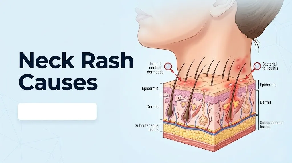
Irritant contact dermatitis does not require prior sensitization. It occurs when a substance directly disrupts the stratum corneum’s integrity, stripping away lipids and keratin proteins that form the skin’s waterproof barrier. Common irritants on the neck include harsh soaps, sweat accumulation in skin folds, synthetic fabric dyes, and repeated rubbing from clothing collars.
According to research published in the Journal of the American Academy of Dermatology, nickel allergy affects approximately 8 to 15 percent of the general population in Western countries, with rates substantially higher in women (up to 20 percent) than in men (approximately 3 percent) due to greater jewelry exposure.
To accurately describe contact dermatitis to your dermatologist:
- Note whether the rash appears in the exact pattern of where jewelry, clothing, or a specific product contacts the skin.
- Record the onset timing: allergic contact dermatitis typically appears 24 to 72 hours after exposure, not immediately on contact.
- Bring the product or jewelry to your appointment, or photograph it specifically.
- List all new products, fabric changes, or jewelry recently introduced in the two weeks before the rash appeared.
- Note whether the rash clears completely when you stop wearing the item or using the product.
Key Takeaway: Allergic contact dermatitis on the neck typically appears 24 to 72 hours after exposure to the trigger (not during exposure), appears exactly where the allergen contacts skin, and clears when the trigger is removed. Nickel from jewelry clasps is the most common cause.
Heat Rash on the Neck: Eccrine Gland Obstruction Explained
Heat rash on the neck, clinically called miliaria rubra, occurs when the ducts of the eccrine sweat glands in the posterior and lateral neck become obstructed by a keratin plug or bacterial biofilm, trapping sweat within the duct and forcing it to extravasate into the surrounding dermis or epidermis, where it triggers a local inflammatory response.
The eccrine sweat glands are distributed throughout the neck skin and produce sweat directly onto the skin surface through long, coiled ducts that ascend through the dermis and epidermis. When the duct becomes blocked (most commonly by skin surface bacteria, particularly Staphylococcus epidermidis, whose polysaccharide biofilm obstructs the duct opening in hot, humid conditions), the pressure from continued sweat production forces sweat sideways through the duct wall into the surrounding tissue.
The result is tiny red papules or vesicles that produce a characteristic prickling or burning sensation with heat, sometimes called “prickly heat.” The inflammatory response comes from the sweat itself acting as an irritant in the dermis, rather than from any pathogen. This is why heat rash resolves completely when the person cools down and the sweat production decreases: the obstruction clears, normal sweating resumes, and the inflammatory response subsides.
There are two forms relevant to the neck. Miliaria rubra (the most common) involves obstruction in the epidermis, producing red papules with the prickling sensation. Miliaria crystallina involves obstruction at the skin surface, producing tiny, fragile, clear vesicles without inflammation, more common in newborns.
According to Mayo Clinic, heat rash is particularly frequent in infants due to underdeveloped sweat duct maturity, in obese adults where skin folds create consistently hot, humid microenvironments, and in people who are bedridden with fever, where excessive sweating occurs in areas pressed against bedding.
Atopic Dermatitis Causing Neck Rash
Atopic dermatitis causes neck rash through a genetic skin barrier defect combined with a Th2-dominated immune response, producing chronically inflamed, dry, intensely itchy skin at the neck flexural surfaces (the creases where the neck meets the jaw and shoulders).
The core defect involves the filaggrin protein, encoded by the FLG gene in the stratum granulosum of the epidermis. Filaggrin normally cross-links keratin filaments in the outermost skin layer, forming the dense, waterproof stratum corneum. Loss-of-function mutations in FLG (present in approximately 10 to 30 percent of Northern European populations, according to research published in JAMA Dermatology) reduce stratum corneum cohesion, increase transepidermal water loss, and allow environmental allergens to penetrate the skin more easily.
The penetrating allergens activate Langerhans cells and innate immune responses, driving a Th2 inflammatory cascade dominated by interleukin-4 (IL-4), interleukin-13 (IL-13), and interleukin-31 (IL-31). IL-31 specifically activates C-fiber itch receptors in the dermis, producing the severe, chronic pruritus that characterizes atopic dermatitis and distinguishes its itch from the more localized itch of contact dermatitis.
The neck is particularly affected in atopic dermatitis because flexural skin folds trap sweat and allergens, and the constant movement of the neck produces frictional irritation of the already compromised skin barrier. The rash appears as dry, thickened, lichenified (rough, leathery) plaques at the neck crease and lateral neck surfaces.
Atopic dermatitis at the neck in adults warrants evaluation by a board-certified dermatologist, particularly if standard moisturization and low-potency topical corticosteroids are not controlling the flare within 2 weeks, as newer targeted therapies (including dupilumab, which targets the IL-4 and IL-13 pathway) may be appropriate for moderate to severe disease.
Folliculitis on the Neck: Infected Hair Follicles as a Cause
Folliculitis causes neck rash when hair follicles in the posterior cervical hairline or shaved areas become infected, typically by Staphylococcus aureus, after mechanical disruption of the follicular epithelium by friction, shaving, or occlusion.
The posterior neck hairline and the shaved nape area are particularly prone to folliculitis because this zone combines dense hair follicle distribution with constant friction from collars and the mechanical trauma of shaving. When shaving disrupts the follicle opening, bacteria normally present on the skin surface gain access to the follicle canal. Staphylococcus aureus, the most common causative organism, adheres to the follicle wall, multiplies rapidly in the warm, moist environment, and triggers a neutrophilic inflammatory response that produces the characteristic small red papules or pustules centered on individual hair follicles.
A specific form called pseudofolliculitis barbae occurs predominantly in individuals with tightly coiled hair, most commonly Black men shaving the neck and jaw area. In this condition, the curled hair shaft re-enters the skin adjacent to the follicle after shaving, acting as a foreign body rather than requiring bacterial infection. The resulting inflammatory response produces papules, pustules, and eventually hyperpigmented scarring in a pattern that can be mistaken for bacterial folliculitis.
Hot tub folliculitis, caused by Pseudomonas aeruginosa rather than Staphylococcus, produces a neck and trunk rash 12 to 48 hours after exposure to poorly chlorinated water. It resolves without treatment in most immunocompetent adults within 7 to 10 days, though it requires appropriate antibiotic treatment in immunocompromised individuals.
Folliculitis in adults on long-term topical corticosteroids to the neck region can paradoxically worsen, as corticosteroids impair local immune responses and allow Staphylococcal and fungal colonization of the follicle to expand. A primary care physician or board-certified dermatologist should evaluate any folliculitis that does not begin improving within 5 to 7 days of basic hygiene measures.
Key Takeaway: Folliculitis on the posterior neck appears as small red papules or pustules centered on individual hair follicles, not between them. Men with coiled hair patterns are at elevated risk for pseudofolliculitis barbae from shaving, which produces a similar appearance through a different mechanism.
Intertrigo in Neck Skin Folds
Intertrigo causes neck rash in the anterior cervical skin folds through a non-infectious mechanism: skin-to-skin friction and moisture accumulation that macerate and break down the stratum corneum, producing raw, red, weeping skin that can then become secondarily infected by Candida or bacteria.
The anterior neck folds, particularly in individuals with obesity or thick neck anatomy, function like all other intertriginous zones (groin, underarm, beneath the breast): warm, poorly ventilated skin surfaces remain in constant contact, rubbing with every head movement, and trapping sweat without evaporation. The moisture softens and weakens the stratum corneum, while friction physically erodes it. The result is a clearly defined red, sometimes moist or weeping rash confined precisely to the skin fold contact area.
The National Institute of Arthritis and Musculoskeletal and Skin Diseases (NIAMS) notes that secondary Candida infection of intertrigo is common when the moist environment persists, producing a brighter red rash with characteristic satellite pustules (small individual lesions appearing just beyond the edge of the main rash area) that signal fungal involvement rather than pure mechanical maceration.
Intertrigo prevalence increases with obesity because adipose tissue compresses cervical skin folds more firmly, reduces air circulation, and elevates local skin temperature. People who are bedridden or in cervical collars face elevated risk because the collar creates a persistent occluded, warm microenvironment against the anterior neck.
Management of simple intertrigo in healthy adults involves keeping the skin fold dry with absorbent barrier powder or soft fabric, which typically resolves the rash within 1 to 2 weeks. Any neck intertrigo in a person with diabetes warrants prompt evaluation by a primary care physician, as hyperglycemia dramatically accelerates Candida growth in macerated skin and impairs healing.
Seborrheic Dermatitis on the Neck
Seborrheic dermatitis causes neck rash through an inflammatory response to Malassezia species, lipophilic yeasts that colonize sebaceous gland-rich skin and metabolize skin surface lipids into fatty acids that irritate the stratum corneum and trigger keratinocyte release of IL-1 and TNF-alpha.
The neck, specifically the skin at the scalp-neck border and behind the ears, is one of the characteristic sites of seborrheic dermatitis because these areas share the high sebaceous gland density of the scalp, which is Malassezia’s preferred habitat. The organism is not an external contaminant. It is a normal skin commensal that overgrows under specific conditions: increased sebum production, immune dysregulation, or neurological disease.
The resulting rash appears as greasy, yellowish or white scales on a red, mildly itchy background, concentrated at the hairline, behind the ears, and at the neck margin. Unlike atopic dermatitis, seborrheic dermatitis scales are oily rather than dry, and the itch is typically milder than the intense pruritus of eczema.
Seborrheic dermatitis has a well-documented association with Parkinson’s disease, HIV infection, and other neurological conditions, according to research published in the British Journal of Dermatology. In Parkinson’s disease, sebaceous gland output may be elevated due to autonomic nervous system dysregulation, and the reduced physical mobility limits regular bathing and skin care. Any adult developing new or dramatically worsening seborrheic dermatitis at the neck without prior history should have an evaluation that includes neurological and immune status assessment by their primary care physician.
Newborns develop a form of seborrheic dermatitis called cradle cap that can extend to the neck fold. This is driven by maternal androgen-stimulated neonatal sebaceous activity and typically resolves without treatment within the first 6 to 12 months of life.
Fungal Infections Causing Neck Rash: Tinea Versicolor and Ringworm
Fungal infections on the neck cause rash through two distinct organisms: Malassezia furfur (causing tinea versicolor, also called pityriasis versicolor) and Trichophyton or Microsporum species (causing tinea corporis, commonly called ringworm), each producing distinctly different rash appearances through different mechanisms.
Tinea versicolor produces discolored, slightly scaly patches on the neck, chest, and back, ranging from pale (hypopigmented) to pink or light brown depending on skin tone and sun exposure. The organism interferes with normal melanocyte function by producing azelaic acid, a byproduct of its metabolism of skin surface fatty acids, which inhibits the enzyme tyrosinase responsible for melanin production. The result is irregular patches of skin that tan differently from surrounding normal skin.
Tinea corporis (ringworm of the body) produces the characteristic ring-shaped rash: a circular, scaly, red border that expands outward with central clearing as the fungal infection moves peripherally from the initially infected point. The dermatophyte organisms (Trichophyton tonsurans, Microsporum canis) produce keratinase enzymes that digest the keratin in the stratum corneum and hair shafts, triggering an inflammatory response at the advancing edge of infection.
According to the Centers for Disease Control and Prevention, tinea corporis is transmitted through direct contact with infected individuals or animals, contaminated surfaces, or soil. Children who wrestle (tinea gladiatorum) and people with indoor cat exposure (Microsporum canis from cats) are at elevated risk.
Quick Tip:
- Tinea versicolor patches look different from surrounding skin in color but do not form rings. Ringworm forms a ring with a scaly border.
- A KOH (potassium hydroxide) preparation of scraped scale examined under microscope differentiates these fungal causes from each other and from non-fungal rashes within a single office visit.
- Immunocompromised individuals can develop extensive tinea infections that cover large body surface areas and require systemic antifungal therapy managed by a primary care physician or dermatologist.
Key Takeaway: Tinea versicolor produces discolored flat patches with mild scaling (no ring), while ringworm (tinea corporis) produces an expanding ring with a scaly red border and central clearing. A KOH preparation performed in a clinician’s office differentiates these from each other and from eczema within minutes.
Shingles as a Cause of Neck Rash
Shingles (herpes zoster) causes neck rash when varicella-zoster virus (VZV), dormant in the dorsal root ganglia of the cervical spinal cord (specifically the C2, C3, or C4 nerve roots), reactivates and travels along the sensory nerve axons to the skin, producing a painful, blistering rash confined to a single dermatomal strip on one side of the neck.
The varicella-zoster virus that causes chickenpox does not leave the body after the primary infection resolves. It retreats to the sensory nerve cell bodies in the dorsal root ganglia, where it remains latent for decades. When cellular immune surveillance weakens (through aging, illness, stress, or immunosuppression), the virus reactivates, replicates within the ganglion, and then travels antegradely down the sensory nerve axons, erupting as a painful, vesicular rash in the skin territory supplied by that specific nerve root.
Cervical shingles (affecting C2-C4 dermatomes) produces a band of pain, burning, or tingling typically beginning several days before the rash appears, followed by clustered blisters on one side of the neck from the ear to the collarbone. The strict unilateral, dermatomal distribution is the clinical hallmark that distinguishes shingles from all other neck rash causes.
According to the Centers for Disease Control and Prevention, approximately 1 in 3 Americans will develop shingles in their lifetime, with risk increasing sharply after age 50 and in immunocompromised individuals. Adults over 50 are eligible for the recombinant zoster vaccine (Shingrix), which reduces shingles risk by over 90 percent in the years following vaccination, according to CDC published data.
Suspected shingles warrants same-day evaluation by a primary care physician, as antiviral medications (such as valacyclovir) are most effective when started within 72 hours of rash onset. Any suspected shingles affecting the ear canal, eye, or producing facial weakness warrants immediate evaluation by an emergency medicine physician or specialist for possible Ramsay Hunt syndrome.
Lyme Disease Rash on the Neck
Lyme disease causes a neck rash in the form of erythema migrans, an expanding, ring-like red lesion that can appear on any body surface including the neck, typically 3 to 30 days after a tick bite from an infected Ixodes scapularis (black-legged tick) or Ixodes pacificus tick in endemic areas of the United States.
The rash occurs because Borrelia burgdorferi, the bacterium transmitted through the tick’s saliva during feeding, spreads outward from the bite site through the skin. The expanding red border represents the leading edge of the bacterial invasion, where the host’s immune response is active. The central clearing occurs because the immune response in the older, initially infected area has cleared the bacteria locally, while the periphery continues to be invaded.
The classic erythema migrans lesion is at least 5 cm in diameter, expands over days to weeks, and may or may not have the “bull’s-eye” target appearance. According to the CDC, the bull’s-eye pattern occurs in only a minority of Lyme disease cases. Many erythema migrans lesions are uniformly red without central clearing.
Erythema migrans on the neck may go unnoticed because the posterior neck is not easily self-examined and ticks favor warm, moist areas near the hairline. The CDC recommends that anyone in a Lyme-endemic area who develops an expanding red lesion at any body site after potential tick exposure be evaluated by a primary care physician promptly, as antibiotic treatment is most effective when started early in the infection.
Lyme disease serology (ELISA followed by Western blot) is not reliable in the first 2 to 4 weeks of infection because antibody production requires time. A clinical diagnosis based on rash appearance and exposure history is the appropriate basis for initiating treatment in this early window.
Neck Rash Caused by Medications and Drug Reactions
Medications cause neck rash through several distinct mechanisms: photosensitivity reactions on sun-exposed areas, drug hypersensitivity reactions driven by immune activation, and direct irritant reactions from topically applied medications.
Photosensitizing medications are the most frequent drug-related cause of rash specifically on the sun-exposed lateral and posterior neck. These include thiazide diuretics (hydrochlorothiazide), ACE inhibitors (lisinopril, enalapril), tetracycline antibiotics (doxycycline), fluoroquinolones (ciprofloxacin), and certain antifungals. These drugs absorb ultraviolet radiation in the skin and undergo a photochemical reaction that generates reactive oxygen species and free radicals, which damage keratinocytes and dermal collagen. The result is an exaggerated sunburn-like reaction on sun-exposed skin only, with sharp borders corresponding exactly to where clothing blocked the sun.
Systemic drug hypersensitivity reactions (such as drug reaction with eosinophilia and systemic symptoms, or DRESS syndrome) produce widespread rashes that can include the neck, typically beginning on the trunk and face and spreading to the neck and extremities. These reactions involve T-cell-mediated immune activation against drug metabolites and require immediate evaluation.
The American Academy of Dermatology recommends patch testing as the standard diagnostic approach for suspected allergic contact dermatitis from topically applied medications, while systemic drug reactions are typically evaluated through clinical history, blood eosinophil count, liver function tests, and lymphocyte stimulation testing in specialist settings.
Older adults are at particularly elevated risk for drug photosensitivity neck rash because this demographic is more likely to be on multiple photosensitizing medications simultaneously, and age-related dermal thinning reduces the skin’s capacity to neutralize photochemical damage. Any patient taking a new medication who develops a neck rash after sun exposure should contact their prescribing primary care physician within 24 hours to assess whether the medication is the cause.
Neck Rash in Specific Populations: Babies, Older Adults, and Pregnancy
Neck rash causes vary substantially by life stage because the skin’s anatomy, immune function, and hormonal environment change at each developmental and reproductive phase, shifting the most likely cause and the appropriate clinical response.
In infants and newborns, the neck is a primary site for heat rash (miliaria) because neonatal eccrine sweat ducts are immature and obstruct easily, and for seborrheic dermatitis (cradle cap) driven by maternal androgens stimulating neonatal sebaceous gland output. The neonatal stratum corneum is also thinner and less mature than in adults, reducing barrier function and increasing susceptibility to irritant contact reactions from saliva pooling in neck folds, fabric dyes, and laundry detergent residues.
In pregnant women, a specific condition called pruritic urticarial papules and plaques of pregnancy (PUPPP) can include the neck region, though it primarily begins on the abdomen. Pregnancy-related immune shifts also increase susceptibility to yeast overgrowth on the skin, making neck fold Candida intertrigo more common in the third trimester.
In older adults, reduced dermal collagen from decades of estrogen decline (in postmenopausal women) and chronological aging produces thinner, more fragile skin with reduced barrier function. Rashes that would remain localized in younger adults can spread more readily. Seborrheic dermatitis worsens with age and may reflect underlying immune senescence or, in severe cases, underlying neurological disease.
According to the National Institute of Arthritis and Musculoskeletal and Skin Diseases, older adults with darker skin tones may present with neck rash manifestations that appear hyperpigmented rather than red, as the inflammatory response in more melanin-rich skin produces post-inflammatory pigment changes that can obscure the underlying rash pattern.
Any new neck rash in an infant under 3 months, any rash in a newborn accompanied by fever, or any rash that spreads rapidly in a child requires same-day evaluation by a pediatrician or pediatric emergency medicine physician.
Key Takeaway: Neck rash in newborns is most often heat rash or seborrheic dermatitis from maternal androgen stimulation; in older adults it frequently reflects reduced skin barrier function from estrogen loss; both groups have thinner, more vulnerable stratum corneum than healthy young adults and deteriorate faster when a rash is untreated.
Red Flag Neck Rash Symptoms: When to Seek Urgent Care
Certain neck rash presentations indicate potentially life-threatening conditions and require emergency evaluation rather than a scheduled appointment or watchful waiting at home.
A neck rash that appears alongside fever and spreads rapidly with increasing warmth, redness, and swelling can indicate cellulitis progressing toward necrotizing fasciitis, a bacterial infection of the deep fascial planes beneath the skin that can destroy tissue and enter the bloodstream within hours. The neck is a particularly dangerous location for deep soft tissue infections because of its proximity to the airway and its rich cervical lymph node and vascular anatomy.
Call 911 or go to the nearest emergency room immediately if you have a neck rash combined with any of the following:
- Rapidly spreading redness and warmth extending beyond the original rash border over hours
- High fever (above 38.5°C / 101.3°F) combined with neck rash and neck stiffness or severe headache (possible meningococcal meningitis)
- A non-blanching, pinpoint or blotchy purple rash (petechiae or purpura) on the neck or body with any fever (possible meningococcemia or sepsis)
- Difficulty swallowing, swelling in the throat, or stridor (high-pitched breathing sound) with a neck rash
- Rapid worsening of a neck rash in a person who is immunocompromised, diabetic, or on chemotherapy
- Confusion, extreme weakness, or fainting alongside any skin rash and fever
These presentations can indicate meningococcal disease, necrotizing fasciitis, Ludwig’s angina (a deep neck infection), or sepsis with dermatological manifestations. They require emergency medical assessment, not urgent care observation or a phone call.
How Neck Rash Causes Are Identified and Diagnosed
Neck rash causes are identified through a combination of clinical history (including exposure history, timing, and symptom pattern), physical examination of the rash morphology and distribution, and targeted laboratory or in-office testing specific to the suspected cause.
A board-certified dermatologist or primary care physician evaluates neck rash by first characterizing the rash type (macule, papule, vesicle, plaque, or pustule), its distribution (localized, dermatomal, diffuse), and its associated symptoms (itch, burning, pain, fever). This clinical characterization alone narrows the differential diagnosis substantially before any testing.
Targeted diagnostic tests by cause:
- Allergic contact dermatitis: patch testing (standard series applied to back skin for 48 to 72 hours) identifies specific allergens. Patch testing is performed and interpreted by a board-certified dermatologist or allergist with experience in contact dermatitis.
- Fungal infection: KOH preparation of skin scrapings examined under microscope. Wood’s lamp ultraviolet examination (Microsporum species fluoresce green-yellow under Wood’s lamp, though Trichophyton does not fluoresce).
- Shingles: clinical diagnosis is usually sufficient given the dermatomal pattern. Tzanck smear of vesicle fluid or viral culture can confirm when the presentation is atypical.
- Lyme disease: ELISA antibody test followed by confirmatory Western blot for positive or equivocal ELISA results. Note: both tests may be negative in the first 2 weeks of infection.
- Atopic dermatitis: clinical criteria (the Hanifin and Rajka criteria) rather than laboratory testing. Total IgE and specific allergen IgE testing provides additional information in some cases.
- Drug reaction: clinical history of medication timing relative to rash onset, complete blood count for eosinophilia, liver function tests for systemic involvement.
Older adults with a new neck rash of unclear cause who also have systemic symptoms (joint pain, fatigue, dry eyes, or dry mouth) should have an antinuclear antibody (ANA) test and erythrocyte sedimentation rate (ESR) ordered by their primary care physician to screen for autoimmune conditions that can manifest with skin findings.
Neck Rash Causes Related to Systemic Conditions
Several systemic diseases produce neck rash as a skin manifestation of an internal pathological process, including psoriasis, systemic lupus erythematosus (SLE), thyroid disease, and secondary syphilis, each through distinct named mechanisms.
Psoriasis at the neck occurs when the hyperactivated Th1 and Th17 immune pathways drive keratinocyte hyperproliferation, shortening the normal 28-day skin cell turnover cycle to approximately 3 to 4 days. Keratinocytes accumulate faster than they can shed, producing the characteristic thick, silvery-scaled plaques on a red base. The neck and scalp margin are among the most common psoriasis sites because of the Koebner phenomenon: psoriasis preferentially appears at sites of skin friction and trauma.
Systemic lupus erythematosus can produce a rash on the V-neck sun-exposed distribution of the chest and neck, as ultraviolet light triggers flares of lupus skin disease by activating inflammatory pathways in keratinocytes. Any adult with a photodistributed neck and chest rash combined with joint pain, fatigue, hair loss, or mouth sores should be evaluated by a primary care physician with testing including ANA, anti-dsDNA, and complete blood count.
Secondary syphilis can produce a generalized maculopapular rash that includes the neck, typically 2 to 10 weeks after the primary chancre resolves. This rash is classically described as non-itchy, affecting the palms and soles in addition to the trunk and neck, and requires serological testing (RPR and TPHA/TPPA) for confirmation.
Thyroid disease, particularly Hashimoto’s thyroiditis and Graves’ disease, can be associated with skin changes including pretibial myxedema (not typically at the neck) and increased skin sensitivity, but the neck rash in thyroid disease is more often from the thyroid goiter compressing superficial neck skin than from a direct dermatological manifestation. Any neck rash accompanied by fatigue, weight changes, or heat or cold intolerance should include thyroid-stimulating hormone (TSH) testing in the initial workup ordered by a primary care physician.
Frequently Asked Questions About Neck Rash Causes
What is the most common cause of a neck rash?
The most common cause of a neck rash in adults is contact dermatitis, particularly allergic contact dermatitis from nickel in jewelry or fragrance ingredients in cosmetics applied to the neck.
According to the American Academy of Dermatology, nickel allergy affects approximately 8 to 15 percent of the general population and is the most frequent metal contact allergy in North America, with the neck being a primary site because of necklace wear.
Heat rash (miliaria) is the most common neck rash cause in infants and in adults during hot, humid conditions with physical activity.
Can a necklace cause a rash on my neck?
Yes: a necklace can cause a neck rash through nickel contact dermatitis, the most common form of metal allergy, which affects approximately 8 to 15 percent of adults according to the Journal of the American Academy of Dermatology.
The rash appears exactly where the metal clasp or chain contacts the skin, typically as a red, itchy, sometimes weeping band, appearing 24 to 72 hours after wearing the necklace rather than immediately.
If removing the necklace resolves the rash within 1 to 2 weeks and it returns with re-wearing, you likely have nickel sensitization, which can be confirmed by patch testing performed by a board-certified dermatologist.
How do I know if my neck rash is infected?
Signs that a neck rash has become infected include increasing warmth and swelling beyond the rash border, pus or honey-colored crusting at the skin surface, red streaks extending outward from the rash, and worsening pain rather than the itching typical of non-infected rashes.
Fever developing alongside a previously non-febrile rash is the most reliable systemic indicator of bacterial skin infection and warrants same-day evaluation by a primary care physician.
Any infected-appearing neck rash with fever, rapidly spreading redness, or swelling warrants emergency room evaluation rather than watchful waiting, as neck cellulitis can progress to deeper tissue infection quickly.
What does a Lyme disease rash on the neck look like?
A Lyme disease rash on the neck appears as an expanding, oval or circular red lesion that enlarges over days to weeks, reaching at least 5 cm in diameter at the site of a tick bite, which on the neck may be near the hairline.
According to the CDC, the classic “bull’s-eye” target pattern with central clearing occurs in only a minority of erythema migrans cases; many are uniformly red without clearing, which is why all expanding red lesions following tick exposure in endemic areas should be evaluated.
Lyme disease serology may be falsely negative in the first 2 to 4 weeks of infection, so a primary care physician should not withhold treatment pending negative test results when the rash pattern and exposure history are consistent.
Can stress cause a rash on the neck?
Stress does not directly cause neck rash through a single named mechanism, but it triggers or worsens several rash conditions by elevating cortisol from the adrenal cortex, which reduces filaggrin expression in the epidermis and impairs the skin barrier, making existing conditions like atopic dermatitis and psoriasis flare more readily.
Cortisol also reduces Langerhans cell surveillance and alters the Th1/Th2 balance in the skin, which can unmask latent skin inflammatory conditions or allow varicella-zoster virus reactivation (shingles) in susceptible individuals.
A rash appearing consistently during periods of high stress, particularly in someone with a history of eczema or psoriasis, warrants evaluation by a primary care physician or dermatologist who can confirm the diagnosis and assess whether the stress response is worsening the underlying condition.
When should I go to the ER for a neck rash?
Go to the emergency room immediately for a neck rash with fever and rapidly spreading redness, a non-blanching purple or red spotted rash (petechiae) on the neck or body with fever and headache, or any difficulty swallowing or breathing associated with a neck rash.
These presentations can indicate meningococcal disease, necrotizing fasciitis, or Ludwig’s angina, all of which require emergency surgical or antibiotic treatment and can be fatal without rapid intervention.
A neck rash with fever and neck stiffness in particular is a potential meningococcal emergency: call 911 immediately rather than driving to an urgent care clinic.
The practical message from this clinical review is straightforward: the location of the rash on the neck, its appearance (blistering vs. scaly vs. red bumps), its timing relative to exposure or activity, and whether it is accompanied by fever or systemic symptoms are the four variables that most reliably narrow the cause.
If your neck rash has persisted for more than two weeks, is spreading, or has not responded to removing the obvious trigger (jewelry, product, or heat source), schedule an appointment with a board-certified dermatologist. Ask specifically about patch testing if you suspect a contact allergen, and bring photographs of any products you use on your neck.
The most important action you can take right now is to note whether your rash is blanching (turning white when you press on it) or non-blanching (staying red). Non-blanching rash with any fever is a genuine emergency. All other patterns allow you time to seek appropriate care at the right level.