Ankle Swelling Causes: What Makes Your Ankles Swell 2026
Ankle swelling causes range from entirely benign (prolonged sitting, pregnancy, summer heat) to immediately life-threatening (deep vein thrombosis with pulmonary embolism risk, decompensated heart failure, preeclampsia), and identifying which category applies to your situation depends on a small number of clinically useful observations: whether one ankle is swollen or both, whether the swelling pits under finger pressure or does not, whether it is accompanied by shortness of breath, and how long it has been present. No single cause produces ankle swelling in isolation, but understanding the mechanism behind each one allows you to place your experience within the right clinical framework.
The American Heart Association estimates that more than 6 million Americans are living with heart failure, the most common serious systemic cause of bilateral ankle edema. Separately, the National Heart, Lung, and Blood Institute reports that deep vein thrombosis affects approximately 900,000 Americans annually, making unilateral ankle swelling one of the most important symptoms to evaluate promptly for its DVT and pulmonary embolism implications.
This article covers the full range of ankle swelling causes with the physiological mechanism behind each, explains the two most clinically useful self-assessment observations (bilateral vs. unilateral and pitting vs. non-pitting), addresses population-specific causes in women and older adults, and specifies exactly which presentations require emergency evaluation rather than a scheduled appointment.
Ankle Swelling Causes: Overview and How Edema Forms
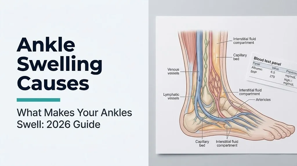
Ankle swelling (also called peripheral edema or ankle edema) occurs when fluid moves from the bloodstream into the surrounding interstitial tissue and accumulates faster than the lymphatic system can return it to circulation, producing the visible and palpable swelling around and below the ankle joint.
The ankle is one of the body’s most dependent regions, meaning gravity pulls fluid downward toward it during upright activity. Every person experiences minor fluid shifts toward the ankles during the day. The reasons some people develop persistent or progressive ankle swelling while others do not come down to a disruption in the balance of forces that normally maintain fluid within blood vessels.
Every cause of ankle swelling, from heart failure to pregnancy to medication side effects, works through one of four basic pathways: too much outward pressure pushing fluid from capillaries into tissue, too little inward protein-osmotic pull keeping fluid inside vessels, impaired lymphatic drainage removing the fluid that does accumulate, or increased capillary leakiness allowing protein to escape into the interstitium and pull more fluid out. The specific cause determines which pathway is disrupted. Understanding this allows a doctor (and an informed patient) to narrow the cause list rapidly from dozens of possibilities to a handful of likely candidates.
According to MedlinePlus (U.S. National Library of Medicine), ankle edema is one of the most frequently reported symptoms prompting physician consultation, and the clinical evaluation focuses first on determining whether the swelling is acute or chronic, bilateral or unilateral, painful or painless, and associated with systemic symptoms such as shortness of breath or fatigue.
Older adults face a compounded vulnerability to ankle swelling because multiple contributing mechanisms are often present simultaneously: reduced cardiac reserve, lower baseline serum albumin from age-related reduced hepatic synthetic function, venous valve incompetence from decades of venous wall stress, and multiple medications with edema as a side effect. What appears as a single symptom may reflect three concurrent causes in a 75-year-old patient.
How Starling Forces Explain All Ankle Swelling Causes
Starling forces are the four physical pressures across the capillary wall that together determine whether fluid stays inside blood vessels or moves into the surrounding tissue, and every named cause of ankle swelling disrupts at least one of these forces.
The four forces, described by British physiologist Ernest Starling in 1896, are: capillary hydrostatic pressure (Pc), which pushes fluid outward from capillaries into tissue; plasma oncotic pressure (πp), which pulls fluid inward due to the osmotic effect of plasma proteins, primarily albumin; interstitial hydrostatic pressure (Pi), which resists further fluid entry into the interstitium; and interstitial oncotic pressure (πi), which draws fluid outward from capillaries due to proteins in the interstitial space. Normally, the net balance favors slight outward filtration at the arterial end of capillaries and slight inward reabsorption at the venous end, with the lymphatics draining whatever net excess accumulates.
Think of the capillary wall like a sieve over a drain. Starling forces determine the pressure head pushing fluid through the sieve. When any force disrupts the balance, the sieve can no longer handle the load, and fluid pools in the surrounding tissue, gravitating to the ankles because that is where it settles lowest.
Heart failure, venous insufficiency, and DVT all raise capillary hydrostatic pressure (Pc), forcing more fluid outward. Kidney disease and liver disease both reduce plasma oncotic pressure (πp) by lowering albumin, removing the osmotic “pull” that keeps fluid inside vessels. Lymphedema impairs lymphatic drainage so that even normal filtration cannot be adequately cleared. Inflammation from cellulitis or allergic reactions increases capillary permeability, letting plasma proteins escape into the interstitium and raising interstitial oncotic pressure (πi), pulling even more fluid outward.
The American Heart Association states that sodium and water retention in heart failure is driven by the activated renin-angiotensin-aldosterone system (RAAS), which further expands plasma volume, raising capillary hydrostatic pressure and compounding the edema from elevated venous pressure alone.
Pitting Edema vs. Non-Pitting Edema: What the Difference Means
Pitting edema and non-pitting edema are distinguished by whether pressing a finger firmly against the swollen ankle for five to ten seconds leaves a visible pit or depression when the pressure is released, and this distinction directly indicates the type of fluid accumulated and which category of cause is most likely.
Pitting edema forms when protein-poor fluid (called a transudate) accumulates in the interstitium. Because the fluid has low protein content, it flows freely and redistributes when compressed, leaving a pit that refills slowly over seconds to a minute. Pitting edema is characteristic of causes that increase capillary hydrostatic pressure or decrease plasma oncotic pressure: heart failure, venous insufficiency, nephrotic syndrome (low albumin from protein loss through diseased kidneys), liver cirrhosis (low albumin from reduced hepatic synthesis), DVT, and medication-induced edema.
Non-pitting edema forms when protein-rich fluid (an exudate) or abnormal structural deposits fill the interstitium. The high protein content causes the fluid to gel and the tissue to develop increased matrix stiffness that resists compression and does not displace. Non-pitting edema is characteristic of lymphedema (disrupted lymphatic drainage allows protein-rich lymph to accumulate, eventually triggering fibrotic remodeling of the interstitial matrix) and myxedema (hypothyroid edema, where thyroid hormone deficiency causes glycosaminoglycans to deposit in the dermis and subcutaneous tissue, creating a firm, rubbery swelling that does not pit).
According to Cleveland Clinic clinical guidance on edema evaluation, the distinction between pitting and non-pitting edema is one of the two most clinically useful physical examination findings in determining the likely cause, alongside the bilateral versus unilateral pattern. A physician will assess both observations within the first minutes of a clinical encounter for ankle swelling.
Inflammatory edema from cellulitis (skin infection) can initially present as non-pitting because the inflammatory exudate is protein-rich. As the infection progresses, the area typically becomes red, warm, tender, and possibly streaked, which distinguishes it from non-inflammatory edema types.
Key Takeaway: Press a finger firmly into your swollen ankle for 10 seconds. If a pit remains when you lift your finger, that is pitting edema, pointing toward cardiac, venous, renal, or hepatic causes. If no pit forms and the tissue is firm or rubbery, non-pitting edema suggests lymphedema or thyroid disease.
Bilateral vs. Unilateral Ankle Swelling: Why the Distinction Matters
Whether both ankles are swollen (bilateral) or only one ankle is swollen (unilateral) is the single most clinically useful initial observation for determining the most likely cause of ankle swelling and the appropriate urgency of evaluation.
Bilateral ankle swelling almost always reflects a systemic cause: a condition that affects the entire body’s fluid balance simultaneously. Both legs and both ankles fill with fluid because the problem is not localized to one limb. Heart failure, chronic kidney disease, liver cirrhosis, nephrotic syndrome, hypoalbuminemia from malnutrition, medications, and the general venous insufficiency of advanced age all produce bilateral swelling. These conditions require physician evaluation but, in most cases, allow time for a scheduled appointment unless accompanied by respiratory symptoms.
Unilateral ankle swelling, meaning swelling of only one ankle without a corresponding change in the other, is a distinctly different clinical picture. A condition affecting only one limb is almost always a localized cause: deep vein thrombosis (DVT) is the most urgent concern; other causes include cellulitis of the lower leg, a Baker’s cyst behind the knee that has ruptured, lymphedema from a localized lymph node problem (surgery, radiation, or obstruction on that side), or a local injury such as an ankle sprain or fracture. DVT-associated unilateral swelling can progress to pulmonary embolism, which is potentially fatal, making any new unilateral ankle swelling without an obvious injury cause a same-day evaluation priority.
The National Heart, Lung, and Blood Institute states that unilateral lower extremity swelling with tenderness, warmth, and redness in a patient with risk factors for DVT (recent surgery, prolonged immobility, prior clot, active cancer) should be evaluated for DVT on the same day of symptom onset using the validated Wells DVT clinical decision score combined with a D-dimer blood test and, if indicated, compression Doppler ultrasonography.
Drivers and long-haul travelers who develop unilateral ankle swelling after extended seated travel should not dismiss the finding as purely positional, because extended immobility is a recognized DVT risk factor.
Heart Failure and Ankle Swelling
Heart failure causes bilateral ankle swelling by raising the hydrostatic pressure within the capillaries of the lower legs to the point where fluid is forced out of the capillary lumen into the interstitial tissue faster than the lymphatics can return it, with the edema worsened by kidney-mediated sodium and water retention.
In right-sided heart failure, the right ventricle cannot pump blood efficiently into the pulmonary arteries. Blood backs up into the right atrium, then into the superior and inferior vena cava, then into the venous system of the entire body. This elevated central venous pressure transmits backward into the venules and capillary beds of the legs and ankles, raising capillary hydrostatic pressure and driving fluid outward into the interstitium. The resulting edema is bilateral, pitting, and classically worst in the late afternoon and evening after a full day of upright activity has allowed gravity to pool fluid at the ankles.
The RAAS is simultaneously activated in heart failure because the kidneys interpret reduced cardiac output as reduced effective circulating volume. The juxtaglomerular apparatus releases renin, triggering the conversion of angiotensinogen to angiotensin II to aldosterone from the adrenal cortex. Aldosterone acts on the principal cells of the renal cortical collecting duct to increase sodium reabsorption and water retention, further expanding plasma volume and compounding the edema. According to the American Heart Association’s 2022 heart failure guidelines, bilateral ankle edema is present in approximately 60% of patients with symptomatic heart failure, making it one of the most prevalent clinical findings in this condition.
Patients taking loop diuretics (furosemide, named for clinical context) for heart failure may notice that their ankle swelling is partially but not completely controlled, reflecting ongoing RAAS activation despite fluid removal. Men with heart failure tend to develop more pronounced ankle edema at lower ejection fraction thresholds than women, possibly reflecting sex-based differences in venous compliance and RAAS responsiveness that are documented in cardiology research.
Chronic Venous Insufficiency as a Cause of Ankle Swelling
Chronic venous insufficiency (CVI) causes bilateral ankle swelling (sometimes asymmetric, with one side worse than the other) by allowing blood to pool in the veins of the lower legs due to incompetent venous valves, which raises local capillary hydrostatic pressure and forces fluid into the surrounding tissue.
The venous valves of the lower extremity leg veins, including the great saphenous vein and the deep veins (posterior tibial and popliteal), normally close between heartbeats to prevent retrograde blood flow downward under gravity. When these valves become incompetent (from prior DVT that damaged the valves, from pregnancy-related venous distension, from prolonged standing, or from genetic predisposition), blood refluxes downward during standing. Venous pressure in the ankle and lower leg rises above normal values. This elevated local venous pressure raises capillary hydrostatic pressure, producing the characteristic pitting edema of CVI.
Clinicians generally observe that CVI-related edema follows a characteristic daily pattern: the ankles are least swollen in the morning after overnight horizontal rest (which reduces venous pooling) and most swollen by late afternoon or evening after prolonged upright activity. This is a practically useful observation for distinguishing CVI edema from cardiac edema, which also worsens with upright activity but is often accompanied by additional symptoms like shortness of breath and fatigue.
According to data published in the Journal of Vascular Surgery, chronic venous insufficiency affects an estimated 40% of adults in the United States to varying degrees, making it one of the most prevalent causes of ankle swelling in the general adult population. Women develop CVI at higher rates than men, with pregnancy-related venous distension and the effects of progesterone on venous wall smooth muscle tone both contributing to increased venous reflux risk in women who have had multiple pregnancies.
Long-term CVI progresses through identifiable clinical stages, from simple varicose veins and ankle edema to skin changes including lipodermatosclerosis (hardening and brown discoloration of the skin above the ankle from hemosiderin deposits) and eventually venous leg ulcers, which represent the end stage of untreated venous hypertension in the lower leg.
Kidney Disease and Ankle Swelling
Kidney disease causes bilateral ankle swelling primarily by allowing albumin to leak into the urine through a damaged glomerular filtration membrane, which reduces plasma oncotic pressure and allows fluid to shift from the bloodstream into the interstitium throughout the body, with the ankles most visibly affected due to gravity.
The kidneys’ glomeruli are highly specialized filtration units whose glomerular basement membrane and associated podocytes (foot-cell processes) normally prevent large proteins like albumin from passing into the urine. In nephrotic syndrome (ICD-10 N04), damage to this filtration barrier from conditions including diabetic nephropathy, membranous nephropathy, minimal change disease, or focal segmental glomerulosclerosis causes massive albumin loss in the urine (proteinuria). Serum albumin levels fall below normal (a condition called hypoalbuminemia), reducing the osmotic force that normally pulls fluid back from the interstitium into capillaries. Fluid accumulates in tissues throughout the body, producing the characteristic bilateral pitting edema of nephrotic syndrome.
The National Kidney Foundation states that proteinuria above 3.5 grams per day in adults is the defining feature of nephrotic syndrome, and ankle edema is present in the vast majority of patients at diagnosis. A 24-hour urine protein collection or a spot urine protein-to-creatinine ratio is the diagnostic test used to quantify protein loss.
In advanced chronic kidney disease (CKD) even without nephrotic-range proteinuria, reduced GFR impairs the kidney’s ability to excrete sodium and water, expanding fluid volume and producing edema through a mechanism similar to the RAAS-driven sodium retention seen in heart failure. Patients with CKD stage 4 or 5 (GFR below 30 mL/min/1.73m²) frequently develop ankle swelling as part of the clinical picture of fluid overload.
Diabetic patients should be aware that diabetic nephropathy is the most common cause of nephrotic syndrome in adults in the United States, and ankle swelling in a person with long-standing poorly controlled type 2 diabetes should prompt a urinalysis and albumin-to-creatinine ratio measurement at the earliest opportunity.
Key Takeaway: Ankle swelling with foamy urine (which can indicate excess protein in the urine) is a combination that suggests kidney disease as the cause and warrants a urine protein test at a primary care physician appointment within days, not weeks.
Liver Disease and Ankle Swelling
Liver disease, particularly cirrhosis (ICD-10 K74.6), causes bilateral ankle swelling through two concurrent mechanisms: reduced hepatic synthesis of albumin (lowering plasma oncotic pressure and allowing fluid to shift out of vessels) and the development of portal hypertension (which raises pressure in the venous system draining the intestines and compounds fluid accumulation).
The liver is responsible for synthesizing virtually all of the body’s circulating albumin. When cirrhosis progressively replaces functional hepatocytes with fibrous scar tissue, albumin synthesis falls. Serum albumin levels below approximately 2.5 g/dL are associated with clinically visible edema. The reduced plasma oncotic pressure allows fluid to move from blood vessels into the interstitium throughout the body, including the ankles.
Portal hypertension develops in cirrhosis because the fibrotic liver tissue obstructs blood flow through the portal vein (which carries nutrient-rich blood from the intestines to the liver). Elevated portal venous pressure causes the development of ascites (fluid accumulation in the peritoneal cavity), which compounds the clinical picture and further reduces effective circulating blood volume, triggering additional RAAS activation and sodium retention.
Liver disease-related ankle edema is therefore the result of three simultaneous mechanisms: low albumin, sodium retention from RAAS activation (triggered by reduced effective circulating volume from both ascites and low oncotic pressure), and in some cases portal hypertension affecting lower body venous drainage. The combination produces bilateral pitting edema that is often worse in the feet and ankles but accompanied by visible abdominal distension from ascites.
Men develop alcoholic liver cirrhosis at higher rates than women for equivalent alcohol consumption levels, reflecting sex-based differences in hepatic alcohol metabolism. Women with autoimmune hepatitis (a non-alcohol-related cause of cirrhosis) are disproportionately affected compared to men, meaning the underlying cause of the liver disease differs by sex even when the resulting edema presentation is similar.
Deep Vein Thrombosis (DVT) and Ankle Swelling
Deep vein thrombosis (DVT) causes ankle swelling in one leg only by partially or completely blocking venous blood flow out of the leg through the affected deep vein, raising local capillary hydrostatic pressure distal to the thrombus and driving fluid into the surrounding tissue.
DVT most commonly forms in the deep veins of the calf (posterior tibial veins, peroneal veins), the popliteal vein behind the knee, or the femoral vein in the thigh. When a clot partially or completely obstructs the vein, blood cannot drain normally from the leg below the clot. Venous pressure rises distal to the occlusion, raising capillary hydrostatic pressure, and the resulting edema is confined to the affected leg, producing the characteristic unilateral swelling of DVT.
The specific warning signs that distinguish DVT-related swelling from benign causes are: swelling of only one leg without any injury to explain it, warmth and redness of the skin over the swollen area, and calf tenderness when the foot is flexed upward (a clinical examination finding called Homan’s sign, though this has limited sensitivity and specificity). The classic DVT patient, according to the NHLBI, is one who has had recent prolonged immobility (surgery, long-haul flight, hospitalization), has cancer, is pregnant, or has a prior history of DVT.
The reason DVT requires same-day evaluation is not the leg swelling itself but the risk that the clot will fragment and travel through the venous circulation to the pulmonary arteries, causing pulmonary embolism (PE). Large pulmonary emboli obstruct enough of the pulmonary arterial circulation to cause acute right heart failure, which can be rapidly fatal. According to the NHLBI, pulmonary embolism is associated with approximately 60,000 to 100,000 deaths annually in the United States.
Call 911 immediately if ankle swelling in one leg is accompanied by sudden chest pain, difficulty breathing, rapid heart rate, or coughing blood, as these symptoms indicate that pulmonary embolism may already be occurring.
Medications That Cause Ankle Swelling
Several commonly prescribed medication classes cause bilateral ankle swelling as a direct pharmacological side effect, with dihydropyridine calcium channel blockers (such as amlodipine, named for clinical context) being the most frequently implicated class in clinical practice.
Dihydropyridine calcium channel blockers lower blood pressure by relaxing the smooth muscle of arterial walls (arterioles), reducing peripheral vascular resistance. This arteriolar dilation increases blood flow into the capillary bed of the lower legs without a matching dilation of the venous side (venules). The result is increased capillary hydrostatic pressure in the legs, which drives fluid into the interstitial tissue. The mechanism is not fluid overload from salt retention but an imbalance in vasodilation between the arterial and venous sides of the capillary bed. The edema is typically bilateral, pitting, and appears within weeks of starting the medication.
Other medication classes with documented ankle swelling as a side effect include:
- NSAIDs (ibuprofen, naproxen, named for clinical context): reduce renal prostaglandin synthesis, impairing the kidneys’ ability to excrete sodium and water, expanding plasma volume
- Corticosteroids (prednisone, named for clinical context): promote renal sodium retention through mineralocorticoid receptor activity, expanding extracellular fluid volume
- Thiazolidinediones (pioglitazone, named for clinical context, used for type 2 diabetes): activate peroxisome proliferator-activated receptor gamma (PPARγ) in the renal tubule, increasing sodium reabsorption and fluid retention
- Hormonal contraceptives containing estrogen: can increase sodium retention and venous distensibility through estrogenic and progestogenic mechanisms
- Pregabalin and gabapentin (named for clinical context, used for nerve pain and seizures): cause peripheral edema through incompletely understood mechanisms that may involve calcium channel modulation in peripheral tissues
Patients who develop new ankle swelling within days to weeks of starting a new medication should bring the medication name, dose, and start date to their primary care physician, along with a description of the swelling pattern, to determine whether a medication change is appropriate.
Key Takeaway: If your ankles began swelling within weeks of starting a new blood pressure medication, particularly amlodipine or another calcium channel blocker, the medication is a highly probable cause, and a discussion with your prescribing physician about switching to an alternative class may resolve the edema without any other treatment.
Pregnancy and Ankle Swelling
Pregnancy causes ankle swelling in most pregnant women through multiple simultaneous physiological mechanisms: expansion of plasma volume, increased venous pressure from the growing uterus compressing the inferior vena cava, and the effects of progesterone on venous wall tone, all of which promote fluid accumulation in the lower legs.
During a normal pregnancy, plasma volume expands by approximately 40 to 50% to supply the placenta and developing fetus. This expanded circulating volume increases capillary hydrostatic pressure throughout the venous circulation. The uterus, as it grows through the second and third trimesters, progressively compresses the inferior vena cava (the large vein returning blood from the lower body to the heart) when the pregnant woman is in the supine (lying on back) or upright position. This compression reduces venous return from the lower legs, raising venous pressure and capillary hydrostatic pressure in the ankle and lower leg region.
Progesterone, produced in large quantities by the placenta throughout pregnancy, relaxes smooth muscle throughout the body, including the smooth muscle of venous walls. This reduces venous tone, allowing veins to distend and pool more blood in the lower extremities, compounding the edema from the other mechanisms.
Physiological pregnancy edema is bilateral, typically worse in the third trimester, worsens with prolonged standing or sitting, and improves with rest and leg elevation. It resolves spontaneously after delivery as the uterine compression resolves and excess plasma volume is excreted.
However, preeclampsia (ICD-10 O14) is a serious pregnancy complication that includes sudden-onset ankle and facial swelling (indicating rapid fluid retention from the systemic vascular endothelial dysfunction of preeclampsia) combined with elevated blood pressure (at or above 140/90 mmHg on two readings), proteinuria, and sometimes headache and visual changes. According to the American College of Obstetricians and Gynecologists (ACOG), preeclampsia occurs in approximately 5 to 8% of pregnancies and can progress to seizures (eclampsia) and maternal death without prompt treatment. Any pregnant woman with rapidly worsening ankle swelling, especially with headache, visual disturbance, or elevated blood pressure, requires same-day obstetric evaluation.
Lymphedema as a Cause of Ankle Swelling
Lymphedema (ICD-10 I89.0) causes ankle swelling when the lymphatic vessels responsible for draining protein-rich interstitial fluid from the lower leg are damaged, absent, or obstructed, allowing lymph to accumulate in the interstitial space and, over time, trigger progressive fibrotic changes in the surrounding tissue.
The lymphatic system of the lower leg consists of an initial network of blind-ended lymphatic capillaries in the skin and subcutaneous tissue that collect interstitial fluid and transport it through progressively larger collecting vessels to the inguinal lymph nodes, and eventually through the thoracic duct back into the venous circulation at the left subclavian vein junction. When this pathway is interrupted, lymph accumulates in the interstitium.
Lymphedema is classified as primary (from congenital abnormalities of lymphatic vessel development, which may present in adolescence or young adulthood) or secondary (from acquired damage to lymphatic vessels). Secondary lymphedema is more common in the United States, where the most frequent causes are cancer surgery (particularly lymph node dissection in the groin for melanoma or gynecological cancers) and radiation therapy that damages inguinal or pelvic lymph nodes. Worldwide, filariasis (infection by the parasitic worm Wuchereria bancrofti, which obstructs lymphatic vessels) is the most common cause of lower extremity lymphedema.
The physical characteristics of lymphedema differ from cardiac or venous edema. The swelling is typically non-pitting (or minimally pitting in early stages), does not resolve overnight with leg elevation as dramatically as venous edema does, and progresses over months to years toward a firm, woody texture as interstitial fibrosis develops. A positive Stemmer sign (inability to pinch a fold of skin at the base of the second toe because the skin is too thick and indurated) is a clinical examination finding specific to lymphedema.
Women who have undergone pelvic lymph node dissection for gynecological cancers are at substantial long-term risk for lower extremity lymphedema, with incidence rates varying widely depending on the extent of dissection and whether radiation was used.
What Causes Ankle Swelling in Older Adults
Ankle swelling in older adults is caused most frequently by a combination of chronic venous insufficiency from years of venous valve deterioration, age-related reduction in serum albumin from declining hepatic synthetic function, increased medication burden with multiple edema-causing drugs acting simultaneously, and reduced cardiac reserve that may produce subclinical heart failure before a formal diagnosis is established.
No single cause dominates in older adults precisely because multiple contributing mechanisms coexist. A 72-year-old woman taking a calcium channel blocker for hypertension and an NSAID for arthritis, with mild CKD (eGFR of 48 mL/min/1.73m²) and chronic venous insufficiency from varicose veins, may have three or four simultaneous causes of ankle swelling, none of which is severe enough on its own to explain the full degree of edema but whose combined effect is readily visible.
Age-related decline in albumin is often overlooked as a contributing factor because it falls within the “normal” laboratory range in many older adults but sits at the lower end of normal. Albumin that is clinically adequate in a 30-year-old may be insufficient to maintain plasma oncotic pressure when combined with the additional venous pressure and medication effects present in an older person.
According to a review published in the Journal of the American Geriatrics Society, ankle edema in community-dwelling adults over age 65 is present in an estimated 20 to 30% of this population, with the majority of cases reflecting multiple concurrent causes rather than a single diagnosable condition. A geriatrician or primary care physician evaluating ankle swelling in this population should review every current medication for edema-causing potential before attributing the finding to systemic disease alone.
Older adults with ankle swelling should inform their primary care physician about every medication including over-the-counter NSAIDs, which are widely used for arthritis pain and frequently contribute to ankle edema without the patient or physician connecting the two.
What Causes Ankle Swelling in Females
Ankle swelling in females is caused by the same systemic conditions that affect both sexes (heart failure, venous insufficiency, kidney disease, liver disease, medications), but also by several female-specific hormonal mechanisms: the effects of estrogen and progesterone on venous tone and sodium retention during the menstrual cycle, the complex physiological changes of pregnancy, and the hormonal transitions of perimenopause and postmenopause.
During the luteal phase of the menstrual cycle (approximately days 14 to 28), progesterone levels rise. Progesterone relaxes venous smooth muscle, allowing veins to distend and pool more blood in the lower extremities. Many premenopausal women notice mild ankle and lower leg swelling in the week before menstruation that resolves with the onset of menstruation and the subsequent fall in progesterone. This physiological cyclical edema does not require investigation if it follows the menstrual cycle reliably and resolves completely between cycles.
Hormonal contraceptives containing estrogen increase venous thrombosis risk (estrogen-containing oral contraceptives raise DVT risk approximately two to four-fold compared to non-users, per data reviewed by the American College of Obstetricians and Gynecologists) and can cause mild sodium retention and ankle edema. Women who develop ankle swelling after starting combined oral contraceptive pills should discuss this with their obstetrician-gynecologist, both to consider a medication change and to assess their DVT risk.
Postmenopausal women on hormone replacement therapy containing estrogen may experience fluid retention and ankle edema as a side effect of the estrogenic component. Women who have undergone surgical menopause (bilateral oophorectomy) at a younger age and are on estrogen replacement at higher physiological replacement doses may be at particular risk.
Females also develop CVI at higher rates than males due to the cumulative effects of multiple pregnancies on venous valve function and the general effect of female sex hormones on venous compliance throughout the reproductive years.
Key Takeaway: In a premenopausal woman, ankle swelling that appears reliably in the week before menstruation and resolves completely after it begins is most likely cyclical hormonal edema and does not require urgent investigation. Swelling that does not resolve between cycles, or that is new and unilateral, warrants physician evaluation.
When Ankle Swelling Is Serious: Red Flags and Emergency Signs
Certain ankle swelling presentations indicate potentially life-threatening underlying conditions and require immediate emergency evaluation. Do not wait to see if these resolve on their own.
Call 911 or go to the nearest emergency room immediately if ankle swelling is accompanied by any of the following:
- Shortness of breath, difficulty breathing at rest, or inability to lie flat: these symptoms with bilateral ankle edema indicate possible decompensated heart failure with pulmonary edema (fluid in the lungs), which requires emergency hospital management
- Sudden chest pain, rapid heartbeat, or coughing blood in the context of new unilateral ankle swelling: these symptoms indicate possible pulmonary embolism from DVT and represent a life-threatening emergency requiring CT pulmonary angiography and anticoagulation without delay
- New unilateral ankle swelling with warmth, redness, and tenderness in the calf or ankle area after surgery, a long flight, or prolonged bed rest: this presentation meets the threshold for same-day DVT evaluation at an emergency department or same-day urgent care with Doppler ultrasound capability
- Ankle swelling in a pregnant woman with headache, visual changes, or blood pressure at or above 140/90 mmHg: this combination meets ACOG diagnostic criteria for preeclampsia and requires immediate obstetric evaluation at the nearest facility with obstetric emergency capability
- Ankle swelling with fever, spreading skin redness, and warmth that is progressing over hours: this indicates possible cellulitis (skin infection) that may be spreading and requires evaluation for antibiotic treatment and possible hospital admission if the infection involves the lymphatics
These presentations indicate conditions including pulmonary embolism, decompensated heart failure, preeclampsia, and rapidly spreading cellulitis, each of which requires emergency medical assessment, not a scheduled appointment or a phone call to an advice nurse.
Frequently Asked Questions About Ankle Swelling Causes
What is the most common cause of ankle swelling?
The most common cause of ankle swelling in otherwise healthy adults is prolonged sitting or standing, which raises venous pressure in the lower legs through gravity-dependent venous pooling and is not a sign of disease.
In adults with underlying health conditions, chronic venous insufficiency and medication side effects (particularly calcium channel blockers and NSAIDs) are among the most frequently identified causes in primary care practice.
Bilateral ankle swelling that persists despite overnight rest and leg elevation warrants evaluation by a primary care physician to rule out systemic causes including heart failure, kidney disease, and liver disease.
Can ankle swelling be a sign of heart failure?
Yes, bilateral ankle swelling is one of the most recognizable clinical signs of heart failure, present in approximately 60% of patients with symptomatic heart failure according to the American Heart Association.
The swelling results from elevated venous pressure caused by the failing heart’s inability to pump effectively, combined with sodium and water retention from renin-angiotensin-aldosterone system activation in response to reduced cardiac output.
Ankle swelling from heart failure is characteristically bilateral, pitting, worst in the late afternoon and evening, and often accompanied by shortness of breath with activity, orthopnea (difficulty breathing when lying flat), and fatigue.
How do I know if my ankle swelling is from a blood clot?
Ankle swelling from a DVT (deep vein thrombosis, or blood clot in a deep vein) is almost always unilateral, meaning only one ankle is swollen.
The swollen leg is often warm to the touch and may be tender to pressure along the calf, and the swelling typically appears after a period of reduced mobility such as a long flight, surgery, or bed rest.
Any new unilateral ankle swelling without an obvious injury explanation, particularly in someone with DVT risk factors, warrants same-day evaluation with a D-dimer blood test and Doppler ultrasound to rule out DVT before the clot has any chance to fragment and reach the lungs.
Why do my ankles swell at the end of the day but not in the morning?
Ankles that swell progressively through the day and are back to normal after sleeping overnight exhibit dependent edema, reflecting fluid that accumulates in the tissue during upright activity under the influence of gravity and drains back into circulation when the legs are horizontal during sleep.
This pattern is characteristic of benign causes including prolonged standing or sitting, early chronic venous insufficiency, and medication-induced edema, all of which produce gravity-dependent fluid shifts that reverse with recumbency.
A bilateral dependent edema pattern that has gradually worsened over months, or that now requires more than one night’s rest to fully resolve, warrants evaluation by a primary care physician to assess cardiac, renal, and venous causes.
What medications cause ankle swelling as a side effect?
The medication class most frequently causing ankle swelling is dihydropyridine calcium channel blockers (such as amlodipine), which cause arteriolar dilation that increases lower leg capillary pressure without matching venodilation.
Other classes that commonly produce ankle swelling include NSAIDs (which impair renal prostaglandin production and reduce sodium excretion), corticosteroids (which cause sodium retention through mineralocorticoid receptor activity), thiazolidinediones (pioglitazone for diabetes), hormonal contraceptives containing estrogen, and pregabalin or gabapentin used for nerve pain or seizures.
Any patient who develops new ankle swelling within weeks of starting a new medication should bring the complete medication list to their primary care physician appointment and specifically ask whether the new medication or any combination of current medications could be contributing.
When should I go to the ER for ankle swelling?
Go to the emergency room immediately (or call 911) if ankle swelling is accompanied by shortness of breath, chest pain, or the inability to lie flat, as these symptoms suggest decompensated heart failure or pulmonary embolism.
New unilateral ankle swelling with warmth, redness, and calf tenderness after surgery or prolonged immobility also warrants emergency evaluation for DVT on the same day of symptom onset.
Ankle swelling in pregnancy combined with headache, visual changes, or blood pressure at or above 140/90 mmHg requires immediate obstetric emergency evaluation for preeclampsia, without waiting for a routine appointment.
Closing
The most practical action step from this article is to make two observations before your next physician appointment: note whether both ankles are swollen or just one, and press a finger firmly into the swelling for ten seconds to determine whether it pits. Those two data points will help your primary care physician narrow the cause within the first minutes of your visit.
Bring a list of every medication you take (including over-the-counter NSAIDs and pain relievers) to that appointment, along with a description of when the swelling is worst, whether it resolves overnight, and whether it has been getting progressively worse. That history is as diagnostically useful as any blood test.
If you have unilateral ankle swelling without a clear injury cause, or bilateral swelling with any shortness of breath, make that appointment today rather than next week.