Dowager’s Hump Causes: Why It Develops and What to Do
Dowager’s hump causes range from reversible postural muscle imbalance to irreversible structural changes in the vertebral bones, and identifying which cause is driving the deformity determines whether physical therapy, bone density treatment, or surgical evaluation is the appropriate path. The condition develops when the upper thoracic or cervicothoracic spine curves forward beyond its normal range, creating the visible rounded prominence at the base of the neck.
According to the National Institute of Arthritis and Musculoskeletal and Skin Diseases (NIAMS), osteoporosis alone affects approximately 10 million adults in the United States, and vertebral compression fractures from osteoporosis are among the leading causes of progressive spinal deformity in older adults. Yet postural kyphosis driven by prolonged forward-loaded sitting posture now affects people decades younger than the name “dowager’s” historically implied.
This article covers every clinically recognized cause of dowager’s hump, the specific anatomical and physiological mechanism by which each cause produces the deformity, the populations most affected by each cause, how doctors evaluate and classify the condition, and the specific warning signs that indicate something more serious is happening to the spine.
Dowager’s Hump Causes: What the Condition Is and Why It Develops
A dowager’s hump develops when the normal thoracic kyphosis of the spine increases beyond its physiological range, producing a visible, rounded prominence at the cervicothoracic junction.
The human thoracic spine normally curves forward (posteriorly convex) between approximately 20 and 40 degrees, measured by the Cobb angle on a standing lateral spinal radiograph. When this angle exceeds 40 to 45 degrees in adults, clinicians classify it as hyperkyphosis. The common term “dowager’s hump” refers specifically to the visible soft tissue and bony prominence that develops as the upper thoracic vertebrae and overlying musculature become increasingly anteriorly flexed.
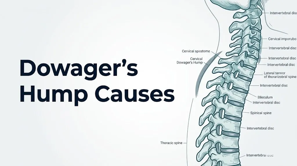
The deformity sits at the junction of the cervical and thoracic spine, most prominently at C7 and T1, where the last cervical vertebra meets the first thoracic vertebra. As the thoracic spine rounds forward, the head translates anteriorly to maintain a forward-facing gaze. This forward head position increases the effective load on the posterior cervical structures and creates a mechanical situation that worsens progressively without intervention.
Three distinct anatomical and physiological mechanisms drive this deformity in adults: postural loading with progressive muscle imbalance and ligamentous stretch, loss of vertebral bone density causing compressive fractures and anterior body collapse, and developmental endplate irregularity during adolescent growth (Scheuermann’s disease) producing wedge-shaped vertebral bodies. Each mechanism requires a different clinical approach.
Key causes overview:
- Postural kyphosis from prolonged forward flexion and muscle imbalance
- Osteoporotic vertebral compression fractures
- Scheuermann’s kyphosis from adolescent vertebral endplate irregularity
- Secondary osteoporosis from medications or systemic disease
- Sarcopenia and age-related paraspinal muscle loss
- Prolonged sedentary posture with device use
People who have had one osteoporotic vertebral compression fracture face an approximately fivefold increased risk of a subsequent fracture, according to data published in Osteoporosis International, making early bone density evaluation particularly important in the progression of this condition.
What Does a Dowager’s Hump Look Like and How Is It Defined
A dowager’s hump appears as a rounded, often firm prominence at the base of the neck and top of the upper back, typically most visible when a person is viewed from the side.
The visible hump has two components. The bony component reflects the altered curvature of the upper thoracic vertebrae, with the spinous processes of T1 through T4 or T5 becoming more prominent as the vertebral column rounds forward. The soft tissue component reflects both the repositioning of the overlying fat pad at the cervicothoracic junction and the increased bulk of the posterior cervical paraspinal muscles that chronically contract in an attempt to resist forward head migration.
Clinically, the Cobb angle is the gold standard measurement for quantifying kyphosis severity. It is measured on a standing lateral full-spine radiograph by drawing lines along the superior endplate of the most tilted superior vertebra and the inferior endplate of the most tilted inferior vertebra in the kyphotic segment, then measuring the angle formed by perpendiculars to those lines. The American Academy of Orthopaedic Surgeons (AAOS) recognizes a Cobb angle exceeding 40 degrees as the threshold for clinically significant thoracic kyphosis in adults, though many clinicians use 50 degrees as a threshold for active management.
A practical self-observation: stand sideways in front of a mirror. A visible rounding at the upper back with the head positioned noticeably forward of the shoulders, so that the ear is in front of the shoulder rather than directly over it, indicates a measurable increase in thoracic kyphosis that warrants evaluation.
| Cobb Angle Range | Classification | Clinical Significance |
|---|---|---|
| 20 to 40 degrees | Normal adult thoracic curve | No clinical concern |
| 40 to 60 degrees | Mild hyperkyphosis | Postural therapy appropriate; monitor for progression |
| 60 to 75 degrees | Moderate hyperkyphosis | Medical evaluation; specialist referral warranted |
| Greater than 75 degrees | Severe hyperkyphosis | Surgical evaluation; pulmonary and neurological risk |
Adolescents and young adults with apparent dowager’s hump deserve specific attention because their presentation is more likely to represent Scheuermann’s kyphosis (structural, progressive) rather than simple postural rounding (functional, often reversible).
Postural Kyphosis: The Most Common Cause of Dowager’s Hump
Postural kyphosis is the most common cause of dowager’s hump in adults, produced by sustained forward-flexed positioning that progressively alters the load distribution across the thoracic vertebral column.
Normal upright posture requires the head (weighing approximately 10 to 12 pounds) to balance directly over the thoracic and lumbar spine, with the ear in line with the shoulder, hip, knee, and ankle when viewed from the side. For every inch the head translates forward from this neutral position, the effective load on the lower cervical and upper thoracic spine increases by approximately 10 pounds, according to biomechanical data reviewed in the journal Spine. At a 3-inch forward head translation, the cervicothoracic junction is absorbing an effective load of 40 pounds or more with every hour of sustained posture.
Think of the spine like a fishing rod held horizontally at one end. The rod is manageable when held at its base. But as the free end tips forward, the mechanical demand at the holding point multiplies rapidly. The cervicothoracic junction is that holding point, and sustained forward head position is the rod tipping further forward with every hour of unrelieved sitting.
Over months and years, this asymmetric loading according to Wolff’s Law (bone remodels according to the mechanical stresses placed upon it) drives progressive anterior remodeling of the upper thoracic vertebral bodies. Disc height reduces anteriorly. The posterior longitudinal ligament and ligamentum nuchae gradually elongate under sustained tensile load, losing their capacity to resist further flexion. The result is a curve that begins as functionally reversible and becomes structurally fixed.
Postural kyphosis is particularly prevalent in adults who spend 6 or more hours daily using computers or smartphones with sustained forward neck flexion. People in occupations requiring prolonged forward-facing work (surgeons, dentists, laboratory technicians, writers) carry elevated risk due to the cumulative daily forward-loading exposure.
Quick Tip:
- Postural kyphosis in its early stages is distinguished from structural kyphosis by its reversibility: when you voluntarily stand tall and extend the spine, the curve flattens. Structural kyphosis does not flatten with voluntary correction.
- The chin-tuck exercise (retracting the chin gently toward the neck, lengthening the suboccipital space) directly activates the longus colli and longus capitis, the deep cervical flexors that are most inhibited in forward head posture.
- For adults over 65, postural kyphosis often coexists with osteoporotic changes, making the distinction between pure postural and structural causes a job for standing lateral spinal radiographs, not visual inspection alone.
How Forward Head Posture and Muscle Imbalance Cause Dowager’s Hump
Forward head posture drives dowager’s hump by creating a predictable pattern of muscle lengthening, weakening, and adaptive shortening across the cervical and upper thoracic regions, progressively shifting mechanical load onto the anterior vertebral bodies.
The muscles most directly involved form two opposing groups. The deep cervical flexors (longus colli, longus capitis, and the anterior scalene muscles) are responsible for holding the head in neutral position against gravity. In sustained forward head posture, these muscles are placed in a chronically elongated position and progressively lose their capacity to generate sufficient tension. At the same time, the suboccipital muscles (rectus capitis posterior major and minor, obliquus capitis superior and inferior) and sternocleidomastoid adaptively shorten, pulling the occiput posteriorly and the chin forward in a compensatory pattern.
In the thoracic region, the lower trapezius and rhomboid major and minor are responsible for retracting the scapulae and extending the upper thoracic spine. In forward-flexed posture, these muscles are chronically lengthened and become inhibited. The opposing muscles, primarily the pectoralis minor and anterior serratus, adaptively shorten and pull the shoulders forward and the thoracic spine into further kyphosis. The result is a classic upper crossed syndrome pattern first described by physiatrist Vladimir Janda: tight upper trapezius and levator scapulae opposing weak deep cervical flexors and lower trapezius, crossing at the cervicothoracic junction.
According to a 2019 review published in the Journal of Physical Therapy Science, forward head posture with a craniovertebral angle (measured on lateral photograph) below 50 degrees is associated with measurably greater cervical spine compressive load and faster progression of cervicothoracic kyphosis compared to individuals with a craniovertebral angle above 50 degrees.
People with hypermobility conditions, including those with generalized hypermobility spectrum disorder or Ehlers-Danlos syndrome (hypermobile type), are at elevated risk for postural kyphosis because their posterior ligamentous structures offer less passive resistance to the forward-loading pattern described above.
Key Takeaway: Postural kyphosis and forward head posture cause dowager’s hump through a specific muscle imbalance: weak deep cervical flexors and lower trapezius combined with shortened pectorals and upper trapezius progressively shift load onto the anterior vertebral bodies, and this pattern can be specifically targeted with a physical therapist’s guidance before structural vertebral changes make it irreversible.
Osteoporosis and Vertebral Compression Fractures as Causes
Osteoporosis causes dowager’s hump when reduced bone mineral density in the thoracic vertebral bodies allows anterior compressive loads to collapse the front of one or more vertebral bodies, permanently altering the spinal curvature angle.
Osteoporosis (ICD-10: M81) is defined by the National Osteoporosis Foundation (NOF) as a bone mineral density T-score of minus 2.5 or below on dual-energy X-ray absorptiometry (DEXA scanning). In this state, the trabecular bone within the vertebral body has lost enough density that forces of normal daily living, getting out of a chair, sneezing, or even sustained upright posture, can produce a vertebral compression fracture (ICD-10: M80). These fractures collapse the anterior vertebral body while the posterior wall often remains intact, creating a wedge-shaped vertebra.
Each wedge-shaped vertebral body adds approximately 10 to 20 degrees to the thoracic Cobb angle, according to data reviewed by the AAOS. A person who sustains three consecutive upper thoracic vertebral compression fractures at T5, T6, and T7 may develop a Cobb angle exceeding 60 degrees purely from the fracture-induced wedging, independent of any muscle imbalance or postural factor.
Crucially, vertebral compression fractures from osteoporosis are frequently painless or produce only brief, mild back pain that is misattributed to muscle strain. The progressive kyphotic deformity may be the first visible sign that fractures have occurred. The NOF estimates that approximately two-thirds of vertebral compression fractures in the United States go clinically undiagnosed at the time they occur.
Women over 50 with a height loss of more than 1.5 inches compared to their peak adult height, or men over 70 with similar height loss, should ask their primary care physician about lateral thoracic spine radiography to identify previously unrecognized vertebral compression fractures as a cause of their developing kyphosis.
Scheuermann’s Disease: A Cause of Dowager’s Hump in Younger Adults
Scheuermann’s disease (ICD-10: M42.0) causes dowager’s hump in adolescents and young adults through a structural irregularity in vertebral endplate ossification that produces anterior vertebral body wedging during the growth period.
Named after Danish radiologist Holger Scheuermann who described it in 1921, Scheuermann’s disease is the most common cause of structural kyphosis in adolescents, with an estimated prevalence of 1 to 8 percent of the adolescent population, according to the AAOS. The condition is diagnosed radiographically when three or more consecutive thoracic vertebrae each show anterior wedging of 5 degrees or more. The total kyphotic Cobb angle typically exceeds 45 degrees at diagnosis.
The vertebral endplates in Scheuermann’s disease develop irregular ossification, and Schmorl’s nodes (herniations of the nucleus pulposus of the intervertebral disc upward or downward through the endplate into the vertebral body) are a characteristic radiographic finding. These endplate defects concentrate mechanical stress on the anterior portion of the vertebral body during the growth spurt, driving greater bone formation posteriorly than anteriorly, producing the anterior wedging that creates the rigid kyphotic curve.
Unlike postural kyphosis, Scheuermann’s kyphosis does not flatten when the person attempts voluntary spinal extension. The rigidity of the curve distinguishes it clinically from postural rounding. This distinction matters because Scheuermann’s kyphosis often requires orthopedic brace management during active growth phases and may require surgical correction when the Cobb angle exceeds 70 to 75 degrees.
Scheuermann’s kyphosis has a male predominance, affecting approximately 2 to 7 males for every female case, according to the AAOS. The precise reason for this sex difference remains unclear, but differences in growth rate, physical activity patterns, and potentially hormonal regulation of endplate ossification during the adolescent growth spurt have been proposed.
Medication-Induced Bone Loss as a Cause of Dowager’s Hump
Several commonly prescribed medication classes reduce bone mineral density through specific pharmacological mechanisms, increasing the risk of vertebral compression fractures and the progressive kyphosis they produce.
Glucocorticoid-induced osteoporosis is the most common form of secondary osteoporosis in the United States. According to clinical guidelines published in Osteoporosis International, long-term glucocorticoid use (any dose for 3 months or more, or higher doses for shorter periods) suppresses osteoblast differentiation and proliferation through direct effects on osteoblast progenitor cells, while simultaneously upregulating RANK-L expression, driving excess osteoclast activity and bone resorption. Trabecular bone in the vertebral bodies is lost faster than cortical bone. Vertebral fracture risk increases substantially even at what are considered low daily doses of prednisone (5 mg or more per day).
Other medication classes that reduce bone mineral density include:
- Anticonvulsant medications (phenytoin, phenobarbital, carbamazepine): induce hepatic cytochrome P450 enzymes that accelerate vitamin D catabolism, reducing 25-hydroxyvitamin D availability for calcium absorption, leading to secondary hyperparathyroidism and increased bone resorption
- Proton pump inhibitors (PPIs): reduce gastric acid, impairing ionized calcium absorption in the duodenum; observational data published in JAMA Internal Medicine found an association between long-term PPI use and increased vertebral fracture risk, though causation is not confirmed
- Androgen deprivation therapy (ADT) in prostate cancer treatment: suppresses testosterone production, reducing its aromatization to estrogen in peripheral tissues; the resulting estrogen deficiency in men on ADT produces a rate of bone mineral density loss comparable to that seen in early postmenopausal women
- Aromatase inhibitors in breast cancer treatment: block peripheral estrogen synthesis, producing rapid bone mineral density loss in premenopausal and postmenopausal women
Adults taking any of these medications for more than 3 consecutive months should discuss baseline DEXA scanning with their primary care physician to establish bone mineral density prior to fracture risk accumulation.
Key Takeaway: Medication-induced bone loss from glucocorticoids, anticonvulsants, and androgen deprivation therapy is a direct and underrecognized cause of the vertebral compression fractures that drive dowager’s hump, and people on these drugs long-term need proactive bone density monitoring, not reactive treatment after fractures have already altered spinal curvature.
Aging, Sarcopenia, and Progressive Spinal Loading
Aging contributes to dowager’s hump through two parallel and interacting pathways: progressive loss of bone mineral density and progressive loss of paraspinal muscle mass, each independently worsening thoracic kyphosis.
Sarcopenia is the age-related loss of skeletal muscle mass, strength, and function. The erector spinae muscles (spinalis, longissimus thoracis, and iliocostalis) are the primary paraspinal extensors that counteract the forward flexion gravity places on the thoracic spine with every minute of upright posture. When these muscles lose mass and contractile capacity with age, the mechanical resistance they provide against gravitational forward flexion decreases proportionally.
A 2020 study published in Spine found that in adults over 65, paraspinal muscle cross-sectional area on MRI correlated inversely with thoracic Cobb angle: the less paraspinal muscle mass present, the greater the measured kyphosis. This relationship was independent of bone mineral density, suggesting that muscle loss contributes to kyphosis progression even without vertebral fractures occurring.
The interplay between sarcopenia and bone loss is compounding. As kyphosis progresses from reduced muscle support, gravitational loading on the anterior vertebral bodies increases. If bone mineral density is already reduced, this increased anterior loading elevates fracture risk. A single vertebral fracture then further increases the Cobb angle, which further increases anterior loading, creating a self-reinforcing cycle.
Older adults with limited weight-bearing physical activity face accelerated sarcopenia through disuse atrophy of the paraspinal extensors and through reduced mechanical loading stimulus on vertebral trabecular bone. The American Academy of Orthopaedic Surgeons recommends that adults over 50 engage in regular weight-bearing activity specifically to preserve both paraspinal muscle mass and vertebral bone density.
Men over 70 experience a particularly rapid intersection of both sarcopenia and declining testosterone-to-estrogen signaling that accelerates both muscle and bone loss simultaneously, making this age group among the highest-risk for progressive kyphosis even without a history of falls or recognized fractures.
Dowager’s Hump Causes in Postmenopausal Women
Postmenopausal women face the highest population-specific risk for osteoporotic dowager’s hump because the decline in estrogen production at menopause removes the primary hormonal brake on bone resorption.
Estrogen (produced by ovarian granulosa cells throughout the reproductive years) regulates bone remodeling by suppressing the expression of RANK-L (receptor activator of nuclear factor kappa-B ligand) on osteoblasts and stromal cells. RANK-L binds to the RANK receptor on osteoclast precursors, driving their maturation and activation. When estrogen levels fall at menopause, RANK-L expression rises, osteoclast activity accelerates, and bone resorption outpaces bone formation. The vertebral trabecular bone, which has high surface area and rapid turnover rate, is lost faster than cortical bone in the first 5 to 10 years after menopause.
The National Osteoporosis Foundation estimates that one in two women over 50 will experience an osteoporosis-related fracture in their lifetime. Vertebral compression fractures are the most common of these fractures, affecting approximately 25 percent of women over 70, according to data from the Journal of Bone and Mineral Research. Each fracture adds to the progressive kyphotic curve.
Beyond bone loss, postmenopausal women face changes in muscle fiber composition, with estrogen playing a role in maintaining type II (fast-twitch) muscle fiber mass in the paraspinal extensors. Estrogen deficiency accelerates paraspinal muscle loss through mechanisms that overlap with the general sarcopenia pathway but occur earlier and more rapidly in women than in age-matched men.
Women in early menopause (ages 50 to 60) who notice any new back pain, height loss, or visible change in upper back posture should request evaluation by their primary care physician, including DEXA scanning if not recently performed, and a discussion of whether standing lateral spine radiography is warranted to detect silent vertebral fractures before significant kyphosis develops.
Dowager’s Hump Causes in Men
Dowager’s hump in men develops through different predominant pathways than in women, with Scheuermann’s kyphosis, ankylosing spondylitis, and long-term corticosteroid use representing the most clinically important causes in this population.
Scheuermann’s kyphosis affects males at approximately 2 to 7 times the rate of females, making it the leading structural cause of fixed thoracic kyphosis in young and middle-aged men who present with a dorsal hump. Men diagnosed with Scheuermann’s kyphosis in adolescence who did not receive orthopedic management during the growth phase frequently present in their 30s and 40s with back pain superimposed on the pre-existing fixed structural kyphosis.
Ankylosing spondylitis (ICD-10: M45), a seronegative spondyloarthropathy, produces progressive spinal kyphosis in adults through a distinct inflammatory mechanism. Chronic inflammation at the vertebral body corners (Romanus lesions), discovertebral junctions, and costovertebral joints drives progressive bony bridging and eventual fusion of the vertebral column into a rigid, kyphotic bamboo spine configuration. Ankylosing spondylitis affects males approximately 2 to 3 times more frequently than females and most commonly presents between ages 20 and 40. A 2021 systematic review in Rheumatology found that untreated ankylosing spondylitis produces a mean Cobb angle increase of approximately 0.7 to 1.2 degrees per year.
Men receiving long-term androgen deprivation therapy for prostate cancer represent a growing population at risk for rapid bone mineral density loss and consequent vertebral compression fractures producing kyphosis. As noted in the medication causes section, the estrogen deficiency produced by ADT in men drives bone loss at rates comparable to early postmenopausal women.
Men who develop new upper back rounding or a visible cervicothoracic hump alongside morning spinal stiffness lasting more than 30 minutes, reduced chest expansion, or buttock pain should ask their primary care physician about evaluation for ankylosing spondylitis with HLA-B27 testing and sacroiliac joint imaging, as early diagnosis significantly alters the disease course.
Key Takeaway: Dowager’s hump in men is more likely to reflect Scheuermann’s kyphosis, ankylosing spondylitis, or medication-related bone loss than simple postural change, and the inflammatory cause in particular requires early rheumatology evaluation because effective disease-modifying treatment substantially reduces long-term spinal deformity.
Risk Factors That Make Dowager’s Hump More Likely
Multiple risk factors independently and additively increase the probability of developing dowager’s hump, and many interact with each other in ways that accelerate the deformity beyond what any single factor would produce alone.
Risk factors for dowager’s hump:
- Female biological sex, particularly after menopause: elevated bone resorption from estrogen withdrawal drives vertebral compression fracture risk
- Age over 50 in women, age over 70 in men: combined bone mineral density and paraspinal muscle mass decline
- Low bone mineral density (T-score between minus 1.0 and minus 2.5, classified as osteopenia) before fractures occur: indicates elevated fracture risk
- Prolonged sedentary posture with sustained forward neck flexion: cumulative mechanical loading on anterior vertebral bodies
- Low body weight or low body mass index: reduced mechanical loading on bones reduces the remodeling stimulus for bone maintenance; adipose tissue also aromatizes androgens to estrogen, so very low body fat reduces circulating estrogen in women
- Vitamin D deficiency (serum 25-OH vitamin D below 20 ng/mL): impairs intestinal calcium absorption, driving secondary hyperparathyroidism and bone resorption
- Long-term corticosteroid therapy (any dose for 3 or more months)
- Family history of osteoporosis or vertebral fractures: genetic components of peak bone mass attainment
- History of anorexia nervosa or other caloric restriction syndromes: chronic caloric restriction suppresses the hypothalamic-pituitary-gonadal axis, reducing sex hormone production and bone mineral density
- Previous vertebral compression fracture: fivefold increased risk of subsequent fracture
- Smoking: associated with reduced bone mineral density through multiple mechanisms including reduced calcium absorption and earlier menopause onset
- Chronic alcohol use: toxic to osteoblasts at the cellular level through direct ethanol effects on bone-forming cell differentiation
According to the National Osteoporosis Foundation, the combination of low body weight, family history of fracture, and current corticosteroid use places an individual in the highest risk category for vertebral fracture-driven kyphosis regardless of DEXA scan T-score.
When Dowager’s Hump Signals a Serious Spinal Condition
A dowager’s hump becomes a medically serious problem when the degree of kyphosis is sufficient to compress the spinal cord, restrict pulmonary function, or accelerate with new symptoms that suggest acute vertebral injury or neurological compromise.
The thoracic spinal cord runs within the thoracic spinal canal, bounded anteriorly by the posterior surfaces of the vertebral bodies and posteriorly by the laminae and ligamentum flavum. As thoracic kyphosis increases beyond 70 to 75 degrees (Cobb angle), the spinal cord is placed under increasing anterior tension as it stretches over the apex of the kyphotic curve. At extreme angles, this tension can produce thoracic myelopathy: dysfunction of the spinal cord producing weakness or spasticity of the lower limbs, sensory disturbances below the level of the compression, and in severe cases, loss of bowel or bladder control.
The respiratory consequences of severe thoracic kyphosis are equally serious. The thoracic cage houses the lungs, and its normal expansion during inspiration depends on costovertebral joint mobility and rib elevation. As thoracic kyphosis progresses, the costovertebral joints are compressed and restricted, reducing the chest wall’s capacity to expand during inspiration. A 2017 study published in Respiratory Medicine found that adults with a thoracic Cobb angle exceeding 65 degrees had measurably reduced forced vital capacity (FVC) compared to age-matched controls, with the reduction correlating with kyphosis severity. At Cobb angles exceeding 100 degrees, respiratory compromise can become severe enough to require ventilatory support.
New onset of any of the following in a person with known kyphosis warrants immediate evaluation by a primary care physician with same-day orthopedic spine or neurology referral:
- Any new weakness of the arms or legs
- Numbness or tingling below the level of the hump
- Difficulty with balance or gait changes
- Sudden worsening of the deformity with severe acute back pain (may indicate a new acute vertebral compression fracture)
- Loss of bowel or bladder control
How Doctors Diagnose the Cause of Dowager’s Hump
Doctors diagnose the specific cause of dowager’s hump through a structured sequence of clinical history, physical examination, radiographic imaging, and laboratory testing, with each step narrowing the differential between postural, osteoporotic, inflammatory, and developmental causes.
Step 1: Clinical history. Age of onset (childhood or adolescent onset favors Scheuermann’s or postural; adult-onset over 50 favors osteoporotic or sarcopenic; onset with back pain in a young man favors ankylosing spondylitis), duration of the deformity, rate of progression, associated symptoms (back pain, neurological symptoms, morning stiffness), medication history (corticosteroids, anticonvulsants, ADT, PPIs), family history of osteoporosis or spondyloarthropathy, and measurement of current versus historical height.
Step 2: Physical examination. The clinician assesses whether the kyphosis is flexible (flattens with voluntary extension or when lying prone) or rigid (does not change position). A flexible curve suggests postural or early Scheuermann’s; a rigid curve suggests structural Scheuermann’s, osteoporotic fracture, or ankylosing spondylitis. The Adams forward bend test is performed to assess for concurrent scoliosis. Neurological examination assesses lower limb strength, sensation, reflexes, and gait.
Step 3: Radiographic imaging. A standing lateral full-spine radiograph is the first-line imaging study for quantifying the Cobb angle. Anterior vertebral body wedging, Schmorl’s nodes, and vertebral body height loss are identified on plain radiographs. MRI of the thoracic spine is indicated if neurological symptoms are present, to evaluate spinal cord compression, disc herniation, and cord signal changes.
Step 4: Bone density testing. DEXA scanning measures bone mineral density at the lumbar spine and femoral neck. A T-score of minus 2.5 or below confirms osteoporosis. The NOF recommends DEXA screening for all women aged 65 and older, and for younger postmenopausal women with risk factors.
Step 5: Laboratory evaluation. Serum 25-hydroxyvitamin D, calcium, phosphorus, parathyroid hormone (PTH), complete metabolic panel, and bone turnover markers (serum CTX and P1NP) characterize the bone remodeling status. In men or younger patients, inflammatory markers (CRP, ESR) and HLA-B27 testing evaluate for ankylosing spondylitis.
Can a Dowager’s Hump Be Reversed or Corrected
Whether a dowager’s hump can be reversed or corrected depends entirely on which type of kyphosis is present: postural kyphosis has the greatest reversibility potential, while structural kyphosis from vertebral compression fractures or Scheuermann’s disease has limited to no reversal potential without surgical intervention.
Postural kyphosis, particularly in younger adults and in cases where significant structural vertebral changes have not yet occurred, can be meaningfully reduced through targeted physical therapy that directly addresses the specific muscle imbalance described earlier. A program supervised by a physical therapist specializing in spinal rehabilitation typically focuses on strengthening the deep cervical flexors (longus colli, longus capitis), lower trapezius, and thoracic extensor muscles, while stretching the adaptively shortened pectoral minor, sternocleidomastoid, and upper trapezius. A 2021 systematic review in the Archives of Physical Medicine and Rehabilitation found that supervised exercise programs incorporating thoracic extension, shoulder retraction, and deep cervical flexor strengthening reduced measured kyphosis angles by 4 to 10 degrees in adults with postural hyperkyphosis over 12 to 24 weeks.
Osteoporotic kyphosis cannot be reversed once vertebral compression fractures have produced permanent anterior body collapse. However, treatment of the underlying osteoporosis with bone density-building therapy (managed by a primary care physician, endocrinologist, or rheumatologist) can halt further fractures and prevent additional kyphosis progression. For selected patients with acute painful vertebral compression fractures, vertebroplasty and kyphoplasty are minimally invasive procedures performed by interventional radiologists or spine surgeons that inject bone cement into the fractured vertebral body to stabilize it, though their effect on restoring vertebral height is limited.
Scheuermann’s kyphosis in adolescents with a Cobb angle between 45 and 60 degrees and remaining skeletal growth may respond to thoracolumbar orthosis (brace) management, which can limit further curve progression during the growth phase. Curves exceeding 70 to 75 degrees with rigid deformity or neurological compromise are typically managed surgically by a board-certified orthopedic spine surgeon with posterior spinal instrumentation and fusion.
Adults over 60 with established structural kyphosis from any cause who are not surgical candidates can be managed with supervised physical therapy to maintain paraspinal muscle strength and slow further progression, combined with bone density optimization to prevent additional vertebral fractures.
Key Takeaway: Postural kyphosis caught before structural vertebral changes occur is genuinely addressable with targeted physical therapy, but once vertebral compression fractures from osteoporosis have permanently altered the vertebral architecture, the structural component of the deformity cannot be reversed without surgical procedures, making early bone density screening and early posture intervention the only truly effective prevention strategy.
When to Seek Medical Evaluation for Dowager’s Hump
Certain features of a developing or established dowager’s hump indicate that medical evaluation should not be delayed, because the underlying cause or its complications may require treatment that is time-sensitive.
| Presentation | Urgency Level | Recommended Action |
|---|---|---|
| Visible hump that flattens with voluntary extension, no pain, no neurological symptoms | Routine | Primary care physician referral for assessment; physical therapy evaluation |
| Visible hump that does not flatten, no neurological symptoms, no fracture history | Routine to soon | Primary care physician within 4 to 6 weeks; standing lateral spinal radiograph |
| Acute severe upper or mid-back pain with sudden worsening of hump, especially in person over 60 | Soon, same week | Primary care physician or urgent care within days; lateral spine radiograph for acute vertebral compression fracture |
| New onset weakness, numbness, or tingling in arms or legs with existing kyphosis | Urgent, same day | Primary care physician or emergency department the same day; spinal MRI |
| Loss of bowel or bladder control with kyphosis | Emergency | Call 911 or go to the nearest emergency room immediately |
| Severe back pain with fever and kyphosis | Urgent | Emergency department evaluation; concern for spinal infection (vertebral osteomyelitis) |
Adults who have lost more than 1.5 inches of height compared to their peak, who have a family history of osteoporosis, who are taking long-term corticosteroids, or who are postmenopausal women over 65 or men over 70 should ask their primary care physician about DEXA scanning even in the absence of visible kyphosis, because vertebral compression fractures can occur silently before the deformity becomes visible.
The specific referral pathway depends on the suspected cause. Postural kyphosis warrants referral to a physical therapist with spinal rehabilitation expertise. Osteoporotic kyphosis warrants evaluation by a primary care physician for DEXA scanning and bone density management, with potential referral to a board-certified endocrinologist or rheumatologist. Scheuermann’s kyphosis in adolescents and young adults warrants referral to a board-certified orthopedic surgeon. Suspected ankylosing spondylitis warrants referral to a board-certified rheumatologist.
Emergency Symptoms: When to Call 911 or Go to the ER
Certain symptoms associated with severe or rapidly progressive kyphosis require immediate emergency evaluation. Do not wait to see if these resolve on their own.
Call 911 or go to the nearest emergency room immediately if you experience:
- Sudden loss of the ability to move your legs or arms: this may indicate acute spinal cord compression from a fractured or dislocated vertebra at the apex of the kyphosis
- Loss of bowel or bladder control with any spinal deformity: loss of sphincter control indicates spinal cord or cauda equina compromise and is a surgical emergency
- Sudden severe back pain with new inability to bear weight: may indicate an acute vertebral compression fracture with unstable components requiring emergency imaging and stabilization
- Progressive weakness in both legs over hours to days combined with existing kyphosis: ascending weakness may indicate expanding spinal cord compression that worsens without urgent surgical decompression
- High fever with severe back pain and rigidity in a person with known spinal deformity: raises concern for vertebral osteomyelitis (spinal bone infection), which can cause rapid vertebral destruction and cord compression
These presentations can indicate acute spinal cord compression, cauda equina syndrome, vertebral fracture instability, or spinal infection, all of which require emergency medical assessment in a hospital setting, not a scheduled appointment or telephone consultation.
Frequently Asked Questions About Dowager’s Hump Causes
What is the main cause of a dowager’s hump?
The most common cause of dowager’s hump in the general adult population is postural kyphosis driven by prolonged forward head position and the specific muscle imbalance it produces.
In adults over 60, particularly postmenopausal women, osteoporotic vertebral compression fractures that permanently collapse the front of the upper thoracic vertebral bodies become the dominant cause of progressive kyphotic deformity.
In adolescents and young adult males, Scheuermann’s disease producing structural anterior vertebral body wedging is the primary structural cause.
Can you get rid of a dowager’s hump once it forms?
Whether a dowager’s hump can be reduced depends on whether the underlying cause has produced permanent structural changes to the vertebral bones or whether the deformity remains functionally postural.
Postural kyphosis without vertebral body changes can be measurably reduced through supervised physical therapy targeting the deep cervical flexors and thoracic extensors, with research published in the Archives of Physical Medicine and Rehabilitation showing 4 to 10 degree Cobb angle improvements over 12 to 24 weeks of targeted exercise.
Structural kyphosis from vertebral compression fractures cannot be reversed without surgical procedures, making early intervention and bone density preservation the most effective approach.
Why do older women get a hump on the back of their neck?
Older women develop a hump at the back of their neck primarily because estrogen loss at menopause removes the main hormonal brake on bone resorption, leading to progressive trabecular bone loss in the thoracic vertebral bodies.
As bone mineral density declines, the anterior portions of the upper thoracic vertebral bodies become vulnerable to compression fractures from normal daily loading, and each fracture adds permanent forward curvature to the thoracic spine.
The National Osteoporosis Foundation reports that one in two women over 50 will experience an osteoporosis-related fracture in their lifetime, and vertebral compression fractures are the most common type.
Can poor posture alone cause a dowager’s hump?
Yes, sustained poor posture with forward head position can cause a dowager’s hump, particularly in younger and middle-aged adults who spend many hours daily in forward-flexed positions.
The mechanism involves progressive weakening of the deep cervical flexors and lower trapezius combined with adaptive shortening of the pectoral and sternocleidomastoid muscles, shifting mechanical load onto the anterior vertebral bodies where, over time, asymmetric loading drives permanent vertebral remodeling according to Wolff’s Law.
In adults without osteoporosis, postural kyphosis can take years to decades to produce a fixed deformity, meaning early targeted physical therapy can interrupt the progression before structural changes become irreversible.
Is a dowager’s hump dangerous?
A mild to moderate dowager’s hump is not immediately dangerous but carries risks that worsen with progression, including chronic pain, restricted spinal mobility, and reduced quality of life.
Severe hyperkyphosis with a Cobb angle exceeding 75 degrees can compress the thoracic spinal cord, restrict lung function by limiting thoracic cage expansion, and increase fall risk due to altered center of gravity, all of which become serious health concerns.
A dowager’s hump that is progressing rapidly, causing neurological symptoms such as limb weakness or numbness, or producing loss of bowel or bladder control requires immediate emergency evaluation.
What type of doctor should I see about a dowager’s hump?
A primary care physician is the appropriate first contact for any adult who notices a new or worsening kyphotic deformity, as they can order the initial diagnostic workup including standing lateral spinal radiographs and DEXA scanning.
Depending on the underlying cause identified, referral to a physical therapist with spinal rehabilitation expertise addresses postural causes; referral to a board-certified endocrinologist or rheumatologist addresses osteoporotic or inflammatory causes; and referral to a board-certified orthopedic spine surgeon is appropriate for structural Scheuermann’s kyphosis, severe kyphosis with Cobb angle above 75 degrees, or any kyphosis with neurological compromise.
An adolescent or young adult with a rigid, fixed thoracic hump should see a board-certified orthopedic surgeon specifically experienced in pediatric and adolescent spinal deformity.
Dowager’s hump is not a single condition with a single cause. The visible deformity at the cervicothoracic junction can reflect years of postural loading without treatment, silent vertebral fractures from undiagnosed osteoporosis, a structural developmental condition from adolescence, or progressive inflammation from ankylosing spondylitis. Each cause requires a different response.
The most actionable step you can take right now: stand sideways in front of a mirror. If your ear is noticeably in front of your shoulder, ask your primary care physician about a physical therapy evaluation and, if you are over 50, about DEXA scanning. If your hump appeared or worsened suddenly with new back pain, request a standing lateral spinal radiograph to rule out an acute vertebral compression fracture before attributing the change to posture.
If you develop any weakness, numbness, or loss of bowel or bladder control alongside your kyphosis, that is a different situation entirely. Go to the nearest emergency room. Spinal cord compromise from severe kyphosis is a time-sensitive emergency, and the window for effective intervention narrows quickly.