Dead Tooth Symptoms: Signs, Stages, and When to Act
Dead tooth symptoms include tooth discoloration, pressure or pain that may come and go, an unpleasant taste or smell, gum swelling, and eventually a visible pimple-like bump on the gum if an abscess has formed. What makes these symptoms clinically distinctive is that the pain pattern is often paradoxical: some people experience severe pain early, others feel nothing at all, and both presentations can represent the same underlying pathological process.
A dead tooth, clinically called a non-vital tooth, occurs when the dental pulp undergoes irreversible necrosis. According to the American Association of Endodontists (AAE), pulp necrosis is one of the most common consequences of untreated deep dental decay and traumatic dental injury. Left without treatment, a non-vital tooth becomes a persistent source of bacterial infection that can spread beyond the jaw into the neck and, in rare but well-documented cases, the airway and mediastinum.
This article covers every recognized symptom of a dead tooth with the physiological explanation behind each one, stages them from earliest warning to late emergency, addresses how symptoms differ in children versus adults and in people with underlying health conditions, and gives you a clear framework for deciding exactly how urgently you need dental or medical care.
What Is a Dead Tooth and How Does Pulp Necrosis Occur
A dead tooth is a tooth in which the dental pulp, the soft connective tissue filling the interior of the tooth, has undergone irreversible pulp necrosis after its blood supply through the apical foramen was compromised or destroyed.
Think of the dental pulp as a living core inside what appears to be an inert hard shell. The pulp is not just nerves. It contains odontoblasts (dentin-producing cells), fibroblasts, capillary networks, lymphatic vessels, and the terminal branches of the trigeminal nerve that supply sensory function to the tooth. All of this living tissue is supplied by one narrow arterial branch entering the tooth through the apical foramen at the root tip. That entry point is the Achilles heel of the tooth’s entire vascular architecture.
When bacterial infection from deep decay, a crack, or a traumatic injury overwhelms pulpal defense mechanisms, or when trauma disrupts the apical blood supply directly, the pulp becomes ischemic. Cells die in a predictable sequence: odontoblasts at the periphery die first, then fibroblasts, then the vascular endothelium, and finally the sensory nerve fibers. The pulp space then becomes a warm, nutrient-rich, anaerobic environment ideal for bacterial colonization.
Anaerobic bacteria including Fusobacterium nucleatum, Porphyromonas gingivalis, and Peptostreptococcus species colonize the necrotic pulp through exposed dentinal tubules. They produce volatile sulfur compounds (responsible for the characteristic foul odor) and lipopolysaccharide (LPS), which diffuses through the apical foramen into the surrounding periapical bone, triggering the inflammatory cascade that produces periapical periodontitis.
The American Dental Association notes that pulp necrosis can follow untreated caries, traumatic injury, or even severe periodontal disease that compromises the apical blood supply from the root’s outer surface.
Individual variation note: Patients with type 2 diabetes mellitus show impaired pulpal microvascular response to infection due to diabetic microangiopathy affecting the small-caliber vessels supplying the apical neurovascular bundle. This can accelerate the progression from pulpitis to pulp necrosis in individuals with poorly controlled blood glucose.
Dead Tooth Symptoms: The Complete Clinical Picture
The complete symptom picture of a dead tooth includes variable pain (often severe early and absent later), visible tooth discoloration, pressure sensitivity, a persistent foul taste or odor, gum swelling, and in later stages a visible abscess or draining sinus tract.
The reason these symptoms don’t always appear together, or appear in a predictable order, is that they reflect different stages of the same pathological process. Early symptoms reflect the transition from inflamed pulp to necrotic pulp. Later symptoms reflect bacterial spread from the necrotic pulp into periapical tissues.
| Symptom | Stage | Mechanism | Urgency |
|---|---|---|---|
| Severe, spontaneous toothache | Early (pulpitis to early necrosis) | C-fiber and A-delta activation by inflammatory mediators | Urgent: see dentist within 24 to 48 hours |
| Pain that has completely resolved | Middle (established necrosis) | Complete neuronal death in necrotic pulp | Still requires evaluation: painless necrosis is not safe necrosis |
| Gray, yellow, or black tooth discoloration | Middle to late | Hemoglobin breakdown products in dentinal tubules | Schedule dentist appointment within days |
| Foul taste or smell | Middle to late | Volatile sulfur compounds from anaerobic bacteria | Schedule dentist appointment within days |
| Gum swelling or tenderness near the tooth | Late | Periapical abscess extending toward gingiva | Urgent: see dentist same day |
| Visible pimple-like bump on gum (parulis) | Late (chronic abscess) | Draining sinus tract from periapical abscess | See dentist within 24 hours |
| Facial swelling, fever, difficulty swallowing | Emergency | Fascial space infection, possible Ludwig’s angina | Emergency room immediately |
Individual variation note: Older adults over 65 may have a calcified or partially calcified pulp due to lifelong secondary dentin deposition. In these patients, the pulp chamber and root canal system are narrowed, which can reduce but not eliminate the pain response during early pulp necrosis, potentially masking early-stage symptoms.
Dead Tooth Pain: Why It Hurts and Why It Sometimes Stops
Dead tooth pain follows a clinically characteristic pattern: severe, often spontaneous pain in the early phase when the pulp is inflamed, followed by complete or near-complete resolution of pain when full necrosis is established, and then a potential return of pain if periapical abscess formation occurs.
This paradox confuses many people. A tooth that was screaming with pain last week has gone quiet. It’s easy to assume the problem has resolved. It has not. What has happened is that the A-delta nerve fibers in the pulp, which produce sharp, well-localized pain in response to thermal and mechanical stimuli, were the first neurons to be destroyed by the ischemic necrosis process. The C-fiber nociceptors, which produce the dull, throbbing, poorly localized pain of inflammation, may continue to fire briefly during the transition period. Once the entire pulp is necrotic and no living neural tissue remains, pain from the pulp itself ceases.
The absence of pain does not mean the infection has stopped. The necrotic pulp continues to harbor bacteria. Bacterial products continue to diffuse through the apical foramen. The periapical inflammatory process continues and may intensify silently for weeks or months.
Pain can return with clinical features that differ from the original pulpal pain. Periapical periodontitis produces a characteristic percussion sensitivity: the tooth becomes tender when tapped directly on the biting surface because the inflamed periodontal ligament fibers around the root apex are being stimulated. Unlike pulpal pain, this pain is localized to the tooth and surrounding gum and worsens with biting pressure.
The Journal of Endodontics has documented in multiple clinical studies that the absence of pain response to pulp vitality testing is the most reliable single clinical indicator of pulp necrosis, more reliable than the presence or absence of spontaneous pain history.
Individual variation note: People taking long-term non-steroidal anti-inflammatory drugs (NSAIDs) such as ibuprofen or naproxen for chronic pain conditions may have a blunted early pain response to acute pulpitis. This can make the transition from reversible pulpitis to irreversible pulpitis and necrosis occur without the warning pain that would normally prompt dental evaluation.
Dead Tooth Color Change Explained
A dead tooth changes color to gray, yellow-brown, or black because the breakdown products of hemoglobin from lysed red blood cells in the necrotic pulp diffuse into the dentinal tubules and permanently stain the dentin from the inside out.
This internal discoloration is different from external staining from coffee, tea, or tobacco. External staining affects only the enamel surface and can be polished or bleached from the outside. Internal discoloration from pulp necrosis is deposited within the dentin, which constitutes the bulk of the tooth’s visible structure, and cannot be removed by surface treatments.
The chemical process works like this: when pulp tissue dies, red blood cells in the pulp’s capillary network lyse. Hemoglobin breaks down to heme, which is further degraded to iron sulfide compounds. These iron sulfide molecules are small enough to penetrate the dentinal tubules (microscopic channels approximately 0.5 to 4 micrometers in diameter that run from the pulp surface through the dentin to the dentin-enamel junction). Once deposited in the dentin, they produce a gray to black discoloration that progressively darkens over months.
Early necrosis may produce only a mild yellow-gray tinge, particularly in teeth with thick enamel. Over time, as more hemoglobin breakdown products accumulate, the color deepens toward dark gray or black. The shade progression is not immediate; it typically develops over weeks to months after pulp death.
The American Association of Endodontists identifies internal tooth discoloration as one of the most common presenting findings in non-vital teeth, particularly following traumatic dental injury where the apical blood supply was acutely disrupted.
Individual variation note: Discoloration develops more slowly and appears less pronounced in teeth with thicker enamel, which is more common in younger adults and in molars. Adolescents and young adults may notice only a subtle gray tinge initially, which darkens over the following year if the tooth is left untreated.
Key Takeaway: A tooth that stops hurting after days of severe pain has not healed itself; it has likely completed pulp necrosis, and the internal bacterial colonization and periapical inflammation continue silently until a dentist intervenes.
Early Signs of a Dead Tooth
The earliest recognizable signs of a dying tooth are severe, spontaneous, and often temperature-sensitive pain that persists well after the thermal stimulus is removed, combined with increasing sensitivity to biting pressure.
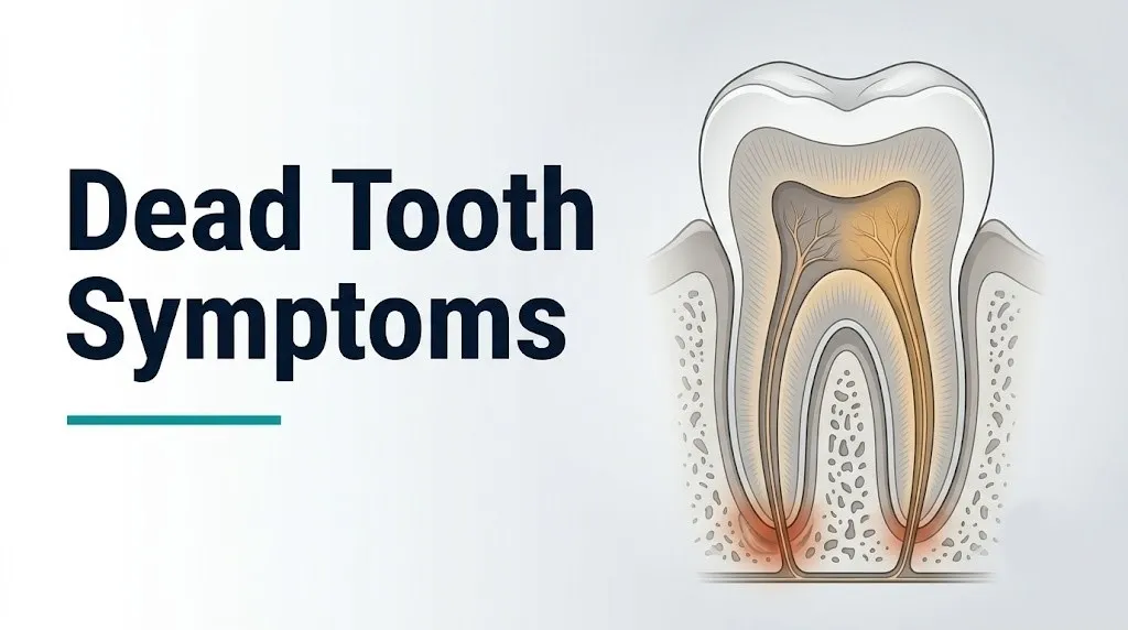
These early signs reflect the transition from reversible pulpitis (inflammation that can resolve without permanent damage) to irreversible pulpitis (inflammation that has overwhelmed the pulp’s vascular capacity and will progress to necrosis). The clinical distinction matters: reversible pulpitis produces pain that resolves within seconds of removing a cold stimulus. Irreversible pulpitis produces pain that lingers for 30 seconds or more after the stimulus is gone.
Early observable signs to monitor before seeing a dentist:
- Spontaneous toothache that wakes you at night, with no obvious triggering stimulus
- Severe pain to cold that lingers for 30 seconds or longer after the cold source is removed (this distinguishes irreversible from reversible pulpitis)
- Pain that is initially sharp and well-localized (A-delta fiber activation) but transitions toward dull, throbbing, and poorly localized (C-fiber dominance as inflammation intensifies)
- A tooth that has recently received a deep filling, crown preparation, or has a visible crack line: all of these represent insults that can push a borderline pulp into irreversible pulpitis
- Very mild sensitivity to biting, which reflects early spread of inflammation from the pulp to the periapical periodontal ligament fibers
According to the International Endodontic Journal, the combination of lingering cold sensitivity (more than 30 seconds) and spontaneous pain predicts irreversible pulpitis with high clinical accuracy in prospective diagnostic studies, making these the two key early signs to communicate to a dental provider.
Individual variation note: Some individuals have dentinal hypersensitivity from gum recession that produces cold sensitivity without any pulp pathology. A dentist can distinguish these by applying a refrigerant spray directly to the isolated tooth: in dentinal hypersensitivity, the pain resolves immediately. In irreversible pulpitis, it persists.
Dead Tooth Smell and Bad Taste in the Mouth
A persistent foul smell or bad taste associated with a specific tooth is produced by volatile sulfur compounds (VSCs), primarily hydrogen sulfide and methyl mercaptan, generated by the anaerobic bacteria colonizing the necrotic pulp space.
The smell of a necrotic tooth is clinically distinctive. It is not the general bad breath of poor oral hygiene, which is distributed across the mouth and worsens through the day. The odor from a dead tooth is more localized and often reported as particularly foul, sometimes described as a sulfurous, rotten, or earthy smell. Patients sometimes notice it most when pressing the tongue against the affected tooth or when a draining abscess opens and releases purulent material into the oral cavity.
The bacteria responsible are the same gram-negative anaerobes that colonize the necrotic pulp space. Fusobacterium nucleatum is one of the most prolific VSC producers among endodontic pathogens, according to microbiological studies published in the Journal of Endodontics. These compounds are not simply unpleasant: they are also biologically active, capable of disrupting the epithelial barrier of the gingival sulcus and contributing to local tissue breakdown.
A bad taste without a draining sinus tract may indicate that bacterial products and breakdown metabolites are diffusing from the necrotic pulp through a microscopic crack or through the gingival sulcus of a deeply periodontally involved tooth. A metallic or pus-like taste with sudden relief of pressure usually indicates that an abscess has spontaneously drained, which reduces acute pressure but does not resolve the underlying infection.
Quick Tip:
- Keep a note of whether the bad taste or smell is localized to one area of the mouth or generalized, as this helps the dentist identify the likely source tooth before examination
- Do not use mouthwash to mask the symptom before your dental appointment, as the taste can provide diagnostic information
- Patients wearing orthodontic appliances may have difficulty isolating the source tooth; inform your orthodontist or dentist of the localized nature of the taste or smell
Individual variation note: People with xerostomia (dry mouth) from medications such as antihistamines, antidepressants, or antihypertensives have reduced salivary flow, which normally dilutes and neutralizes VSCs. Reduced saliva makes odor from a necrotic tooth more pronounced and may make the patient more acutely aware of the taste than someone with normal salivary flow.
Dead Tooth Swelling and Gum Symptoms
Gum swelling near a dead tooth reflects the extension of periapical inflammation and infection from the root apex through the alveolar bone toward the gingiva, and it represents a clinically more advanced stage than pain or discoloration alone.
When bacterial toxins from the necrotic pulp diffuse through the apical foramen into periapical bone, they trigger a localized immune response. Macrophages, neutrophils, and osteoclasts are recruited to the periapical area. Prostaglandin E2 production drives osteoclast-mediated alveolar bone resorption, creating the radiolucent lesion visible on periapical radiographs as a dark halo around the root tip. As the infection enlarges, it erodes through the alveolar bone cortex and tracks toward the mucosal surface of the gum.
The gum swelling associated with a dead tooth has specific characteristics that distinguish it from gum disease (periodontitis):
- It tends to be localized directly adjacent to the affected tooth rather than generalized across multiple teeth
- The swelling feels firm initially (cellulitis phase) and becomes fluctuant (soft, fluid-filled) when an abscess cavity forms
- Pressing on the swelling may produce a sensation of pressure relief or may cause purulent discharge into the mouth
- The overlying gum may be red, shiny, and tender to touch
Swelling that extends beyond the gum and onto the cheek, under the chin, or down the neck has crossed from a dental-level infection into a fascial space infection. This transition represents a clinical emergency requiring immediate evaluation, not a dental appointment scheduled for next week.
The Mayo Clinic identifies spreading facial swelling from dental infection as a medical emergency because the fascial spaces of the head and neck are anatomically connected and allow rapid spread of infection toward the airway and mediastinum.
Individual variation note: Patients taking corticosteroids chronically (for conditions such as asthma, rheumatoid arthritis, or inflammatory bowel disease) may show blunted gum swelling and reduced localized pain response despite significant underlying infection, because corticosteroids suppress the prostaglandin-mediated inflammatory response. These patients may present with more advanced infection than the external examination suggests.
Key Takeaway: Gum swelling that extends visibly beyond the tooth and gum area onto the cheek, floor of the mouth, or neck is not a dental appointment situation: it is an emergency room situation, because the infection is tracking through fascial spaces toward the airway.
Periapical Abscess Symptoms From a Dead Tooth
A periapical abscess from a dead tooth produces throbbing pain that is worse with biting or lying down, localized gum swelling that may be fluctuant, systemic symptoms including fever and malaise, and visible bone destruction detectable on dental radiographs.
A periapical abscess forms when the accumulation of pus at the root apex is contained within a walled-off cavity in the alveolar bone. The pressure inside this cavity drives the characteristic throbbing pain of dental abscess, which is intensified by increased blood pressure during physical exertion and by the dependent positioning of the head during sleep, both of which increase perfusion pressure at the infected site.
The paradox of periapical abscess pain is that the necrotic tooth itself produces no pain (the pulp is dead), but the surrounding living periodontal ligament and alveolar bone are intensely inflamed by the bacterial products diffusing from the root apex. The pain is coming from the tooth’s neighborhood, not from the tooth itself. This is why the pain is described as aching and pressure-like, localizable to the jaw rather than the specific tooth, and worsened by tapping the tooth but not by thermal stimuli.
Key symptoms that distinguish a periapical abscess from uncomplicated pulp necrosis:
- Return of moderate to severe pain after a pain-free period of days to weeks
- Constant, throbbing pain that is not triggered by a specific stimulus
- Tenderness when biting on the affected tooth (percussion sensitivity of the periodontal ligament)
- Swelling of the gum adjacent to the root tip, sometimes with a fluctuant quality
- Fever (temperature above 38°C / 100.4°F), indicating systemic bacterial involvement
- Swollen lymph nodes under the jaw or in the neck on the same side as the affected tooth
Individual variation note: Patients who are immunocompromised from HIV infection, chemotherapy, or organ transplant immunosuppression may not mount a normal fever or significant gum swelling response despite severe periapical abscess, because their immune system cannot produce the inflammatory reaction that drives these signs. In this population, the absence of swelling or fever does not indicate a less serious infection.
Dead Tooth Fistula and Sinus Tract
A dental fistula, also called a sinus tract or parulis, is a small, pimple-like bump on the gum surface through which the periapical abscess drains, and its appearance signals that the infection has been present long enough to erode a channel through alveolar bone to the mucosal surface.
A sinus tract forms when the pressure inside a periapical abscess exceeds the resistance of the alveolar bone and mucosal tissue. Pus follows the path of least resistance through the bone, bursts through the cortical plate, and creates a draining channel to the oral mucosal surface. The small bump visible on the gum is called a parulis: a mucosal elevation at the external opening of the sinus tract.
The clinical significance of a sinus tract is twofold. First, its presence confirms that a chronic periapical abscess exists and has been present long enough to produce bone erosion and a stable drainage channel. Second, the temporary drainage of pus through the sinus tract may substantially reduce acute pain, creating the misleading impression that the infection is resolving. It is not resolving; the source of infection in the necrotic pulp remains, and the sinus tract will continue to drain until the source is treated.
Endodontists use a clinical technique called gutta-percha tracing to identify which tooth a sinus tract is associated with: a small flexible cone (gutta-percha point) is gently inserted into the sinus tract opening, and a periapical radiograph is taken to visualize the tract’s path back to the offending root apex. This is the standard diagnostic approach when the source tooth is not obvious from clinical examination alone.
Quick Tip:
- A sinus tract on the gum that appears and disappears repeatedly over weeks is not a minor recurring irritation; it is chronic abscess drainage signaling an untreated necrotic tooth
- Do not attempt to squeeze or pop the bump; this does not accelerate drainage and risks introducing oral flora into the deeper tissue
- Sinus tracts associated with back teeth (molars and premolars) can sometimes open on the palatal gum or buccal mucosa rather than on the labial gum, so examine the full gum surface around the suspected tooth
Individual variation note: In children with primary teeth (baby teeth), a draining sinus tract from a necrotic primary molar can be located not on the gum near that tooth but at a site that appears anatomically distant, because the roots of primary molars are positioned adjacent to the developing permanent tooth germs. The infection can sometimes mimic a skin lesion on the chin or jaw, leading to misdiagnosis as a dermatological condition before the dental source is identified.
Late Stage Dead Tooth Symptoms
Late stage dead tooth symptoms include severe recurring pain at the periapical site, visible facial asymmetry from swelling that has extended beyond the gum, systemic signs of infection including fever, malaise, and cervical lymphadenopathy, and in the most severe cases, difficulty opening the mouth, swallowing, or breathing.
By the time a dead tooth has reached the late stage, the periapical infection has typically been progressing for weeks to months. The chronic granuloma or cyst that forms around the root apex continues to enlarge, eroding progressively more alveolar bone. At this stage, the bone destruction visible on periapical radiographs is often extensive, and the tooth may be non-restorable.
Late-stage systemic signs that indicate spread beyond the tooth and jaw:
- Fever above 38°C (100.4°F) with no other obvious cause, combined with jaw or facial pain
- Swelling under the jaw or in the submandibular region, which indicates infection has spread into the submandibular space
- Swelling of the floor of the mouth, which can push the tongue upward (a warning sign of Ludwig’s angina)
- Trismus: difficulty or inability to fully open the mouth (normal opening is 35 to 55 mm between upper and lower incisors), indicating involvement of the masseter or pterygoid muscles
- A muffled or “hot potato” voice quality, indicating pharyngeal or parapharyngeal space involvement
- Any difficulty breathing or swallowing
These late-stage systemic signs are not dental office presentations. They are emergency department presentations.
According to a retrospective case series published in Oral Surgery, Oral Medicine, Oral Pathology, Oral Radiology, the average time from onset of dental pain to hospital admission for serious odontogenic space infections is approximately 5 to 7 days, underscoring how rapidly an untreated periapical abscess can progress to a life-threatening complication.
Individual variation note: Elderly patients and patients with chronic kidney disease have impaired immune responses and reduced febrile response to infection. An older adult with a late-stage dental infection may present with systemic deterioration, confusion, or reduced appetite without a fever that would normally alert a clinician to an infectious process.
Dead Tooth Symptoms in Children
Dead tooth symptoms in children follow the same pathological mechanism as in adults but differ in clinical presentation and urgency because the wider apical foramen of primary teeth and developing permanent teeth allows faster spread of infection, and because periapical infection from a primary tooth can permanently damage the developing permanent tooth germ beneath it.
Children’s primary teeth have proportionally larger pulp chambers and wider apical foramina than adult permanent teeth. This means bacterial products from a necrotic primary pulp can diffuse more rapidly into periapical tissues, and the infection can extend more quickly to the alveolar bone and beyond. The developing permanent tooth germ lies directly below the roots of the primary molar in the mandible and maxilla. Periapical infection from a necrotic primary molar exposes the permanent tooth bud to bacterial toxins and inflammatory mediators that can cause:
- Enamel hypoplasia (defective enamel formation) on the crown of the developing permanent tooth
- Turner’s hypoplasia: a specific pattern of enamel defect named after its association with periapical infection from primary teeth
- Delayed eruption of the permanent successor
- In severe cases, structural damage to the permanent tooth bud requiring extraction
Signs in children that a primary tooth may be non-vital:
- A tooth that was previously painful and has gone quiet, combined with a new gum bump (parulis) near the root tip
- Swelling of the gum or cheek near a tooth that received prior trauma (a fall, impact from a toy, or sport injury)
- A primary tooth that appears grayish or darker than adjacent teeth
- A child who stops chewing on one side without explaining why
Parents should bring a child to a pediatric dentist immediately, not wait for the next scheduled checkup, if any of these signs are observed. Pediatric dentists are the appropriate primary provider for primary dentition evaluation; they can refer to a pediatric endodontist if root canal treatment of the primary tooth (pulpectomy) is indicated.
Key Takeaway: A dead primary tooth is not a minor concern because the infection is inevitable and the child “will lose the tooth anyway”; the periapical infection from a necrotic primary tooth can permanently damage the developing permanent tooth beneath it.
Dead Tooth Symptoms After Trauma
Dead tooth symptoms following dental trauma may not appear until weeks to months after the injury, because traumatic disruption of the apical blood supply often produces a slow ischemic necrosis rather than an immediate acute pulpitis.
Traumatic dental injuries are classified by the International Association of Dental Traumatology (IADT), with tooth luxation injuries (where the tooth is partially displaced in its socket), intrusion injuries (where the tooth is driven deeper into the socket), and avulsion injuries (where the tooth is completely knocked out) carrying the highest risk of subsequent pulp necrosis.
The mechanism differs from decay-related necrosis. In traumatic injury, the apical blood vessels supplying the pulp may be severed or stretched at the apical foramen from the impact. The pulp does not die immediately because the disruption of blood flow is incomplete in many cases. Over days to weeks, ischemic changes accumulate: odontoblasts die first, then fibroblasts, then the vascular endothelium, and finally the sensory nerves. The pulp dies without the dramatic acute pain of bacterial pulpitis because there is no bacterial activation of the inflammatory cascade in the early ischemic phase.
What to monitor after any dental trauma:
- Check the tooth color every two weeks for the first three months after injury: any gray or dark shift should trigger a dental evaluation
- Report any percussion sensitivity (tenderness when tapping the tooth) to a dentist, as this may indicate early periapical periodontitis from ischemic necrosis
- Attend the follow-up appointments recommended by the IADT guidelines, which specify periapical radiograph monitoring at 6 to 8 weeks, 6 months, and 1 year after significant traumatic injury
- Do not assume that a tooth that felt fine in the days immediately after trauma will remain healthy: delayed pulp necrosis after trauma can occur months later
- Inform the treating dentist about any prescribed medications taken at the time of trauma, particularly blood thinners or NSAIDs, which can affect pulpal blood flow assessment
Individual variation note: Young patients between ages 7 and 14 with incompletely formed root apices (open apices) have a wider apical foramen and richer apical blood supply, giving their traumatized teeth a greater capacity for pulp revascularization (spontaneous recovery of pulp vitality after ischemic injury). A dentist may elect to monitor rather than immediately treat a traumatized immature tooth for this reason. This approach is not appropriate for fully formed adult teeth with closed apices.
Dead Tooth Symptoms After a Root Canal
Symptoms after a root canal treatment can indicate either a successfully treated non-vital tooth that is healing normally, or a failing root canal with persistent or recurrent infection, and distinguishing between these two requires specific clinical and radiographic assessment.
After a technically successful root canal, the necrotic pulp tissue has been removed from the pulp chamber and root canals, the canal walls have been cleaned and shaped, and the space has been sealed with an inert filling material (gutta-percha) and sealer. A tooth that has had a successful root canal is dead by definition: it no longer contains living pulp tissue and has no nerve supply for sensation. Some patients are surprised to find that a root-canal-treated tooth feels different from adjacent teeth: slightly less sensitive to changes in temperature, and occasionally a subtle change in the perception of biting pressure.
Symptoms after a root canal that suggest normal healing (typically resolving within 1 to 2 weeks post-treatment):
- Mild to moderate soreness when biting on the treated tooth, from inflammation in the periapical periodontal ligament that was disturbed during instrumentation
- Mild gum tenderness around the treated tooth
- Gradual fading of any pre-existing swelling
Symptoms that suggest a failing or failed root canal (requiring re-evaluation by an endodontist):
- Pain or swelling that returns after a period of apparent resolution, typically weeks to months after treatment
- Persistence of a sinus tract or parulis beyond 3 months post-treatment
- No radiographic reduction in the size of a pre-existing periapical lesion at the 6-month follow-up radiograph (normally, a periapical lesion should show visible reduction in size within 6 months as the body resorbs the healed area)
- New swelling or pain in a tooth that was previously asymptomatic after root canal treatment
The American Association of Endodontists states that the success rate of primary root canal treatment for teeth with pre-existing periapical periodontitis is approximately 85 percent at four-year follow-up, confirming that a minority of cases require retreatment or surgical endodontic intervention (apicoectomy).
Individual variation note: Patients taking bisphosphonate medications (including alendronate, zoledronic acid) for osteoporosis or cancer-related bone metastases face an increased risk of medication-related osteonecrosis of the jaw (MRONJ) if a treated tooth is subsequently extracted. For these patients, maximizing the success of root canal treatment to avoid extraction is particularly important. An endodontist should be aware of bisphosphonate use before planning treatment.
When Dead Tooth Symptoms Require Emergency Evaluation
Certain symptoms associated with a dead tooth require immediate emergency evaluation. Do not wait to see if these resolve on their own.
A dead tooth that has progressed to a spreading fascial space infection can produce Ludwig’s angina, descending necrotizing mediastinitis, cavernous sinus thrombosis, or septicemia: all potentially fatal complications that require emergency surgical and medical intervention.
Call 911 or go to the nearest emergency room immediately if you experience:
- Swelling of the floor of the mouth that is lifting the tongue upward, indicating sublingual space involvement and imminent airway compromise
- Difficulty swallowing (odynophagia or dysphagia) combined with jaw or neck swelling, indicating pharyngeal or parapharyngeal space involvement
- Difficulty breathing, any sensation of airway narrowing, or stridor (a high-pitched sound during breathing), indicating supraglottic edema or parapharyngeal space infection near the airway
- Trismus (inability to open the mouth more than approximately 20 mm) combined with fever and neck or jaw swelling, indicating pterygoid or masseteric space infection with systemic involvement
- Fever above 38.5°C (101.3°F) combined with visible facial or neck swelling on the same side as a known or suspected dental infection
- A muffled or “hot potato” voice quality, indicating the pharyngeal space is being compressed by swelling
- Rapid onset swelling of the neck below the jawline, which can indicate spread into the danger space with risk of descending mediastinitis
- Confusion, chills, or feeling severely unwell out of proportion to the visible dental findings, which may indicate early sepsis from odontogenic bacteremia
These presentations require emergency medical assessment at a hospital with oral and maxillofacial surgery capabilities, not an urgent care center or dental office.
How Dentists Diagnose a Dead Tooth
Dentists diagnose a dead tooth through a combination of pulp vitality testing, percussion and palpation examination, and periapical radiography, with cone beam computed tomography (CBCT) used in ambiguous cases.
The diagnostic process typically unfolds as follows:
- Clinical history: The dentist asks about the onset, character, and duration of pain; history of trauma to the tooth; prior dental treatment; and any systemic health conditions that affect immune function or bone metabolism
- Visual examination: Assessment of tooth color (internal discoloration), any visible swelling of adjacent gum tissue, any visible crack lines, and examination of the gingival sulcus for signs of periodontal involvement
- Percussion test: The dentist gently taps the biting surface of the suspected tooth and adjacent teeth with a dental mirror handle. A dead tooth with periapical periodontitis produces a distinct, positive percussion response: tenderness that is greater than on adjacent teeth, indicating inflammation of the periodontal ligament fibers around the root apex
- Palpation test: The dentist presses on the gum overlying the root apex. Tenderness here confirms periapical tissue involvement
- Electric pulp test (EPT): A small electrical current applied to the tooth surface stimulates sensory nerve fibers in the pulp. A vital tooth produces a tingling or mild sharp sensation. A non-vital (dead) tooth produces no response at any current level. A negative EPT response is a primary indicator of pulp necrosis.
- Cold test: A cotton pellet or paper point soaked in refrigerant spray is applied to the tooth. Absence of any cold sensation confirms non-vitality and is often used alongside the EPT for corroboration
- Periapical radiograph: The most clinically essential imaging tool. A radiolucent (dark) halo around the root apex on the radiograph indicates bone destruction from periapical periodontitis, confirming established disease. Widening of the periodontal ligament space at the apex is an earlier radiographic sign.
- CBCT imaging: Used when the periapical radiograph is inconclusive, when the tooth has multiple roots with complex anatomy, or when surgical endodontic planning is needed. CBCT provides three-dimensional visualization of the root, surrounding bone, and any associated lesion.
The American Association of Endodontists recommends that all patients with suspected non-vital teeth be evaluated by a board-certified endodontist when the clinical presentation is complex or when initial treatment has not resolved symptoms.
Individual variation note: Pregnant women requiring dental radiographs for a suspected non-vital tooth should be reassured that modern dental radiographs with digital sensors and thyroid collar shielding deliver extremely low radiation doses. The American College of Obstetricians and Gynecologists confirms that necessary dental radiographic examination during pregnancy is safe and should not be deferred if the clinical situation warrants it.
Key Takeaway: A negative response to both the electric pulp test and the cold test in the same tooth is the strongest combined clinical evidence that a tooth is non-vital; a dentist who finds this combination will almost certainly recommend endodontic evaluation rather than watchful waiting.
Treatment Options for a Dead Tooth
Treatment for a dead tooth is either root canal treatment (endodontic therapy), which preserves the tooth structure while eliminating the necrotic pulp and periapical infection, or tooth extraction, which removes the entire tooth and its associated infection; the choice depends on the degree of remaining tooth structure, bone support, and the patient’s overall dental health.
Root canal treatment for a necrotic tooth involves:
- Accessing the pulp chamber by drilling through the crown
- Removing all necrotic pulp tissue from the pulp chamber and root canal system using progressively larger files
- Irrigating the canal system with sodium hypochlorite solution to kill residual bacteria and dissolve organic debris
- Shaping the canal walls for a predictable seal
- Filling the cleaned and shaped canal system with gutta-percha and sealer to prevent bacterial recolonization
- Restoring the crown of the tooth, typically with a dental crown, to protect the now-brittle tooth structure from fracture
For teeth with successful root canal treatment but persistent internal discoloration, internal bleaching (the walking bleach technique) involves placing a sodium perborate bleaching agent inside the sealed pulp chamber to oxidize the hemoglobin breakdown products staining the dentin from within.
Tooth extraction is appropriate when the tooth is not restorable due to extensive decay, fracture below the bone level, or severe bone loss, or when the patient cannot afford root canal treatment and crown restoration.
After extraction of a tooth with a periapical abscess, the socket is irrigated and debrided. Systemic antibiotic therapy is typically prescribed for 5 to 7 days when systemic signs of infection are present or when the patient is immunocompromised, according to clinical guidelines from the American Dental Association.
Individual variation note: Patients with end-stage renal disease on hemodialysis have altered drug pharmacokinetics that affect the dosing and selection of antibiotics used for post-extraction dental infection management. An oral and maxillofacial surgeon or endodontist treating these patients should coordinate antibiotic prescribing with the patient’s nephrologist.
Frequently Asked Questions About Dead Tooth Symptoms
Can a dead tooth hurt?
A dead tooth can hurt, particularly during the transition from acute pulpitis to pulp necrosis in the early stage, and again if a periapical abscess forms in the later stage.
The pulp necrosis phase itself may produce no pain because the sensory nerve fibers have been destroyed, but pain returns when periapical inflammation from bacterial toxins diffusing through the apical foramen stimulates the living periodontal ligament and surrounding bone.
Any tooth that has stopped hurting after a period of intense pain should still be evaluated by a dentist, because painlessness does not mean the infection has resolved.
What does a dead tooth look like?
A dead tooth typically appears gray, yellow-brown, or black compared to adjacent teeth, because hemoglobin breakdown products from lysed red blood cells in the necrotic pulp have diffused into the dentinal tubules and stained the dentin from the inside.
The discoloration develops gradually over weeks to months after pulp death and cannot be removed by surface polishing or whitening toothpaste because it is internal, within the dentin itself.
A dentist can confirm whether discoloration is from internal necrosis or from external staining (coffee, tea, tobacco) by examining the tooth surface and performing pulp vitality testing.
How do I know if my tooth is dead or just infected?
A dead tooth and an infected tooth are not mutually exclusive; a dead tooth is almost always infected, because necrotic pulp tissue becomes colonized by anaerobic bacteria that drive the periapical infection.
The clinical distinction that matters is between a tooth with localized infection (necrotic pulp with periapical periodontitis contained within the jaw) and a tooth whose infection has spread into the fascial spaces of the head and neck.
A licensed dentist or endodontist can confirm pulp necrosis through the electric pulp test and cold test, and can assess the extent of infection through clinical examination and periapical radiography.
Can a dead tooth go away on its own?
A dead tooth cannot resolve without treatment; the necrotic pulp does not regenerate, and the bacterial colonization of the pulp space does not self-sterilize.
A periapical abscess may temporarily drain through a sinus tract, reducing acute pain and creating the false impression of resolution, but the infection source in the necrotic pulp remains and will continue to drive periapical bone destruction.
Without root canal treatment or extraction, a dead tooth will progress to a larger and more destructive periapical lesion, and may eventually produce a spreading fascial space infection.
How long does it take for a tooth to die?
The time from initial pulp insult to complete pulp necrosis varies widely, from days in the case of acute traumatic vascular disruption to weeks or months in the case of slowly progressing bacterial invasion from deep dental decay.
According to the International Endodontic Journal, the rate of pulp necrosis depends on the virulence and quantity of the infecting bacteria, the adequacy of the pulp’s vascular defense response, and individual immune factors.
There is no reliable way to predict the timeline in a specific individual, which is why prompt dental evaluation of any tooth with prolonged pain or cold sensitivity is clinically appropriate rather than a wait-and-see approach.
When is a dead tooth a dental emergency?
A dead tooth becomes a dental emergency when the associated infection has spread beyond the confines of the tooth and alveolar bone into the surrounding fascial spaces of the face and neck.
Specific emergency signs include spreading facial or neck swelling, fever above 38.5°C (101.3°F), difficulty swallowing or breathing, inability to fully open the mouth combined with systemic signs, or a muffled voice quality, any of which indicate a potentially life-threatening space infection requiring emergency room evaluation immediately.
For a non-vital tooth without these signs, evaluation by a dentist or endodontist within 24 to 48 hours is appropriate rather than emergency room presentation.
Closing
The symptoms of a dead tooth follow a predictable biological sequence once you understand the underlying anatomy: from the early severe pain of irreversible pulpitis, to the deceptive quiet of established necrosis, to the return of pressure and swelling as periapical infection expands into surrounding bone. Every stage has observable signs, and every stage has a corresponding clinical action.
The most important practical takeaway is this: a tooth that stops hurting is not a tooth that has healed. Get it evaluated by a dentist or endodontist within days, not weeks.
If you’re experiencing spreading facial swelling, fever, difficulty swallowing, or any breathing change alongside jaw pain, that is not a dental appointment situation. Go to the nearest emergency room immediately. Those symptoms describe a fascial space infection that can threaten the airway within hours, and that distinction between a dentist visit and an emergency room visit is the single most clinically important thing this article can tell you.