Tight Psoas Symptoms: The Complete Clinical Guide 2026
Tight psoas symptoms extend well beyond lower back pain, and that distinction matters if you are trying to figure out what your body is actually doing. The psoas major is the only muscle connecting your lumbar spine directly to your lower limb, which means when it shortens or strains, the symptom pattern can reach your groin, your thigh, your pelvis, and even your digestive tract.
According to the National Institute of Arthritis and Musculoskeletal and Skin Diseases (NIAMS), lower back pain affects an estimated 80% of adults at some point, and hip flexor dysfunction, including tightness of the psoas and iliacus, is among the most consistently underidentified musculoskeletal contributors to that statistic. The psoas is rarely the first structure examined, which is why so many people with chronic lower back pain and hip pain continue cycling through treatment without resolution.
This article covers the full clinical picture of tight psoas symptoms, from the most common presentations to the atypical ones that most health content never mentions. It explains the anatomical mechanism behind each symptom, distinguishes a chronically tight psoas from an acute psoas strain and a psoas tear, identifies who is at elevated risk, and tells you precisely when a symptom requires professional evaluation rather than home management.
Tight Psoas Symptoms: What They Are and Why They Happen
Tight psoas symptoms are the physical and neurological effects produced when the psoas major and its companion muscle the iliacus shorten adaptively or become acutely overloaded, altering the mechanics of the lumbar spine, the hip joint, the pelvis, and the nearby nerve structures.
The range of symptoms is wider than most people expect. Pain in the lower back and hip is the most recognizable presentation, but the psoas also generates referred pain into the groin and anterior thigh, postural changes visible in the shape of the lower back, and in some cases neurological symptoms in the distribution of the nerves that run directly through or alongside the muscle itself.
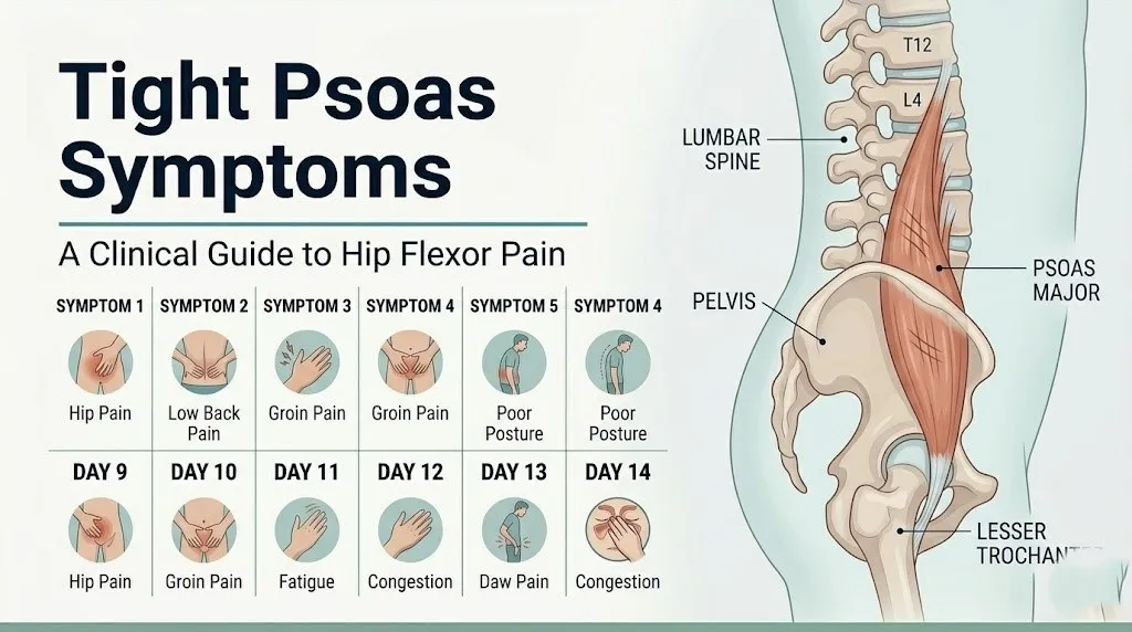
The primary symptom categories associated with psoas tightness:
- Deep, aching lower back pain, typically worse after sitting for extended periods
- Hip flexor pain at the front of the hip or into the groin, especially with standing from a seated position
- Anterior pelvic tilt producing an exaggerated lumbar arch and lower back stiffness
- Referred pain into the anterior thigh, groin, or inner thigh in the distribution of the femoral or genitofemoral nerve
- Difficulty fully straightening the hip when standing or walking (hip flexion contracture)
- Snapping or clicking sensation in the front of the hip with movement (internal coxa saltans)
- Pelvic floor tension and in some individuals altered bowel or bladder function
- In severe or traumatic cases, acute severe hip and groin pain with loss of hip flexion strength
Symptoms generally follow a spectrum from mild adaptive tightness, manageable with targeted physical therapy, through to acute strain or complete tear requiring imaging and specialist evaluation. Identifying where on that spectrum you are sitting is the most practically useful thing this article can help you do.
What Is the Psoas Muscle and the Iliopsoas Complex?
The psoas major is a long, thick, fusiform (spindle-shaped) muscle that originates from the anterior surfaces and transverse processes of the T12 through L5 vertebrae and descends through the posterior abdominal wall, passing beneath the inguinal ligament to join the iliacus muscle and insert together on the lesser trochanter of the femur. This shared distal tendon is the iliopsoas tendon, and together the two muscles form the iliopsoas complex, the body’s most powerful hip flexor.
Think of the psoas major like a thick rope running from your mid-back to the top of your inner thigh, passing through your pelvis. When that rope is the right length, it holds your lumbar spine and hip in balanced alignment. When it is too short, it pulls both ends of the rope toward each other, dragging your lumbar vertebrae forward and tilting your pelvis down at the front. Everything attached to those structures has to compensate.
The iliacus originates separately from the iliac fossa (the inner concave surface of the iliac bone) and the anterior sacroiliac ligament, making it a pure hip flexor without the spinal attachment of the psoas major. This anatomical distinction matters clinically: symptoms arising from the iliacus alone tend to be more focal to the hip and groin, while symptoms arising from the psoas major involve the lumbar spine as well.
A third muscle, the psoas minor, is present in approximately 50% of the population. It plays a minor postural role and rarely produces isolated symptoms, but its presence or absence affects the overall tension distribution within the iliopsoas region.
The lumbar plexus (L1-L4 nerve roots) runs within the substance of the psoas major muscle belly itself. This anatomical fact is the key to understanding why psoas tightness can produce nerve symptoms far from the hip.
Tight Psoas Lower Back Pain
Tight psoas lower back pain is produced by the anterior traction force the shortened psoas major exerts on the L1-L5 vertebral bodies and transverse processes, increasing lumbar lordosis, compressing the posterior intervertebral facet joints, and narrowing the intervertebral foramina through which spinal nerve roots exit.
The pain typically presents as a deep, dull ache in the lower lumbar region, often worse after prolonged sitting and initially better with movement, then worsening again with sustained activity. It is frequently described as feeling “stuck” or stiff rather than sharp. Rising from a chair often triggers a recognizable moment of discomfort as the shortened psoas resists the transition from flexion to extension.
The mechanism is specific. The psoas major attaches to the anterior aspects of the vertebral bodies from T12 to L5. When chronically shortened, it applies a constant forward-pulling moment on those vertebrae, which increases the compressive load on the posterior elements of the lumbar spine: the facet joint capsules, the ligamentum flavum, and the posterior longitudinal ligament. A 2020 study published in the Journal of Orthopaedic and Sports Physical Therapy found that hip flexor tightness measured by the modified Thomas test was independently associated with increased lumbar extension muscle activity at rest, suggesting that a tight psoas forces the lumbar extensors to work harder just to maintain neutral posture.
Individuals with pre-existing lumbar disc disease at L4-L5 or L5-S1 experience a compounding effect. The increased lordosis from psoas tightness adds posterior disc compression to a structure that may already have reduced disc height or annular integrity. Back pain in this group is frequently more severe and slower to improve than in individuals with no pre-existing disc pathology.
Quick Tip:
- Lower back pain from a tight psoas is typically worse after sitting more than 60 minutes and in the first few minutes of walking after standing up.
- It often improves temporarily with walking and returns with sustained standing or heavy loading.
- Individuals over age 50 with both a tight psoas and known osteoarthritis of the hip may experience back pain that is partially from the hip and partially from the psoas; a physical therapist or physiatrist can distinguish these contributions through clinical examination.
Tight Psoas Hip and Groin Pain
Tight psoas hip and groin pain occurs at the anterior hip joint and along the course of the iliopsoas tendon as it passes over the iliopectineal eminence (the bony ridge between the iliac bone and pubic bone) and inserts on the lesser trochanter of the femur.
The anterior hip pain from a tight psoas is characteristically felt deep in the groin or at the front of the hip crease, often described as a deep ache that transitions to a sharper sensation with specific movements: rising from a chair, climbing stairs, lifting the knee toward the chest, or transitioning from lying to standing. This is the direct mechanical effect of a shortened iliopsoas tendon being placed under sudden tensile load when the hip moves from flexion into extension.
Groin pain in the distribution of the genitofemoral nerve (L1-L2), which exits through the anterior surface of the psoas major, is a less recognized but clinically documented feature of psoas hypertonicity. This nerve provides sensory innervation to the inner thigh and in males to the scrotal skin, and in females to the labial skin via its genital branch. Research published in Clinical Orthopaedics and Related Research has documented genitofemoral nerve entrapment and irritation in individuals with iliopsoas pathology, producing a symptom pattern that can be misattributed to a hernia, testicular pathology, or gynecological condition.
A snapping or clicking sensation at the front of the hip during hip flexion and extension is called internal coxa saltans, or internal snapping hip syndrome. It is produced when the thickened iliopsoas tendon flicks over the iliopectineal eminence with hip movement. The American Academy of Orthopaedic Surgeons (AAOS) notes that internal snapping hip is often painless but becomes painful when combined with underlying iliopsoas bursitis or tendinopathy. If the snap is consistently painful, evaluation by a board-certified orthopedic surgeon or sports medicine physician is warranted.
Key Takeaway: Groin or inner thigh pain in someone with a known tight psoas may not be a hernia or reproductive issue; it may be genitofemoral nerve irritation from psoas hypertonicity, and this distinction requires clinical examination to establish.
Tight Psoas Anterior Pelvic Tilt and Posture Changes
Anterior pelvic tilt produced by a tight psoas is a postural adaptation in which the front of the pelvis tips downward and the back tips upward, increasing the lumbar lordosis (the inward curve of the lower back) and tilting the torso slightly forward at the hips.
The mechanical cause is direct. The psoas major’s proximal attachment at T12-L5 means that when it shortens, it pulls the lumbar vertebrae anteriorly and inferiorly. The iliacus, attaching along the inner surface of the iliac bone, simultaneously pulls the anterior pelvis downward. The combined effect rotates the pelvis forward on the femoral heads, producing the characteristic posture: a pronounced inward lower back curve, a protruding lower abdomen, and buttocks that appear to stick out posteriorly.
This postural shift has mechanical consequences beyond cosmetic appearance. The sacroiliac joint (SI joint) is placed under asymmetric shear loading when anterior pelvic tilt is sustained. Many people with chronic sacroiliac joint pain have co-occurring psoas tightness, though the causal relationship between the two is bidirectional and the evidence base is observational rather than from controlled trials.
The transversus abdominis, the deep abdominal muscle that functions as the primary anterior counterbalance to the psoas, becomes functionally inhibited in the presence of sustained anterior pelvic tilt. Research published in the British Journal of Sports Medicine has found an association between hip flexor tightness and reduced activation of the deep abdominal stabilizers during functional tasks, suggesting that a tight psoas does not just pull the spine forward but also reduces the opposing muscular control system’s efficiency.
Postmenopausal women with reduced estrogen-related changes in connective tissue extensibility may find that anterior pelvic tilt from psoas tightness is more pronounced and less responsive to stretching alone than in younger adults. A licensed physical therapist with orthopedic specialization can assess pelvic alignment and design a program addressing both the psoas tightness and the secondary transversus abdominis weakness.
Tight Psoas Nerve Symptoms and Referred Pain
Tight psoas nerve symptoms occur because the lumbar plexus (the network of L1-L4 nerve roots) runs directly through the substance of the psoas major muscle belly, not alongside it. When the psoas is hypertonic or acutely swollen from strain, it compresses the nerve roots within it, producing neurological symptoms in the distribution of the nerves that emerge from the plexus.
The femoral nerve (L2-L4) exits from the lateral border of the psoas major and travels into the anterior thigh. Compression or irritation from a tight psoas can produce paresthesia (tingling or numbness) in the anterior thigh and medial leg, reduced quadriceps strength, and sometimes a diminished knee reflex. This symptom pattern can mimic a lumbar disc herniation at L3-L4, and clinically distinguishing the two requires careful physical examination and, in uncertain cases, MRI of the lumbar spine.
The lateral femoral cutaneous nerve (L2-L3) exits the lateral border of the psoas and crosses the iliac fossa before passing beneath the inguinal ligament. Compression of this nerve produces meralgia paresthetica, a condition characterized by burning, tingling, and numbness on the outer surface of the thigh. While the inguinal ligament crossing point is the most common site of compression for this nerve, psoas hypertonicity that narrows the nerve’s passage through the iliac fossa can contribute to the same symptom pattern.
Referred pain from the psoas follows a pattern described in myofascial pain literature as running from the lower back diagonally through the ipsilateral buttock and down the anterior thigh, without following a dermatome precisely. This referral pattern is generated by nociceptor activation in the psoas muscle fascia itself, not by nerve root compression. The distinction between myofascial referred pain and radicular nerve root pain is clinically important: the former does not produce true neurological deficits (weakness, absent reflexes, dermatome-specific numbness), while the latter does.
Tight Psoas and Pelvic Floor Symptoms
A tight psoas can contribute to pelvic floor symptoms through its anatomical proximity to the pelvic floor musculature and its effect on intra-abdominal pressure mechanics, though this relationship is supported by clinical observation and pelvic health physical therapy literature rather than large-scale controlled trials.
The iliopsoas complex runs through the posterior pelvis in close proximity to the levator ani muscle group (the primary pelvic floor muscle complex). The psoas major also lies adjacent to the sympathetic chain ganglia on the anterior surfaces of the lumbar vertebrae (L1-L5), which regulate smooth muscle tone in the pelvic viscera including the bladder and bowel. Clinicians in pelvic floor physical therapy consistently observe that individuals with psoas hypertonicity often present with co-occurring pelvic floor hypertonicity, though whether the psoas causes the pelvic floor tightness or both arise from a common neuromuscular tension pattern remains an open clinical question.
Observable symptoms associated with this connection:
- Urinary urgency or frequency without a urinary tract infection
- Difficulty fully emptying the bladder
- Pelvic pressure or heaviness, particularly with prolonged standing
- Constipation or difficulty with bowel evacuation related to altered pelvic floor muscle coordination
- Dyspareunia (painful intercourse) in females with co-occurring pelvic floor hypertonicity
- Pelvic pain radiating to the inner thigh or perineum
These symptoms, when present alongside hip and lower back pain, should prompt referral to a pelvic floor physical therapist for assessment. A pelvic floor PT can assess iliopsoas tension through internal pelvic examination and determine whether a co-occurring pelvic floor component is present. This is a specifically trained specialty distinct from general orthopedic physical therapy.
Women in their reproductive years are more likely to seek care for these symptoms than men, partly because of the female pelvic anatomy (a wider pelvis creates different angulation of the iliopsoas through the pelvic brim) and partly because dyspareunia is an established clinical trigger for seeking pelvic health evaluation. Men with these symptoms are less likely to connect them to psoas function and may be evaluated for prostate or urological conditions first.
Key Takeaway: Urinary urgency or pelvic pressure alongside hip and lower back pain without a urinary infection found on testing warrants assessment by a pelvic floor physical therapist, not just a urologist or gynecologist.
Weak Psoas Symptoms
Weak psoas symptoms are distinct from tight psoas symptoms and are frequently confused with them, yet they produce an opposite clinical pattern that requires different evaluation and management.
A weak psoas major fails to adequately support the lumbar spine’s neutral alignment during loaded activity. Instead of pulling the lumbar spine into excessive lordosis as a tight psoas does, a weak psoas allows the lumbar spine to flex under load, reducing the protective muscular corset around the L1-L5 vertebral bodies. The result is lumbar instability, difficulty sustaining upright posture under load, and a tendency toward compensatory overactivation of the erector spinae and quadratus lumborum muscles.
Weak psoas symptoms include:
- Difficulty lifting the knee against resistance (hip flexion weakness, particularly above 90 degrees of hip flexion)
- Lumbar flexion under load (the lower back rounds when standing from a squat or picking up an object)
- Inability to maintain a stable single-leg stance without lateral trunk shift
- Fatigue and aching in the lateral lumbar and hip region from compensatory overload of the quadratus lumborum
- A tendency to “hinge” at the lower back rather than the hip during forward bending
- Hip instability during walking or running, visible as lateral pelvic drop (Trendelenburg gait pattern)
Psoas weakness and psoas tightness can coexist. A chronically shortened psoas can simultaneously be weak in its functional range because sarcomere adaptations from sustained shortening reduce the muscle’s capacity to generate force across its full contractile length. This co-occurrence is well-documented in the physical therapy literature and explains why stretching alone does not resolve symptoms in everyone with psoas tightness.
According to clinical guidelines from the American Physical Therapy Association (APTA), hip flexor strength testing should be included in any comprehensive lower back pain assessment, as weakness in this muscle group alters lumbar segmental stability and changes the loading pattern on the posterior spinal elements.
Older adults aged 65 and above show age-related sarcopenia (muscle mass reduction) that affects the psoas major along with other large postural muscles. In this population, psoas weakness contributes to falls risk and difficulty rising from a seated position. A board-certified physiatrist or an orthopedic physical therapist can design a progressive strengthening program specific to this age group’s functional and structural needs.
Psoas Strain Symptoms vs. Chronic Psoas Tightness
A psoas strain and chronic psoas tightness are not the same condition. They differ in mechanism, onset, symptom character, severity, and appropriate management, and confusing them leads to the wrong treatment.
Chronic psoas tightness develops gradually from repeated or sustained hip flexion (sitting, cycling, running) that reduces the muscle’s resting length through adaptive sarcomere shortening. The onset is insidious. There is rarely a specific incident the person can point to. The pain is a deep, dull ache that builds over weeks to months, is worse after prolonged sitting, and is not associated with immediate bruising or swelling.
Acute psoas strain occurs when the psoas major is subjected to a load or force exceeding its tensile capacity, producing partial or complete disruption of muscle fibers. This can happen during a sudden forceful hip flexion (kicking, sprinting, cutting movements in sport) or during an eccentric contraction where the muscle is attempting to decelerate hip extension (the landing phase of a jump). The onset is immediate and identifiable.
| Feature | Chronic Psoas Tightness | Acute Psoas Strain |
|---|---|---|
| Onset | Gradual, over weeks to months | Sudden, during or immediately after a specific activity |
| Pain character | Deep ache, stiffness | Sharp, intense, immediate |
| Pain location | Lower back, hip crease, groin | Anterior hip, groin, and sometimes lower abdomen |
| Swelling or bruising | Absent | Possible in moderate to severe strain |
| Motion restriction | Mild reduced hip extension at end range | Significant loss of hip flexion strength and range |
| Aggravating factor | Prolonged sitting, then standing | Active hip flexion, resisted knee lift |
| Relieving factor | Gentle movement, position changes | Rest and ice in acute phase |
| Management | Physical therapy, stretching, postural correction | Rest, ice, graduated rehabilitation; imaging if severe |
| Timeline to recovery | 4 to 12 weeks with consistent therapy | 6 weeks to 6 months depending on grade |
Research published in the British Journal of Sports Medicine classifies muscle strains by grade: Grade 1 (minor fiber disruption, full strength preserved, tenderness on palpation), Grade 2 (partial tear, moderate strength loss, pain with resisted hip flexion), Grade 3 (complete rupture, complete loss of function, possible palpable defect). Grade 2 and Grade 3 psoas strains require evaluation by a board-certified sports medicine physician or orthopedic surgeon, with MRI to characterize the extent of fiber disruption.
Psoas Tear Symptoms
Psoas tear symptoms are the most severe presentation on the iliopsoas injury spectrum and represent a partial or complete rupture of the psoas major muscle belly or its tendon at the musculotendinous junction or at the insertion on the lesser trochanter.
A complete psoas tear or avulsion produces an acute, severe pain in the groin and anterior hip that is typically described as a pop or tearing sensation at the moment of injury. Immediate loss of hip flexion strength follows. The person is unable to lift the affected knee against any resistance and often cannot maintain a standing position on the affected side due to instability. Bruising may appear in the inner thigh or groin region within 24 to 48 hours as blood tracks along the fascial planes from the torn muscle.
Avulsion fracture of the lesser trochanter of the femur, where the iliopsoas tendon pulls away a fragment of bone at its attachment site, is a specific injury pattern seen most often in skeletally immature adolescents (ages 12 to 17) during rapid acceleration sports (sprinting, soccer, gymnastics). The AAOS notes that lesser trochanter avulsion typically presents as sudden severe groin pain during a sprinting or kicking motion, inability to flex the hip against gravity, and a positive plain radiograph showing the avulsed bony fragment. This injury is rare in adults with fully fused growth plates but documented in elderly patients with metastatic bone disease affecting the lesser trochanter.
Specific psoas tear symptoms requiring imaging evaluation:
- Sudden onset severe groin and anterior hip pain following a specific athletic or traumatic mechanism
- Complete inability to actively flex the hip against gravity or minimal resistance
- A palpable gap or concavity in the anterior hip or inner groin region
- Visible bruising extending from the groin toward the inner thigh within 48 hours of injury
- Lower abdominal pain radiating toward the hip on the same side
Any suspected psoas tear requires MRI of the hip and pelvis, ordered by a board-certified sports medicine physician or orthopedic surgeon. Plain radiographs alone will not reveal a muscle tear, though they will identify an avulsion fracture if one is present. Do not attempt to stretch or load a suspected tear before imaging.
Key Takeaway: Sudden severe groin pain following an athletic mechanism, especially if accompanied by loss of hip flexion strength, is a psoas tear until proven otherwise by MRI and warrants urgent evaluation by a sports medicine physician, not a rest-and-stretch approach at home.
Tight Psoas Symptoms in Athletes
Athletes develop tight psoas symptoms through a different mechanism than sedentary individuals, and the symptom pattern in this population often includes signs that a clinician who primarily sees desk-worker presentations may not immediately recognize.
In runners, repeated hip flexion through thousands of stride cycles per training session loads the psoas major eccentrically during the terminal swing phase of gait (when the hip extends behind the body and the psoas must decelerate that extension before driving the knee forward again). Over time, the psoas adapts to this repeated eccentric demand by increasing its baseline resting tone, a process that reduces its functional extensibility and eventually manifests as anterior hip tightness, reduced stride length on the affected side, and lower back stiffness in the early miles of a run.
In cyclists, the sustained hip flexion position maintained throughout pedaling places the psoas in a shortened position for extended periods, producing the same adaptive sarcomere shortening pattern as prolonged sitting, but with the added load of repeated concentric hip flexion contraction. Hip flexor tightness in cyclists often presents as groin discomfort at the end of long rides and stiffness in the first minutes off the bike.
Soccer players, gymnasts, and dancers are at elevated risk for iliopsoas tendinopathy and internal snapping hip syndrome (coxa saltans interna), both of which produce different symptom patterns from simple tightness: a painful audible or palpable snap at the front of the hip during specific movements (bringing the leg from a flexed, abducted, externally rotated position back to neutral), and localized tenderness along the iliopsoas tendon anterior to the hip joint.
Research published in the British Journal of Sports Medicine found that hip flexor tightness, assessed by the modified Thomas test, was present in a higher proportion of athletes reporting chronic lower back pain compared to athletes without back pain. The association was particularly strong in sports requiring repeated unilateral hip flexion loading (running, swimming, soccer).
Athletes with tight psoas symptoms who are mid-season should be evaluated by a board-certified sports medicine physician to determine whether the training load needs modification and whether an in-season physical therapy program is feasible without risking a progression to acute strain.
Tight Psoas Symptoms in Desk Workers and Sedentary Adults
Desk workers and sedentary adults represent the largest population experiencing tight psoas symptoms, and their presentation is distinct from the athletic population in both the mechanism of tightness and the symptom character.
Prolonged sitting maintains the hip in a flexed position for 6 to 10 or more hours daily. At approximately 90 degrees of hip flexion (a standard seated position), the psoas major is at a shortened resting length relative to its full anatomical range. The National Institute of Arthritis and Musculoskeletal and Skin Diseases (NIAMS) notes that sustained static muscle loading without recovery periods is a primary driver of musculoskeletal adaptive changes, including the sarcomere length changes that underlie chronic hip flexor tightness.
The most characteristic symptom pattern in desk workers is the “standing up stiffness” sign: a pronounced tightness and aching at the front of the hip and in the lower back during the first 30 to 60 seconds of standing after prolonged sitting. The shortened psoas resists the hip extension required for upright standing posture, producing that recognizable moment of pulling sensation at the hip crease.
Additional features specific to this population:
- Lower back pain that is consistently worse at the end of a workday but may improve on weekends with more movement
- Difficulty walking fully upright without a slight forward trunk lean at the hips
- Increased lower back pain when sleeping prone (face down), as this position places the hip into extension and tensionally loads a shortened psoas
- Morning stiffness in the lower back and hips that improves within 30 minutes of moving around
Adults aged 35 to 55 in sedentary occupations who also carry excess body weight may experience a more severe presentation of anterior pelvic tilt from the combined effect of psoas tightness and the anterior weight loading of abdominal adipose tissue. This combination increases lumbar compressive forces and is frequently mismanaged as simple lower back pain without addressing the underlying hip flexor component.
A structured assessment by a licensed orthopedic physical therapist, including the modified Thomas test and hip extension range of motion measurement, can confirm psoas tightness and distinguish it from other sources of low back pain within a single evaluation session.
Tight Psoas Symptoms During Pregnancy
Pregnant women experience a physiologically amplified version of psoas tightness symptoms because of two compounding anatomical changes: the progressive anterior shift of the body’s center of gravity from uterine enlargement, and the hormonally mediated ligamentous laxity produced by relaxin (a hormone secreted by the corpus luteum and placenta that increases joint and ligamentous mobility in preparation for delivery).
As the uterus enlarges across the second and third trimesters, the center of gravity shifts forward. The body compensates by increasing lumbar lordosis, a postural change that places the psoas major under greater mechanical demand. Simultaneously, the pelvic ligaments become more lax under the influence of relaxin, which reduces the passive restraint normally provided by the sacroiliac joint and pubic symphysis. The psoas, as an active hip flexor and lumbar stabilizer, is recruited more intensively to compensate for this reduced passive stability, which accelerates its tendency toward hypertonicity.
Tight psoas symptoms during pregnancy specifically include:
- Deep groin and anterior hip pain that worsens in the second and third trimester
- Lower back pain extending into the buttock that is worse with walking on uneven surfaces
- Difficulty turning over in bed at night from hip flexor discomfort
- Sacroiliac joint pain that co-occurs with hip flexor tightness from altered pelvic mechanics
- Pelvic pressure and pelvic floor heaviness, potentially amplified by combined psoas and pelvic floor hypertonicity
Management of psoas tightness in pregnancy requires modifications. Many standard psoas stretches (such as a low lunge with trunk extension) are not appropriate in the second or third trimester because of increased lumbar load and altered pelvic stability. A licensed physical therapist with perinatal specialization can prescribe pregnancy-safe hip flexor mobility work that addresses psoas tightness without compromising pelvic girdle stability. Pelvic support belts may be recommended as an adjunct.
Postpartum women should note that psoas tightness does not automatically resolve after delivery. Relaxin levels remain elevated during breastfeeding, and the combined demands of infant care (repeated bending, carrying, nursing in flexed positions) can perpetuate the tightness pattern established during pregnancy.
Key Takeaway: Psoas-related hip and back pain during pregnancy requires pregnancy-specific physical therapy assessment, not the same stretching protocols used for non-pregnant adults, because of the altered biomechanics introduced by relaxin-mediated ligamentous laxity.
Tight Psoas Red Flags and When to Seek Care
Tight psoas red flags are symptoms that indicate the psoas region pain is not from simple tightness or strain but from a more serious underlying condition requiring urgent medical evaluation.
The most serious diagnosis in the differential for severe one-sided lower back and hip pain is a psoas abscess. This is a bacterial infection, most commonly caused by Staphylococcus aureus or Mycobacterium tuberculosis in endemic regions, that forms within the psoas major muscle compartment. Psoas abscess is life-threatening without drainage and antibiotic treatment. It presents with fever, night sweats, severe unilateral lower back and flank pain, and often the person walks with the hip flexed on the affected side because extension is agonizing. The Mayo Clinic notes that psoas abscess can arise from direct spread from adjacent infected structures (vertebral osteomyelitis, Crohn’s disease affecting the terminal ileum, renal abscess) or hematogenous seeding in immunocompromised individuals.
Red flag symptoms requiring immediate or urgent medical evaluation:
- Fever above 101 degrees Fahrenheit combined with severe unilateral lower back, hip, or flank pain (possible psoas abscess)
- Unexplained weight loss combined with psoas region pain (possible neoplasm or systemic infection)
- Night pain that wakes from sleep and does not improve with position change (possible malignancy or serious infection)
- Bladder or bowel incontinence or retention combined with lower back and hip pain (possible cauda equina syndrome, a spinal cord emergency)
- Saddle area numbness (perineum, inner thighs, buttocks) combined with back and hip pain (possible cauda equina syndrome)
- Bilateral leg weakness or numbness developing alongside psoas region pain
- A pulsating mass or abnormal pulsation felt in the abdomen alongside lower back pain (possible abdominal aortic aneurysm)
- Psoas region pain following abdominal or retroperitoneal surgery (possible postsurgical complication)
These presentations are not psoas tightness. They are medical emergencies or serious conditions that require evaluation in an emergency department or by a physician the same day.
How a Tight Psoas Is Diagnosed Clinically
A tight psoas is diagnosed clinically through a structured physical examination combining flexibility testing, strength assessment, palpation, and functional movement analysis, with imaging reserved for cases where a tear, avulsion, abscess, or other structural pathology is suspected.
The primary clinical test for psoas and hip flexor length is the Thomas test. The modified version is performed as follows:
To perform and interpret the modified Thomas test:
- The patient sits at the edge of an examination table, then lies back while bringing both knees to the chest.
- One knee is released and allowed to lower toward the table while the opposite knee remains held to the chest, flattening the lumbar spine.
- In a person with normal iliopsoas length, the lowered thigh rests flat on the table surface (approximately 0 degrees of hip extension).
- In a person with psoas or iliopsoas tightness, the lowered thigh rises above the table level, indicating a hip flexion contracture. The angle above horizontal is measured as the degree of tightness.
- If the knee spontaneously extends as the hip lowers, this implicates the rectus femoris (another hip flexor) as a contributing muscle to the tightness rather than the psoas alone.
- Lateral fall of the lowered thigh (hip abduction with lowering) implicates the tensor fasciae latae and iliotibial band rather than the iliopsoas.
A study published in the Journal of Orthopaedic and Sports Physical Therapy reported that the modified Thomas test has good reliability for clinical hip flexor length assessment (intraclass correlation coefficient above 0.80 for experienced examiners), though its sensitivity for identifying the specific contribution of the psoas major versus the rectus femoris requires supplementary clinical observations.
Imaging is added when the diagnosis is uncertain or when red flags are present. Magnetic resonance imaging (MRI) of the hip and pelvis is the most informative modality for visualizing the psoas major, iliopsoas tendon, and iliopsoas bursa. MRI identifies muscle fiber disruption in a psoas strain, fluid within the iliopsoas bursa in bursitis, and soft tissue mass or abscess formation. Ultrasound imaging is increasingly used for real-time dynamic assessment of the iliopsoas tendon in suspected snapping hip syndrome, as it allows visualization of the tendon snap during active movement.
When to See a Doctor for Tight Psoas Symptoms
Most mild to moderate tight psoas symptoms can be initially managed with a targeted physical therapy program, but specific clinical circumstances require professional evaluation before or instead of self-directed management.
| Symptom Pattern | Recommended Provider | Timeframe |
|---|---|---|
| Mild lower back and hip stiffness, no neurological symptoms, no red flags | Licensed orthopedic physical therapist | Within 1 to 2 weeks if no improvement with activity modification |
| Anterior hip pain with snapping, possible bursitis or tendinopathy | Board-certified sports medicine physician or orthopedic surgeon | Within 1 to 2 weeks |
| Acute severe groin/hip pain following athletic activity | Board-certified sports medicine physician | Within 24 to 48 hours; MRI likely needed |
| Anterior thigh numbness, tingling, or quadriceps weakness | Primary care physician or physiatrist | Within 5 to 7 days |
| Urinary urgency or pelvic floor symptoms with hip/back pain | Pelvic floor physical therapist; also primary care for urinalysis | Within 1 to 2 weeks |
| Psoas pain during pregnancy | Licensed physical therapist with perinatal specialization | Within 1 week of symptom onset |
| Fever, night sweats, severe unilateral back/hip pain | Emergency department or primary care physician | Same day |
| Bladder or bowel dysfunction with back and hip pain | Emergency department | Immediately |
When you see a provider for psoas-related symptoms, bring: a description of when the pain started, what specific activities worsen or relieve it, any neurological symptoms you have noticed (numbness, tingling, weakness), any recent changes in bowel or bladder function, and a list of any previous imaging or physical therapy assessments for this region. This information allows a clinician to triage appropriately and avoid redundant testing.
Individuals who have already seen a primary care physician for lower back pain, received a generic diagnosis of “muscle strain,” and not improved after 4 to 6 weeks should specifically request a referral to a licensed physical therapist with orthopedic specialization for a hip flexor length assessment. This test takes approximately 5 minutes and frequently identifies an untreated psoas component that was not assessed in the initial evaluation.
Emergency Symptoms: When to Call 911 or Go to the ER
Certain symptoms associated with the psoas region require immediate emergency evaluation. Do not wait to see if these resolve on their own.
Call 911 or go to the nearest emergency room immediately if you experience:
- Loss of bladder or bowel control combined with lower back and hip pain (possible cauda equina syndrome, a spinal cord emergency requiring surgery within hours to preserve function)
- Inability to urinate combined with lower back and hip pain and lower extremity numbness (possible cauda equina compression)
- Numbness or complete loss of sensation in the saddle area (perineum, inner thighs, buttocks) with back and hip pain
- High fever (above 101 degrees Fahrenheit), severe unilateral lower back and flank pain, and inability to straighten the hip (possible psoas abscess, a life-threatening infection)
- Sudden severe tearing pain in the lower abdomen or back with a pulsating sensation in the abdomen (possible abdominal aortic aneurysm, a vascular emergency)
- Bilateral sudden onset leg weakness or paralysis with back pain following trauma
These presentations can indicate cauda equina syndrome, a retroperitoneal infectious emergency, or a life-threatening vascular event, and they require emergency medical assessment at a hospital, not an urgent care clinic or a next-day physician appointment.
Frequently Asked Questions About Tight Psoas Symptoms
What does a tight psoas feel like?
A tight psoas typically feels like a deep, dull ache in the lower back and a pulling or gripping sensation at the front of the hip or groin, especially when standing up after sitting for an extended period.
The discomfort often includes a sense of stiffness or limited range at the hip during the transition from sitting to standing, and may extend into the anterior thigh or inner groin.
Some individuals also notice that one side feels tighter than the other, producing a slightly uneven or “hitching” gait pattern when walking.
Can a tight psoas cause nerve pain down the leg?
A tight psoas can cause nerve pain in the anterior thigh and medial leg because the femoral nerve (L2-L4) exits directly from the lateral border of the psoas major.
When the psoas is hypertonic or swollen from strain, it can compress the femoral nerve and produce paresthesia, burning, or numbness in the anterior thigh and inner knee area.
This symptom pattern requires evaluation by a primary care physician or physiatrist to distinguish psoas-related femoral nerve irritation from a lumbar disc herniation at L3-L4, which produces a similar distribution of symptoms.
How do I know if my psoas is tight or strained?
A tight psoas produces a gradual onset of deep aching stiffness in the lower back and anterior hip that worsens after prolonged sitting and improves somewhat with movement.
An acute psoas strain produces sudden, sharp, immediate pain during or immediately after a specific activity such as sprinting, kicking, or rapid hip flexion, and is accompanied by a notable loss of hip flexion strength.
A licensed physical therapist can distinguish between these two presentations using the modified Thomas test for flexibility and resisted hip flexion strength testing during a single evaluation session.
Can a tight psoas cause bladder problems?
A tight psoas can contribute to urinary urgency or frequency in some individuals through its anatomical proximity to the pelvic floor musculature and the sympathetic nerve chain on the lumbar vertebral bodies.
This relationship is supported by clinical observation in pelvic floor physical therapy practice rather than large-scale controlled trials, so urinary symptoms should always be evaluated with a urinalysis first to rule out a urinary tract infection.
If the urinalysis is normal and urinary symptoms persist alongside hip or back pain, assessment by a pelvic floor physical therapist is the appropriate next step.
What is the Thomas test and what does it show?
The Thomas test is a clinical assessment of hip flexor length in which the patient lies on an examination table, holds one knee to the chest to flatten the lumbar curve, and releases the other leg to lower toward the table.
If the lowered thigh cannot reach the table surface and rises above it, this indicates a hip flexion contracture, meaning the iliopsoas is shortened and is restricting full hip extension.
The modified Thomas test additionally observes whether the lowered knee extends (implicating rectus femoris tightness) or whether the thigh falls outward (implicating tensor fasciae latae), allowing the clinician to identify which specific hip flexor structure is most restricted.
How long does it take for a tight psoas to loosen up?
A mildly to moderately tight psoas from adaptive shortening typically shows measurable improvement in hip extension range of motion within 4 to 8 weeks of consistent targeted stretching and physical therapy.
Individuals with more chronic tightness (months to years of sustained hip flexion from desk work or athletic training) may require 10 to 16 weeks to achieve a functionally meaningful change in resting muscle length.
An acute psoas strain, depending on grade, requires 6 to 24 weeks of graduated rehabilitation before return to full sport, with timeline assessed by a sports medicine physician based on MRI findings and functional testing.
What to Do With This Information
Tight psoas symptoms have a specific anatomical logic to them. Once you understand that the psoas major connects the lumbar vertebrae directly to the femur, runs the lumbar nerve plexus through its belly, and works in tandem with the pelvic floor, the symptom spread from lower back to groin to thigh to pelvis makes clinical sense. That understanding helps you communicate more clearly with a provider and ask better questions.
If your symptoms match the mild-to-moderate pattern (gradual onset, worse with sitting, better with movement, no neurological changes, no red flags), a licensed orthopedic physical therapist is the right first professional contact. Request a hip flexor length assessment specifically.
If anything in the red flag section applies to you, that changes the priority completely. Bladder or bowel symptoms alongside back and hip pain are a same-day evaluation situation. Do not let a healthcare provider delay that assessment based on a presumption of muscle tightness alone.