Crohn’s Disease Symptoms: Your Complete 2026 Clinical Guide
Crohn’s disease symptoms include persistent abdominal pain, chronic diarrhea, unexplained weight loss, and profound fatigue, but the full clinical picture is far more varied and location-dependent than most people expect. Whether you’re in the early stages of seeking a diagnosis or trying to understand a condition you’ve already been told you have, what’s actually happening in your intestine matters.
According to the National Institute of Diabetes and Digestive and Kidney Diseases (NIDDK), Crohn’s disease affects approximately 780,000 Americans, with new diagnoses occurring most frequently between ages 15 and 35. The Crohn’s and Colitis Foundation reports that Crohn’s disease accounts for roughly half of all inflammatory bowel disease (IBD) diagnoses in the United States, and many patients experience symptoms for years before receiving an accurate diagnosis.
This article covers the full spectrum of Crohn’s disease symptoms, from the first signs that something is wrong, through the digestive and non-digestive manifestations, to the severe presentations that require emergency care. It also addresses how symptoms differ depending on where in the gastrointestinal tract the disease is active, who is affected, and what a confirmed diagnosis actually involves.
What Is Crohn’s Disease and How Does It Cause Symptoms
Crohn’s disease is a chronic inflammatory condition of the gastrointestinal tract in which the immune system mounts an abnormal, self-sustaining inflammatory response against normal intestinal tissue, producing symptoms that range from abdominal cramping and diarrhea to joint pain, skin lesions, and severe malnutrition.
The root of every symptom in Crohn’s disease is transmural inflammation, meaning inflammation that penetrates all layers of the bowel wall: the mucosa, submucosa, muscularis propria, and serosa. This is the defining physiological difference between Crohn’s disease and ulcerative colitis, which involves only the mucosal and submucosal layers. Because Crohn’s inflammation goes all the way through, it creates structural damage, including strictures (narrowings), fistulas (abnormal tunnel-like connections between organs), and abscesses (pockets of infection) that ulcerative colitis typically does not.
The immune mechanism is specific. In genetically susceptible individuals, mutations in genes including NOD2 (which encodes an innate immune receptor that normally detects bacterial cell wall components) impair the body’s ability to contain commensal gut bacteria at the mucosal surface. This triggers activation of CD4+ T-helper lymphocytes, primarily along the Th1 pathway (driven by IL-12 from macrophages, producing interferon-gamma and TNF-alpha) and the Th17 pathway (driven by IL-23 from macrophages, producing IL-17). TNF-alpha activates NF-kB signaling in intestinal epithelial cells, disrupting tight junction proteins and increasing intestinal permeability. The result is a self-amplifying cycle: more bacteria cross the leaky barrier, triggering more immune activation, producing more inflammatory cytokines, and causing more tissue damage.
Think of it this way: in a healthy gut, the intestinal lining functions like a one-way security checkpoint. Nutrients pass through; bacteria are held back. In Crohn’s disease, that checkpoint breaks down, and the immune system responds as if the body is under constant bacterial assault. The inflammatory response never gets the signal to stand down.
Prostaglandins produced by cyclooxygenase enzymes in the inflamed mucosa accelerate intestinal motility and secretion, which is what produces diarrhea. Substance P and calcitonin gene-related peptide, sensory neuropeptides released in the inflamed bowel wall, amplify visceral pain signals through the enteric nervous system and autonomic nervous system. This explains why Crohn’s disease pain can feel disproportionately severe relative to objective inflammation markers in some patients.
| Mechanism | What It Does | Resulting Symptom |
|---|---|---|
| TNF-alpha activation | Disrupts intestinal epithelial tight junctions | Increased intestinal permeability, contributing to diarrhea |
| Prostaglandin E2 | Increases intestinal secretion and smooth muscle contraction | Diarrhea, cramping |
| Transmural inflammation | Damages all bowel wall layers | Strictures, fistulas, abscess formation |
| Th1/Th17 cytokine cascade | Drives chronic mucosal and systemic inflammation | Pain, fatigue, fever, extra-intestinal manifestations |
| Substance P release | Amplifies visceral pain signals | Severe abdominal pain disproportionate to measurable inflammation |
People with IBD often say their symptoms feel unpredictable, which reflects the relapsing-remitting course driven by fluctuations in immune activation rather than a steady, linear worsening.
Early Signs of Crohn’s Disease
The early signs of Crohn’s disease are often subtle and easily attributed to other causes: a stretch of loose stools, cramping after meals, and a vague sense of not feeling well that persists longer than a stomach bug should.
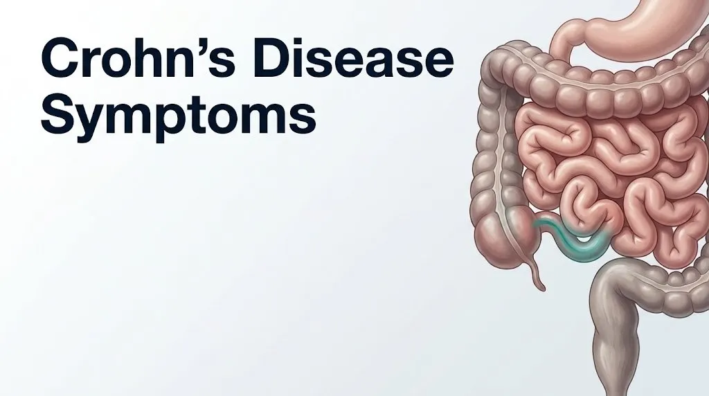
The earliest recognizable symptom in most patients is recurrent abdominal cramping, particularly in the lower right quadrant of the abdomen, which corresponds anatomically to the terminal ileum, the final section of the small intestine. This location is involved in approximately 70 percent of Crohn’s disease cases, according to Montreal classification data. Pain in this region is often mistaken for appendicitis in the early stages, and some patients receive exploratory surgery before Crohn’s disease is identified.
A second early indicator is a change in bowel habits that persists beyond two to four weeks. This is not the brief diarrhea of a viral illness. It’s a pattern of looser, more frequent stools that recurs without a clear infectious cause. According to research published in the journal Inflammatory Bowel Diseases, the average time between first symptom onset and confirmed Crohn’s disease diagnosis is approximately three to four years, in part because early symptoms overlap substantially with irritable bowel syndrome (IBS), food intolerances, and anxiety-related gastrointestinal complaints.
Early-stage Crohn’s disease often includes low-grade fever, which reflects systemic immune activation. A temperature that repeatedly returns to 37.5 to 38.0 degrees Celsius (99.5 to 100.4 degrees Fahrenheit) over weeks, with no identified infectious source, is a pattern that gastroenterologists look for in the diagnostic workup.
Early red flags that should prompt referral to a gastroenterologist rather than watchful waiting:
- Diarrhea or loose stools persisting more than four consecutive weeks
- Abdominal pain recurring in the right lower quadrant without a confirmed alternative diagnosis
- Unintentional weight loss of more than 5 percent of body weight over one to two months
- Blood visible in stool or on toilet tissue beyond a single isolated episode
- Persistent low-grade fever with no identified infectious cause
- Fatigue that interferes with normal daily function and is not explained by sleep disruption or recent illness
For adolescents, an early and often overlooked sign is slowing or stalling of height and weight growth curves, discussed in more detail in the pediatric section below.
Core Digestive Symptoms of Crohn’s Disease
The core digestive symptoms of Crohn’s disease are abdominal pain, diarrhea, nausea, blood in stool, and fever, each produced by a distinct mechanism related to the location and severity of intestinal inflammation.
Abdominal pain in Crohn’s disease is generated by multiple overlapping mechanisms. Inflamed bowel walls have heightened sensitivity to normal intestinal stretch and motility, a phenomenon called visceral hypersensitivity, mediated by sensitized enteric neurons. When the intestine contracts during peristalsis, the inflamed segment generates pain signals that are amplified by substance P release in the inflamed wall. Meals trigger peristalsis, which is why pain frequently worsens after eating.
Nausea occurs through two pathways. In small bowel Crohn’s, inflammation slows intestinal transit and disrupts normal peristaltic coordination, creating functional backing-up of intestinal contents that triggers nausea via the vagus nerve and the vomiting center (area postrema) in the brainstem. Systemic cytokines, particularly IL-1 and IL-6, also act directly on central nervous system receptors to produce nausea independent of the gastrointestinal obstruction component.
Blood in stool reflects mucosal erosion and ulceration. In colonic Crohn’s disease, blood tends to appear mixed with stool or on its surface in visible red streaks. In small bowel Crohn’s, bleeding is more often occult, meaning it is not visible to the naked eye but shows up as iron-deficiency anemia on a blood panel. The American College of Gastroenterology (ACG) notes in its clinical practice guidelines for Crohn’s disease that rectal bleeding is less common in Crohn’s than in ulcerative colitis, occurring in approximately 45 percent of Crohn’s disease patients versus more than 90 percent of ulcerative colitis patients, which reflects the differences in colonic vs. small bowel predominance.
Fever occurs in active Crohn’s disease as a direct result of cytokine-mediated action on the hypothalamic thermoregulatory center. TNF-alpha and IL-1 stimulate prostaglandin E2 production in the hypothalamus, resetting the body’s temperature setpoint upward. A persistent fever above 38.5 degrees Celsius (101.3 degrees Fahrenheit) in a patient with known Crohn’s disease should not be assumed to be disease-related: it may indicate a superimposed infection, abscess formation, or a complication of immunosuppressant therapy, and requires prompt evaluation by the treating gastroenterologist.
Older adults with Crohn’s disease, particularly those on corticosteroid therapy, may not mount a fever even when significant bowel inflammation or secondary infection is present, because corticosteroids suppress the hypothalamic fever response. This means the absence of fever in an elderly patient does not rule out serious active disease.
Key Takeaway: Crohn’s disease symptoms are driven by transmural immune-mediated inflammation, and their pattern, including which symptoms dominate, depends on where in the gastrointestinal tract the inflammation is most active.
Where Is Crohn’s Disease Pain Located
Crohn’s disease pain location depends directly on which segment of the gastrointestinal tract is inflamed, and the anatomical patterns follow predictable regional distributions.
The most common location for Crohn’s disease is the terminal ileum, the final 20 to 30 centimeters of the small intestine before it connects to the large intestine at the ileocecal valve. Pain from terminal ileal inflammation is typically felt in the right lower quadrant of the abdomen, the same region where appendicitis pain localizes. The character is often cramping or colicky, meaning it comes in waves timed to intestinal contractions, and it is frequently worst after eating.
When Crohn’s disease affects the colon (Crohn’s colitis), pain shifts depending on which portion of the colon is involved. Ascending colon disease produces right-sided pain. Transverse colon disease produces mid-abdominal or upper abdominal pain. Descending colon or sigmoid colon disease produces left lower quadrant pain. This regional variability is one reason Crohn’s disease is notoriously difficult to localize on symptom history alone.
Gastroduodenal Crohn’s disease, which affects the stomach and duodenum, produces upper abdominal pain, nausea, and symptoms resembling peptic ulcer disease. This subtype accounts for approximately two to four percent of all Crohn’s disease cases according to Montreal classification data, making it rare but important to recognize because it responds differently to standard Crohn’s therapies.
| Crohn’s Disease Location (Montreal Classification) | Where Pain Is Felt | Associated Symptoms |
|---|---|---|
| Ileal (L1) — terminal ileum | Right lower quadrant | Cramping after meals, possible right lower quadrant mass |
| Colonic (L2) — colon only | Variable: right, central, or left abdomen | Bloody stool more likely than in ileal disease |
| Ileocolonic (L3) — both ileum and colon | Right lower quadrant and diffuse abdominal | Mixed features, most common subtype overall |
| Upper GI (L4) — stomach, duodenum | Epigastric (upper central abdomen) | Nausea, vomiting, early satiety |
| Perianal disease (modifier) | Perianal region, buttock, perineum | Fistula, abscess, severe local pain |
The distinction matters clinically because treatment decisions and surgical planning depend on disease location. A patient whose pain is entirely in the right lower quadrant with an ileal pattern will have a different endoscopic, imaging, and pharmacological management plan than one with colonic Crohn’s producing left-sided pain and rectal bleeding.
Crohn’s Disease Diarrhea and Bowel Changes
Crohn’s disease diarrhea is chronic, recurring, and mechanistically distinct from infection-related or irritable bowel diarrhea in ways that experienced gastroenterologists can often identify before the first scope.
The diarrhea of Crohn’s disease stems from three overlapping mechanisms. First, secretory diarrhea results from cytokine-driven upregulation of chloride and water secretion into the intestinal lumen by inflamed epithelial cells, producing watery, high-volume stool. Second, malabsorptive diarrhea results from impaired nutrient and water absorption in the inflamed small bowel: when terminal ileal disease prevents proper bile salt reabsorption, excess bile salts reach the colon and stimulate colonic secretion, producing a characteristic post-meal urgency and loose stool pattern called bile salt malabsorption diarrhea. Third, inflammatory diarrhea results from ulcerated mucosa releasing mucus, blood, and pus into the stool, particularly in colonic Crohn’s disease.
According to the NIDDK, most patients with active Crohn’s disease have more than three to five loose or watery bowel movements per day during a flare. The frequency alone, however, does not distinguish Crohn’s disease from other causes. The clinical red flags that point toward Crohn’s rather than IBS or a viral gastroenteritis are: persistence beyond four weeks, nocturnal diarrhea (waking from sleep to defecate, which IBS typically does not cause), visible blood or mucus in stool, and diarrhea accompanied by significant unintentional weight loss or systemic fever.
Nocturnal diarrhea is a particularly useful distinguishing feature. The enteric nervous system normally suppresses intestinal motility during sleep. Inflammation in Crohn’s disease disrupts this suppression, generating urgency and bowel movements at night. Patients with IBS very rarely report true nocturnal diarrhea that wakes them from sleep, whereas patients with active Crohn’s disease commonly do.
Patients who have had ileal resection (surgical removal of the terminal ileum due to Crohn’s disease complications) face an additional bowel challenge: without the terminal ileum, bile salt reabsorption is permanently impaired, and ongoing diarrhea related to bile salt malabsorption can persist even when the Crohn’s disease itself is in remission. This is a critical population-specific caveat: diarrhea in a Crohn’s patient after bowel surgery does not automatically indicate active inflammation and requires separate evaluation by their gastroenterologist.
Crohn’s Disease Fatigue and Weight Loss
Fatigue and unintentional weight loss are two of the most debilitating and frequently underappreciated symptoms of Crohn’s disease, and both have specific physiological explanations that go well beyond “feeling run-down.”
Fatigue in Crohn’s disease operates through at least four distinct mechanisms simultaneously. First, systemic cytokine production, particularly TNF-alpha and IL-6, acts on the brain through the blood-brain barrier and through vagal afferent signaling to produce a state of sickness behavior: fatigue, reduced motivation, cognitive slowing, and disturbed sleep. This is the same mechanism by which a severe flu makes you feel exhausted, but in Crohn’s disease the cytokine signal is chronic rather than self-limiting. Second, iron-deficiency anemia from chronic gastrointestinal blood loss reduces oxygen-carrying capacity in the blood, directly impairing cellular energy production. Third, vitamin B12 deficiency develops specifically in patients with terminal ileal Crohn’s disease, because the terminal ileum is the exclusive absorption site for vitamin B12; without it, B12 serum levels fall and contribute to fatigue and neurological symptoms. Fourth, disrupted sleep from nocturnal pain, urgency, and bathroom trips creates accumulated sleep debt.
A 2021 study published in the Journal of Crohn’s and Colitis found that more than 70 percent of Crohn’s disease patients in remission still reported clinically significant fatigue, confirming that fatigue is not simply a product of active inflammation and does not reliably normalize when disease activity is controlled.
Weight loss in Crohn’s disease occurs through three pathways. Malabsorption of nutrients, particularly fats, proteins, fat-soluble vitamins (A, D, E, K), vitamin B12, zinc, and iron, from inflamed small bowel segments directly reduces the body’s usable caloric intake regardless of how much the patient eats. Patients eat less because food reliably triggers abdominal pain and diarrhea, a conditioned behavioral response called food avoidance, which further reduces caloric intake. Chronic systemic inflammation increases the body’s basal metabolic rate through cytokine-driven catabolism, burning more calories at rest than the digestive system can replace.
Children and adolescents with Crohn’s disease are particularly vulnerable: weight loss and caloric deficit during developmental years produces measurable growth failure and can permanently affect final adult height if the disease is not diagnosed and treated early.
Quick Tip:
- If fatigue persists despite Crohn’s disease being in documented remission, ask your gastroenterologist for a panel including serum ferritin (iron stores), serum B12, serum folate, serum vitamin D, and thyroid-stimulating hormone (TSH), since thyroid disease occurs at higher rates in IBD patients.
- If you are losing weight steadily over four or more weeks, quantify the loss (weigh yourself weekly) and report the exact amount and timeline to your gastroenterologist. This is a metric that directly influences treatment escalation decisions.
- Patients on long-term methotrexate for Crohn’s disease are at elevated risk for folate deficiency contributing to fatigue; folic acid supplementation is typically prescribed alongside methotrexate for this reason.
Key Takeaway: Crohn’s disease fatigue is not a secondary inconvenience; it’s driven by chronic cytokine signaling, nutritional deficiencies, and disrupted sleep, and it persists even during remission in the majority of patients.
Crohn’s Disease Flare Symptoms vs. Remission
A Crohn’s disease flare is a period of active, symptomatic intestinal inflammation that contrasts with remission, a state in which inflammatory activity is suppressed and most symptoms are absent or minimal.
During a flare, the inflammatory cascade described above is actively generating mucosal and transmural damage. Flare symptoms typically include a return or worsening of abdominal pain, increased stool frequency, loose or watery stools, blood in stool (particularly in colonic disease), fever, loss of appetite, nausea, and fatigue. The Harvey-Bradshaw Index and the Crohn’s Disease Activity Index (CDAI), two validated clinical scoring tools, quantify flare severity by tracking stool frequency, abdominal pain score, general well-being, presence of complications, and physical examination findings. The CDAI is the research standard; the Harvey-Bradshaw Index is more practical for clinical office use.
Remission does not mean the disease is absent. The underlying immunological dysregulation remains. Some patients in clinical remission (no significant symptoms) still show endoscopic activity, meaning that inflammation visible on colonoscopy is still present despite minimal or no symptoms. This discrepancy between symptoms and endoscopic findings is why the current standard of care, endorsed by the American College of Gastroenterology, is treat-to-target remission: aiming for both clinical remission (symptom control) and endoscopic remission (mucosal healing visible on scope), not symptom control alone.
What a patient feels during remission varies substantially. Some patients in true endoscopic remission feel nearly normal between flares. Others experience persistent functional symptoms, including bloating, looser stools than their pre-disease baseline, and fatigue, even when inflammation markers (CRP, fecal calprotectin) are normal. This persistent symptom pattern during remission is increasingly recognized as post-inflammatory bowel dysfunction, and it may involve persistent changes in enteric nervous system sensitivity and gut motility rather than ongoing mucosal inflammation.
Triggers that can precipitate a flare include smoking (the strongest modifiable risk factor, with research from the New England Journal of Medicine confirming that smokers with Crohn’s disease have more frequent relapses and higher surgery rates than non-smokers), nonsteroidal anti-inflammatory drug (NSAID) use, gastrointestinal infections, significant psychological stress, and antibiotic use disrupting the gut microbiome.
Patients on biologic therapy (anti-TNF agents, vedolizumab, or ustekinumab) may experience attenuated flare symptoms because the cytokine cascade is partially suppressed. This means a “milder-feeling” flare in a patient on biologics can still represent significant endoscopic disease activity. Contact with the treating gastroenterologist when any flare symptoms emerge is the appropriate response, not waiting for symptoms to worsen to a prior pre-treatment severity level.
Crohn’s Disease Perianal Symptoms
Perianal Crohn’s disease is a specific and particularly painful manifestation affecting the anal canal, perianal skin, and surrounding tissues, and it occurs in approximately 25 to 35 percent of all Crohn’s disease patients.
The perianal region becomes involved when Crohn’s disease transmural inflammation extends to the rectum and anal canal. The hallmark of perianal Crohn’s disease is fistula formation: abnormal tunnel-like tracts that form between the anal canal and the perianal skin, the vagina (in women), the bladder, or the buttock. These fistulas arise because the full-thickness bowel wall inflammation erodes through tissue planes, creating paths of least resistance. A fistula opening on the perianal skin appears as a small opening that may drain foul-smelling fluid, blood, or stool.
Perianal abscess is the acute, infected stage that often precedes or accompanies fistula formation. It presents as a tender, fluctuant, red mass near the anus or within the anal canal. This is not a condition to monitor at home. A perianal abscess in Crohn’s disease requires urgent surgical drainage, typically performed by a colorectal surgeon, and failure to drain an abscess promptly risks progression to a complex deep-space infection.
Additional perianal symptoms in Crohn’s disease include:
- Anal fissures: painful tears in the anal mucosa that do not heal normally due to impaired tissue repair from ongoing inflammation; they often appear in unusual anatomical positions (lateral rather than the typical posterior or anterior midline of non-Crohn’s fissures)
- Skin tags: excess perianal tissue folds that develop from repeated cycles of swelling and resolution, which can trap moisture and cause ongoing discomfort
- Rectal stricture: narrowing of the anal canal or distal rectum from fibrotic scarring, causing a sense of incomplete evacuation, straining, and pencil-thin stools
Perianal Crohn’s disease is associated with more aggressive systemic disease behavior and higher rates of surgery. A 2020 study published in Gastroenterology found that patients with perianal disease at diagnosis had a significantly higher rate of surgical intervention within five years compared to those with luminal-only Crohn’s disease.
Women with Crohn’s disease are at risk of rectovaginal fistula, a fistulous tract connecting the rectum to the vagina that causes passage of stool, gas, or mucus through the vagina. This complication requires evaluation by both a gastroenterologist and a colorectal surgeon experienced in IBD management.
Key Takeaway: Perianal Crohn’s disease, including fistulas, abscesses, and anal fissures in unusual locations, is a serious phenotypic variant requiring specialist evaluation by a gastroenterologist and colorectal surgeon, not home management.
Extra-Intestinal Manifestations of Crohn’s Disease
Extra-intestinal manifestations (EIMs) of Crohn’s disease are symptoms occurring outside the gastrointestinal tract and affecting the joints, skin, eyes, liver, and mouth, driven by systemic immune activation rather than local bowel inflammation.
According to the Crohn’s and Colitis Foundation, up to 40 percent of people with Crohn’s disease develop at least one extra-intestinal manifestation. Some EIMs parallel intestinal disease activity (meaning they worsen during flares and improve during remission), while others run an independent course regardless of bowel status.
Joint symptoms are the most common EIM and take two forms. Peripheral arthropathy affects large joints, most often the knees, ankles, and elbows, producing pain, swelling, and stiffness that typically tracks with intestinal disease activity. It does not produce joint destruction (unlike rheumatoid arthritis). Axial arthropathy (including sacroiliitis and ankylosing spondylitis) affects the spine and sacroiliac joints, producing lower back stiffness that is worst after rest and improves with movement, and this form can progress independently of bowel inflammation.
Skin manifestations include two named entities. Erythema nodosum produces tender, red-to-purple nodules on the shins and lower legs, typically measuring one to five centimeters. It generally tracks with intestinal flare activity and resolves as bowel disease improves. Pyoderma gangrenosum is a more severe skin complication producing painful, rapidly enlarging ulcers with irregular, undermined borders, most often on the legs or around a stoma site. Unlike erythema nodosum, pyoderma gangrenosum can progress independent of bowel disease activity and requires specialized dermatological management.
Eye manifestations include episcleritis (superficial inflammation of the white of the eye producing redness and mild discomfort, typically tracks with disease activity) and uveitis (deeper inflammation of the uveal tract inside the eye, producing more severe pain, photophobia, and blurred vision that can progress to vision loss if untreated, and can run an independent course from bowel inflammation). Uveitis in Crohn’s disease requires urgent ophthalmological evaluation, not watchful waiting.
Oral manifestations include aphthous ulcers (canker sores in the mouth), angular stomatitis (cracking at the corners of the mouth from zinc and B12 deficiency), and cobblestone-pattern mucosal changes in the mouth that mirror the intestinal mucosa pattern.
Hepatobiliary manifestations include primary sclerosing cholangitis (PSC), a progressive scarring of the bile ducts that occurs in a subset of Crohn’s and colitis patients. PSC presents with fatigue, right upper abdominal discomfort, and elevated liver enzymes and can progress to liver failure independently of bowel disease control.
| Extra-Intestinal Manifestation | Body Area Affected | Tracks With Bowel Activity | Specialist Needed |
|---|---|---|---|
| Peripheral arthropathy | Knees, ankles, elbows | Yes, typically | Rheumatologist |
| Axial arthropathy / ankylosing spondylitis | Spine, sacroiliac joints | Independent | Rheumatologist |
| Erythema nodosum | Shins and lower legs | Yes | Dermatologist if severe |
| Pyoderma gangrenosum | Legs, peristomal skin | Independent | Dermatologist |
| Episcleritis | White of eye | Yes | Ophthalmologist |
| Uveitis | Internal eye structures | Independent | Ophthalmologist urgently |
| Primary sclerosing cholangitis | Bile ducts, liver | Independent | Hepatologist |
| Aphthous ulcers | Mouth | Tracks with activity | Gastroenterologist |
Crohn’s Disease Symptoms in Women
Crohn’s disease symptoms in women differ from the general clinical picture in several physiologically grounded ways, most notably through menstrual cycle-linked flare patterns, higher rates of extra-intestinal manifestations, and the unique complications of pregnancy.
Women with Crohn’s disease frequently report worsening of symptoms in the days before and during menstruation. This pattern has a clear physiological basis: prostaglandins released during menstruation act on intestinal smooth muscle in the same way they contract the uterine myometrium, accelerating bowel transit, increasing intestinal secretion, and amplifying visceral pain sensitivity through shared prostaglandin E2 and F2-alpha pathways. Research published in Inflammatory Bowel Diseases found that a substantial proportion of women with IBD report pre-menstrual worsening of abdominal pain, diarrhea, and bloating that exceeds their baseline disease activity. Clinicians managing Crohn’s disease in women should specifically ask about this menstrual correlation, because it is often not spontaneously reported.
Extra-intestinal manifestations occur at higher rates in women with Crohn’s disease. A 2019 analysis in Gastroenterology found that women with Crohn’s disease had significantly higher rates of erythema nodosum, aphthous ulcers, and peripheral arthropathy compared to men, a finding attributed in part to sex-based differences in immune regulation, including the effect of estrogen on Th17 cell activity.
Regarding pregnancy, Crohn’s disease activity at the time of conception is the single strongest predictor of pregnancy outcome. According to the American College of Gastroenterology, women who conceive in clinical remission have pregnancy outcomes similar to those without Crohn’s disease. Women who conceive during active disease have significantly higher rates of preterm birth, low birth weight, and disease relapse during pregnancy. Management decisions around biologic therapy during pregnancy require specialist input from both a gastroenterologist and a maternal-fetal medicine (high-risk obstetrics) physician.
Perianal Crohn’s disease in women, particularly rectovaginal fistula, requires evaluation by both a gastroenterologist and a colorectal surgeon with IBD expertise. Obstetric history is also relevant: women who have had episiotomies or perineal tears during delivery have anatomical changes that can complicate fistula assessment and repair.
Crohn’s Disease Symptoms in Children and Teenagers
Crohn’s disease symptoms in children and teenagers include all the adult presentations but add two features that do not occur in adult-onset disease: impaired linear growth and delayed puberty, both of which can become the most prominently visible manifestation before digestive symptoms are even recognized.
Growth failure in pediatric Crohn’s disease results from three converging mechanisms. Chronic intestinal inflammation elevates systemic TNF-alpha and IL-6, which suppress growth hormone-axis signaling at the level of the liver, reducing production of insulin-like growth factor 1 (IGF-1), the primary mediator of skeletal growth. Caloric deficit from food avoidance, malabsorption, and increased basal metabolic rate further reduces substrate available for growth. And corticosteroid therapy, if used repeatedly, directly suppresses bone growth through glucocorticoid effects on growth plate chondrocytes. The American Academy of Pediatrics and the North American Society for Pediatric Gastroenterology, Hepatology, and Nutrition (NASPGHAN) consider unexplained growth deceleration in an otherwise healthy-appearing child a reason for gastroenterological evaluation.
Delayed puberty occurs through a related mechanism: chronic inflammation suppresses the hypothalamic-pituitary-gonadal (HPG) axis through IL-6 and TNF-alpha mediated suppression of gonadotropin-releasing hormone (GnRH) pulsatility. Boys with Crohn’s disease are more commonly affected by delayed puberty than girls, though both sexes can be affected.
The diagnosis of Crohn’s disease is more frequently delayed in children than in adults because abdominal pain and diarrhea in children are common and often attributed to anxiety, dietary intolerance, or IBS. A child with recurrent abdominal pain, declining growth velocity on their growth chart, unexplained anemia, or mouth sores recurring more than three times per year should be referred to a pediatric gastroenterologist for evaluation. A single 30-minute office visit with a primary care physician reviewing growth curves over two to three years can be the pivotal moment in identifying a child who has had undiagnosed Crohn’s disease for years.
Anemia in pediatric Crohn’s disease is particularly common and often the first laboratory abnormality identified. Iron-deficiency anemia from occult gastrointestinal bleeding, combined with anemia of chronic disease from the inflammatory state, produces fatigue and pallor that parents and pediatricians often attribute to dietary iron deficiency without investigating an inflammatory cause.
Key Takeaway: In children with Crohn’s disease, growth faltering and delayed puberty are often the most visible symptoms and should prompt referral to a pediatric gastroenterologist even when digestive symptoms are mild or absent.
Crohn’s Disease Severe Symptoms and Complications
Severe Crohn’s disease produces complications that represent structural failure of the intestinal wall rather than simply worsening inflammation, and these complications are associated with sharply elevated morbidity, need for surgery, and in some cases risk to life.
Intestinal stricture (stenosis) develops when repeated cycles of inflammation and healing produce fibrotic scarring that narrows the bowel lumen. The clinical presentation is bowel obstruction symptoms: postprandial abdominal distension, cramping pain that builds in waves, nausea and vomiting, and eventually inability to pass gas or stool. Strictures can be inflammatory (swollen inflamed tissue) or fibrotic (permanent scarring). This distinction matters because inflammatory strictures may respond to anti-inflammatory therapy, while fibrotic strictures typically require endoscopic balloon dilation or surgical strictureplasty.
Fistula formation (beyond perianal fistulas) can involve the bladder (vesical fistula), producing recurrent urinary tract infections and passage of gas or stool in the urine (pneumaturia or fecaluria); the skin (enterocutaneous fistula), producing external drainage of intestinal contents; or adjacent bowel loops (entero-enteric fistula), producing malabsorption by short-circuiting normal transit.
Intra-abdominal abscess presents with fever, localized abdominal pain and tenderness, an elevated white blood cell count, and in some cases a palpable abdominal mass. This is a surgical emergency requiring drainage, performed percutaneously (through the skin under imaging guidance) or through open or laparoscopic surgery, combined with antibiotic therapy.
Nutritional deficiencies in severe Crohn’s disease can reach a degree of clinical severity requiring hospitalization for parenteral (intravenous) or enteral (tube-fed) nutrition. Specific deficiencies include:
- Vitamin B12 deficiency (terminal ileal disease or after ileal resection): neurological symptoms including paresthesias, subacute combined degeneration of the spinal cord in severe untreated cases
- Vitamin D deficiency: bone density loss (osteopenia or osteoporosis) accelerated by corticosteroid use
- Zinc deficiency: impaired wound healing, skin rash (acrodermatitis), taste changes
- Iron deficiency: severe anemia requiring intravenous iron infusion or, rarely, transfusion
- Fat-soluble vitamin (A, D, E, K) deficiency: from bile salt malabsorption in ileal disease
Severe Crohn’s disease requiring hospitalization is characterized by: more than six bowel movements per day with blood, fever above 37.8 degrees Celsius (100 degrees Fahrenheit), heart rate above 90 beats per minute, hemoglobin below 10.5 g/dL, CRP significantly elevated (above 30 mg/L), and low serum albumin (below 3.0 g/dL) as a marker of protein malnutrition and disease severity. These are the thresholds used by gastroenterologists to define a severe flare requiring inpatient management.
Emergency Symptoms of Crohn’s Disease: When to Call 911 or Go to the ER
Certain symptoms associated with Crohn’s disease require immediate emergency evaluation. Do not wait to see if these resolve on their own.
Call 911 or go to the nearest emergency room immediately if you experience:
- Severe abdominal pain that is sudden in onset, constant rather than cramping, and accompanied by a rigid or board-like abdomen: this pattern suggests intestinal perforation and peritonitis, a life-threatening surgical emergency
- Fever above 38.5 degrees Celsius (101.3 degrees Fahrenheit) with severe abdominal pain and inability to tolerate fluids: suggests an intra-abdominal abscess, severe infectious complication, or toxic megacolon
- Extreme abdominal distension with high fever, rapid heart rate (above 100 beats per minute), and prostration: the classic presentation of toxic megacolon, a rare but immediately life-threatening complication of severe colonic Crohn’s disease requiring emergency intensive care
- Heavy rectal bleeding: defined as passage of large amounts of bright red blood from the rectum, soaking a pad or filling the toilet bowl, indicating significant hemorrhage from an intestinal blood vessel
- Signs of obstruction: inability to pass stool or gas for more than 24 hours, with worsening distension, vomiting, and escalating cramping pain, indicating complete bowel obstruction
- Severe dehydration: inability to keep any fluids down, extreme dryness of mouth and skin, dizziness on standing, dark urine or no urination for eight or more hours, particularly during a severe diarrheal flare
- Sudden severe abdominal pain in a patient who has been taking steroids long-term: corticosteroids can mask peritoneal signs, making perforation less painful than expected; any new sudden severe pain in this population warrants emergency evaluation
These presentations can indicate intestinal perforation, toxic megacolon, life-threatening hemorrhage, or complete bowel obstruction, and they require emergency medical assessment, not an urgent care appointment or a phone call to reschedule a clinic visit.
How Crohn’s Disease Is Diagnosed
Crohn’s disease is diagnosed through a combination of clinical history, laboratory testing, endoscopy with biopsy, and imaging, and no single test alone is sufficient to confirm the diagnosis.
The first step in clinical diagnosis is a thorough history covering symptom duration, character, and pattern; bowel habit changes; weight loss; extra-intestinal symptoms; family history of IBD; and medication history. A blood panel is ordered early and typically includes complete blood count (CBC) (to identify anemia and elevated white blood cell count), C-reactive protein (CRP) and erythrocyte sedimentation rate (ESR) (inflammatory markers), and serum albumin (protein status and disease severity marker). Fecal calprotectin, a protein released by intestinal white blood cells into the stool, is a sensitive marker for intestinal inflammation that can help distinguish Crohn’s disease from functional bowel disorders like IBS.
The cornerstone diagnostic procedure is ileocolonoscopy with segmental biopsies: direct visualization of the colon and terminal ileum through a flexible camera, combined with tissue sampling from multiple segments of the bowel. The pathological finding of non-caseating granulomas (clusters of macrophages forming nodular aggregates without central necrosis) on biopsy is highly specific for Crohn’s disease, though granulomas are found in only 30 to 60 percent of Crohn’s biopsies.
Small bowel imaging is typically performed alongside or following endoscopy. Magnetic resonance enterography (MRE) uses MRI to create detailed images of the small bowel wall, luminal narrowing, fistula tracts, and abscesses without radiation exposure, making it the preferred imaging modality for Crohn’s disease according to the American College of Gastroenterology. Computed tomography enterography (CTE) offers similar information with higher spatial resolution but involves ionizing radiation, making it less ideal for young patients who may need repeated imaging over a lifetime of disease.
How a typical diagnostic process unfolds from first presentation to confirmed diagnosis:
- Primary care physician evaluates persistent diarrhea, abdominal pain, or weight loss. Blood work ordered, including CBC, CRP, and fecal calprotectin.
- If fecal calprotectin is elevated (typically above 150 to 250 micrograms per gram, laboratory thresholds vary) or blood work shows anemia and elevated CRP, referral to gastroenterologist is made.
- Gastroenterologist takes a detailed history and physical exam, reviews blood work, may order additional stool tests to rule out infectious causes (Clostridioides difficile, ova and parasites, stool culture).
- Ileocolonoscopy with biopsies performed to visualize and sample the terminal ileum and colon. Results reviewed with the pathologist.
- If small bowel disease is suspected beyond the terminal ileum, MRE is ordered.
- Diagnosis confirmed when clinical, endoscopic, radiographic, and histological findings are consistent with Crohn’s disease. Montreal classification (location and behavior) assigned.
- Gastroenterologist presents diagnosis, discusses disease behavior classification, and begins discussion of treatment strategy tailored to disease severity and location.
Capsule endoscopy (swallowing a small wireless camera) is used in specific circumstances to visualize small bowel segments beyond the reach of conventional ileocolonoscopy, but is contraindicated when intestinal stricturing is suspected due to the risk of capsule retention.
Crohn’s Disease Symptoms vs. Ulcerative Colitis
The symptoms of Crohn’s disease and ulcerative colitis overlap substantially, but the differences in symptom pattern, anatomical distribution, and complication types can help distinguish the two conditions before diagnostic testing confirms the picture.
Ulcerative colitis (UC) is confined to the colon and rectum, always begins at the rectum, and involves only the mucosa and submucosa rather than the full bowel wall thickness. Rectal bleeding is present in more than 90 percent of UC patients at some point in their disease course, and tenesmus (urgent, painful rectal straining with minimal output) is a hallmark of rectal involvement. Crohn’s disease, by contrast, can occur anywhere from the mouth to the anus, often spares the rectum, and produces rectal bleeding in only about 45 percent of patients.
Perianal disease (fistulas, abscesses, complex perianal fistulas) is strongly associated with Crohn’s disease and very rarely occurs in UC. If a patient has perianal fistulas, the diagnosis is almost certainly Crohn’s disease.
Obstructive symptoms (nausea, vomiting, inability to pass stool, cramping that builds over hours) are associated with Crohn’s disease because transmural inflammation creates strictures. UC does not typically cause strictures because it does not penetrate beyond the submucosa.
| Clinical Feature | Crohn’s Disease | Ulcerative Colitis |
|---|---|---|
| Affected region | Any part of GI tract from mouth to anus | Colon and rectum only |
| Inflammation depth | Transmural (all bowel wall layers) | Mucosal and submucosal only |
| Distribution pattern | Skip lesions (patchy areas of inflammation) | Continuous, begins at rectum |
| Rectal involvement | Often spared | Always involved |
| Rectal bleeding | Present in approximately 45% | Present in more than 90% |
| Perianal disease | Common (25 to 35%) | Rare |
| Stricture formation | Common | Uncommon |
| Fistula formation | Common | Very rare |
| Granulomas on biopsy | Present in 30 to 60% | Absent |
| Small bowel involvement | Common | Never (unless backwash ileitis) |
Approximately 10 to 15 percent of patients with colonic IBD cannot be definitively classified as Crohn’s disease or UC on initial evaluation, and are given the designation indeterminate colitis or IBD unclassified (IBDU). Over time, with further disease behavior and additional endoscopic findings, the classification becomes clearer in most cases.
Key Takeaway: The presence of perianal disease, skip lesions on endoscopy, small bowel involvement, or fistula formation strongly favors Crohn’s disease over ulcerative colitis, but definitive diagnosis requires endoscopy, biopsy, and imaging interpreted by a gastroenterologist.
Frequently Asked Questions About Crohn’s Disease Symptoms
What are the first signs of Crohn’s disease?
The first signs of Crohn’s disease are typically recurring abdominal cramping in the right lower abdomen, persistent loose or watery stools lasting more than four weeks, and low-grade fever without an identified infectious cause.
Weight loss, fatigue, and blood in stool may appear early in some patients but are more common as the disease progresses without treatment.
Anyone experiencing these symptoms for more than four consecutive weeks should seek evaluation by a primary care physician and ask for referral to a gastroenterologist.
Where exactly does Crohn’s disease pain occur?
Crohn’s disease pain most commonly occurs in the right lower quadrant of the abdomen, corresponding to the terminal ileum, which is the most frequently affected intestinal segment.
When Crohn’s disease involves the colon, pain can occur anywhere from the right to left abdomen depending on which colonic segment is inflamed; gastroduodenal Crohn’s produces upper central (epigastric) pain.
Pain location in Crohn’s disease directly reflects disease location, which is why imaging and endoscopy are needed to map the affected segments accurately.
Can you have Crohn’s disease without diarrhea?
Yes, some patients with Crohn’s disease, particularly those with stricturing disease causing partial bowel obstruction, can present with constipation rather than diarrhea because the narrowed bowel restricts stool passage.
Patients with gastroduodenal Crohn’s disease may have predominantly nausea, vomiting, and upper abdominal pain without significant diarrhea.
The absence of diarrhea does not rule out Crohn’s disease, which is one reason endoscopy and imaging are necessary for diagnosis rather than symptom assessment alone.
What does a Crohn’s disease flare feel like?
A Crohn’s disease flare typically feels like a return of worsening abdominal cramping, increased urgency and frequency of bowel movements, renewed fatigue, and in many patients low-grade fever and appetite loss.
Some patients describe flare pain as a constant, gnawing ache with superimposed sharp cramping during bowel activity, while others report a feeling of internal pressure or fullness that is distinct from gas or constipation.
Flare symptoms that include fever above 38.5 degrees Celsius, inability to eat or drink, or severe pain require contact with the treating gastroenterologist the same day, not watchful waiting.
How do you know if Crohn’s disease symptoms are serious?
Crohn’s disease symptoms are serious and require same-day or emergency evaluation when they include fever above 38.5 degrees Celsius with abdominal pain, heavy rectal bleeding, rigid abdomen, inability to pass stool or gas, or signs of severe dehydration.
Signs of toxic megacolon (extreme abdominal distension, high fever, rapid heart rate, extreme weakness) are a medical emergency requiring a call to 911 immediately.
Persistent symptoms that are worsening rather than stable, or any new symptom type not previously experienced, should prompt contact with the treating gastroenterologist the same day.
Is Crohn’s disease pain constant or does it come and go?
Crohn’s disease pain typically comes and goes in a relapsing-remitting pattern, with periods of active flare during which pain is frequent or near-constant, alternating with remission periods during which pain is minimal or absent.
During a flare, pain often worsens after eating and during bowel movements, then partially improves between these triggers, producing a wave-like pattern rather than truly unrelenting pain in most patients.
Pain that becomes truly constant, unresponsive to prior management, and is associated with new features such as rigidity or rebound tenderness is a warning sign of a complication such as abscess or perforation and requires emergency evaluation.
Closing
Crohn’s disease symptoms are not a simple list to check off. They’re the product of a specific, named immunological process playing out in specific anatomical locations, and what you feel depends heavily on where your disease is active, how severe the inflammation is, and what complications have developed over time.
The most important action you can take with this information is to translate it into specific, useful questions for your gastroenterologist: Where exactly is my disease located? What is my disease behavior classification? Am I in clinical remission, and have we confirmed endoscopic remission? Are my current symptoms consistent with a flare or a complication? What symptoms should make me contact you urgently versus go directly to an emergency room?
You now have the clinical framework to understand the answers to those questions. Use it.