Abdominal Strain Symptoms: Your Full Clinical Guide
Abdominal strain symptoms include localized muscle pain that worsens with movement, trunk flexion or rotation, coughing, or sneezing; tenderness on direct pressure over the affected muscle; involuntary muscle spasm; and sometimes bruising that appears 24 to 72 hours after the injury. The specific symptom pattern depends on which of the four abdominal wall muscle layers is involved and whether the strain is Grade I (microtear), Grade II (partial fiber disruption), or Grade III (complete rupture).
Abdominal muscle strains are among the most common soft tissue injuries in both the athletic and general population. The National Institute of Arthritis and Musculoskeletal and Skin Diseases (NIAMS) notes that musculoskeletal soft tissue injuries, including muscle strains, account for tens of millions of emergency and primary care visits annually in the United States, with abdominal and trunk strains representing a meaningful proportion among active adults and manual laborers.
This guide covers the full symptom picture of abdominal strain, explains the physiological mechanism behind each symptom, maps symptoms to specific muscle layers and injury grades, addresses the critical question of distinguishing strain from dangerous intra-abdominal conditions, and identifies exactly when the ER is the right call rather than watchful waiting.
Abdominal Strain Symptoms: The Clinical Overview
Abdominal strain symptoms are produced by physical disruption of the myofibrils within one or more of the four muscles of the anterior abdominal wall, triggering pain, protective spasm, localized tenderness, and sometimes visible bruising in patterns that differ by muscle affected and injury severity.
The characteristic feature that distinguishes abdominal strain from internal organ pain is that muscle strain pain consistently worsens with active contraction or passive stretching of the injured muscle. Pain that intensifies when you tighten your abdomen, sit up from lying, rotate your trunk, or raise your legs while lying flat is pointing toward a muscular source.
Pain from internal organ pathology tends to behave differently: it often remains present at rest, may radiate to distant sites (such as the right shoulder in gallbladder or diaphragm irritation), and is frequently accompanied by nausea, vomiting, fever, or changes in bowel habits that have no parallel in muscle strain.
Core symptoms of abdominal strain at a glance:
- Sharp or aching pain localized to a specific region of the abdominal wall
- Pain that worsens sharply with trunk flexion, rotation, coughing, sneezing, or Valsalva effort
- Tenderness reproducible by pressing directly over the affected muscle
- Involuntary protective muscle spasm in the surrounding fibers
- Stiffness and reluctance to move through full range of trunk motion
- Weakness when attempting resisted abdominal contraction
- Bruising (ecchymosis) appearing hours to days after a Grade II or III strain
- A possible audible or felt “pop” at the moment of Grade III rupture
People taking anticoagulant medications (warfarin, apixaban, rivaroxaban, dabigatran) should be aware that bruising from even a Grade I or II strain may be substantially more extensive and develop more rapidly than in a person not on anticoagulation, and that a rectus sheath hematoma (blood collecting within the rectus sheath due to tearing of the epigastric vessels) is a risk that warrants prompt evaluation in this population.
What Is an Abdominal Strain and Which Muscles Are Involved
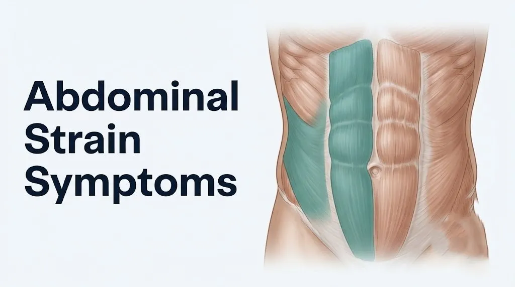
An abdominal strain is a partial or complete tear of one or more of the four muscles forming the anterior abdominal wall, occurring when mechanical force applied to those muscle fibers exceeds their tensile strength.
The four muscles involved, from superficial to deep, are:
| Muscle | Location | Primary Function | Strain Mechanism |
|---|---|---|---|
| Rectus abdominis | Vertical paired columns, pubic symphysis to ribs 5-7 | Trunk flexion, intra-abdominal pressure | Sudden explosive sit-up, heavy lifting with trunk flexion |
| External oblique | Most superficial lateral muscle, ribs 5-12 to linea alba | Trunk rotation toward opposite side, flexion | Twisting movements, sudden rotation under load |
| Internal oblique | Deep to external oblique, thoracolumbar fascia to linea alba | Trunk rotation toward same side, flexion | Same-side rotation under load, powerful expiration |
| Transversus abdominis | Deepest flat muscle, horizontal fibers | Intra-abdominal pressure regulation, core stabilization | Forceful coughing, Valsalva effort, heavy lifting |
All four muscles converge at the linea alba, the tendinous midline raphe. Strain at or near the linea alba can produce midline tenderness that is sometimes mistaken for a hernia or visceral pain.
The muscles are innervated by the intercostal nerves T7 through T12 and the subcostal nerve, which is why abdominal muscle strain sometimes produces a referred pain pattern along the dermatomal distribution of these nerves, creating pain that radiates laterally around the flank or toward the groin. This referred pattern surprises many people because the pain seems to extend beyond the muscle itself.
Athletes and individuals with pre-existing diastasis recti abdominis (separation of the two rectus abdominis bellies at the linea alba) are at higher risk for recurrent rectus abdominis strain because the mechanical disadvantage created by the widened linea alba reduces the force-generating capacity of each rectus belly.
Pulled Stomach Muscle Symptoms: The Core Clinical Picture
The core symptoms of a pulled stomach muscle are acute-onset abdominal wall pain with a definable injury moment, tenderness localized to the muscle rather than a diffuse ache, pain that tracks predictably with muscle use, and protective spasm that makes the abdomen feel stiff or guarded.
Think of an abdominal muscle like a bundle of tightly woven rubber bands. When a handful of those bands snap simultaneously from excessive force, the immediate sensation is sharp and localized. The surrounding intact bands reflexively tighten to protect the injured area. That tightening is the spasm you feel, and it is why the abdomen often feels rigid in the hours after a significant strain.
The pain of an acute strain is typically described as sharp or tearing at the moment of injury, transitioning to a dull, aching soreness within the first few hours as the initial nociceptor signal settles and the inflammatory response establishes. According to the American Academy of Orthopaedic Surgeons (AAOS), abdominal muscle strain pain is characteristically aggravated by activities that increase intra-abdominal pressure, including lifting, bearing down, and coughing.
Key distinguishing features of pulled stomach muscle symptoms:
- Pain with a clear onset moment (a specific lift, twist, cough, or exercise effort)
- Reproducible tenderness at a specific point on the abdominal wall surface
- Worsening with contraction of the affected muscle (crunch motion, leg raise, trunk rotation)
- Partial relief with rest and avoidance of the aggravating movement
- No accompanying fever, nausea, vomiting, or change in bowel habits
- No referred pain to the right shoulder or scapula (which would suggest diaphragm or gallbladder involvement)
Obese individuals and those with high body mass index may have more difficulty localizing their pain precisely, because the extra soft tissue between the skin surface and the abdominal wall muscles reduces the ability to identify point tenderness accurately during self-palpation.
Abdominal Muscle Strain Grades and What Each One Feels Like
Abdominal muscle strains are classified into three grades based on the extent of muscle fiber disruption, and each grade produces a distinct symptom pattern that reflects the degree of structural damage.
Grade I (Mild): Fewer than approximately 5% of muscle fibers are disrupted (microtearing at the sarcomere level). Pain is mild to moderate, typically felt as a sharp ache that worsens with specific movements but does not significantly limit function. There is localized tenderness but no swelling visible to the eye, no bruising, and no loss of strength beyond mild pain-related inhibition. Most Grade I strains do not produce a clearly felt “pop.” They are often attributed to a pulled muscle after exercise.
Grade II (Moderate): A partial-thickness portion of the muscle belly is disrupted. Pain is moderate to severe, clearly felt at the injury moment, and significantly limits the affected movement. Tenderness is more pronounced, and swelling or localized firmness may be palpable within the first 24 hours. Bruising appears in many (though not all) Grade II strains, typically emerging 24 to 48 hours post-injury as intramuscular hematoma tracks to the skin surface. Strength testing (attempting to perform a sit-up or resisted rotation) produces significant pain and measurable weakness.
Grade III (Severe): Complete rupture of the muscle belly or myotendinous junction occurs. A pop or tearing sensation is frequently described at the moment of injury. Pain is severe and immediate. A visible or palpable gap in the muscle may be detectable. Bruising is typically extensive. Functional loss is significant: the person cannot perform the movement controlled by that muscle without severe pain and mechanical failure.
| Grade | Fiber Disruption | Pain Level | Bruising | Functional Loss | Typical Recovery |
|---|---|---|---|---|---|
| I | Less than 5% | Mild to moderate | Rare | Minimal | 1 to 2 weeks |
| II | Partial (5 to 50%) | Moderate to severe | Frequent (24-48 hours delay) | Moderate | 3 to 6 weeks |
| III | Complete (greater than 50%) | Severe | Extensive | Significant | 3 to 6 months or surgical repair |
Research published in the American Journal of Sports Medicine confirms that grade classification guides treatment decisions, with Grade I and most Grade II strains managed conservatively and Grade III tears often requiring surgical evaluation for repair.
Adolescent athletes are at elevated risk for Grade II and III strains because their muscles and tendons grow faster than the myotendinous junction can adapt, creating a period of relative vulnerability at the muscle-tendon interface.
Key Takeaway: The three grades of abdominal strain produce fundamentally different symptom profiles; Grade III tears involve a palpable gap, extensive bruising, and significant functional loss that require urgent orthopedic evaluation rather than home management.
Lower Abdominal Strain Symptoms
Lower abdominal strain symptoms are concentrated below the umbilicus and are most often associated with strain of the lower fibers of the rectus abdominis, the lower portion of the internal oblique, or the transversus abdominis where it attaches near the inguinal ligament and pubic symphysis.
Pain in the lower abdomen from muscle strain is typically sharp, well-localized (the person can often point to a specific spot with one finger), and worsens with activities that engage the lower abdominal muscles: leg raises, resisted hip flexion, sit-ups, and any activity requiring forceful intra-abdominal pressure regulation. Some people describe the sensation as a pulling or dragging pain in the groin or pubic region, which reflects the anatomical proximity of the lower abdominal muscle attachments to the pubic symphysis.
A clinically important mimicking condition in the lower abdomen is athletic pubalgia (also called sports hernia), a syndrome involving injury to the posterior wall of the inguinal canal and its associated structures. Athletic pubalgia produces groin and lower abdominal pain strikingly similar to lower abdominal strain but has a different anatomical basis and requires a distinct treatment approach. A 2021 review published in the British Journal of Sports Medicine found that athletic pubalgia is frequently misdiagnosed as a simple abdominal or groin strain, particularly in male soccer, hockey, and American football athletes.
Lower abdominal strain should also be distinguished from inguinal hernia, which can produce groin and lower abdominal pain that worsens with exertion or Valsalva effort. The distinguishing feature is the presence of a palpable bulge in the inguinal region that appears with straining and reduces when lying flat. Abdominal strain does not produce a hernia bulge.
Quick Tip:
- Point tenderness located directly over the lower rectus abdominis (below the navel, above the pubic bone) and worsening with straight leg raises strongly suggests lower abdominal muscle strain
- Pain centered at the pubic symphysis itself, especially in athletes with bilateral groin discomfort, may indicate osteitis pubis rather than simple muscle strain and warrants evaluation by a board-certified sports medicine physician
- Women with lower abdominal pain should inform their evaluating physician of menstrual cycle timing, as dysmenorrhea can produce lower abdominal cramping that is sometimes confused with musculoskeletal pain
Upper Abdominal Strain Symptoms
Upper abdominal strain symptoms are located above the umbilicus, most often in the epigastric region or the lateral upper quadrants, and typically involve the upper fibers of the rectus abdominis, the external oblique, or occasionally the costal attachments of the internal oblique near the lower ribs.
Pain in the upper abdomen from a muscle strain produces a particularly challenging diagnostic picture because the upper abdomen is also the territory of several important internal structures: the liver (right upper quadrant), the stomach and spleen (left upper quadrant), the gallbladder (right upper quadrant inferior to the liver), and the lower lobes of the lungs above. Pain in this region from a muscle strain must be carefully distinguished from pain with an internal source.
The distinguishing clinical feature of upper abdominal muscle strain is that the pain is sharply reproduced by physical contraction of the overlying muscle and is not reproduced by deep inspiration, eating, position changes that shift intra-abdominal organs, or percussion over the liver or spleen. Upper abdominal strain pain typically worsens with trunk curling (flexion), lateral bending toward the side of the strain, and direct palpation of the muscle belly itself.
People who develop upper right quadrant abdominal pain after exercise or physical effort should specifically note whether the pain has any relationship to meals (suggesting gallbladder involvement) or to breathing deeply (suggesting diaphragmatic or pleuritic involvement), as these patterns point away from muscle strain toward internal organ pathology.
According to the American Academy of Family Physicians (AAFP), any upper abdominal pain accompanied by nausea, vomiting, fever, jaundice, or dark-colored urine warrants same-day evaluation by a primary care physician or emergency medicine physician, regardless of the presumed mechanism.
Individuals with a history of peptic ulcer disease or gastroesophageal reflux disease who develop upper abdominal pain should not assume the pain is muscular without evaluation, because ulcer perforation can present with sudden severe upper abdominal pain that is initially localized and mistaken for a muscle pull.
Rectus Abdominis Strain Symptoms
Rectus abdominis strain symptoms center on the vertical midline of the abdomen, running from just below the sternum to the pubic bone, with the exact pain location depending on which of the muscle’s four tendinous intersections is nearest the injury.
The rectus abdominis runs as a paired band of muscle on either side of the linea alba, enclosed within the rectus sheath (formed by the aponeuroses of the three flat abdominal muscles). Because it is divided by four horizontal tendinous intersections (inscriptiones tendineae), a strain of the rectus abdominis often produces pain that is horizontally banded across the abdomen rather than diffuse, reflecting injury at or near one of these structural divisions.
Pain with rectus abdominis strain is most reliably provoked by trunk flexion: sit-ups, crunches, rising from a supine position, and any movement that draws the sternum toward the pubic symphysis. Athletes who perform heavy Olympic lifting, gymnastics, or rowing frequently strain the rectus abdominis during the eccentric loading phase (the controlled lowering phase) of trunk exercises, where the muscle must generate force while lengthening, placing peak mechanical stress on the sarcomere units.
A specific and serious condition involving the rectus abdominis is rectus sheath hematoma, in which tearing of the epigastric arteries (superior or inferior) within the rectus sheath causes blood to collect inside the enclosed fascial compartment. Rectus sheath hematoma produces pain, tenderness, and a firm palpable mass within the rectus abdominis that does not cross the midline. It is most common in people on anticoagulant therapy, in pregnant women, and in older adults following forceful coughing. It can be mistaken for an abdominal hernia or an abdominal mass of internal origin. Any firm, painful, non-reducing abdominal wall mass should be evaluated by an emergency medicine physician the same day.
The Carnett’s sign is a bedside clinical test used to distinguish abdominal wall pain (including rectus abdominis strain) from visceral pain: with the patient supine, the examiner identifies the point of maximal tenderness, then asks the patient to tense the abdominal wall by raising the head and shoulders. If the pain increases or remains the same with tensing (positive Carnett’s sign), the source is the abdominal wall. If the pain decreases (negative Carnett’s sign), the source is more likely intra-abdominal. Research published in the Journal of Emergency Medicine has found Carnett’s sign to have high sensitivity for distinguishing abdominal wall from visceral pain in emergency department settings.
Oblique Muscle Strain Symptoms
Oblique muscle strain symptoms are located in the lateral abdomen (the flank region), and their characteristic feature is that the pain is most sharply provoked by trunk rotation rather than pure trunk flexion, because the oblique muscles are the primary rotators of the thorax.
The external oblique originates from the outer surfaces of ribs 5 through 12 and its fibers run diagonally downward and inward toward the linea alba and inguinal ligament. Straining the right external oblique (which rotates the trunk to the left) typically produces pain on the right lateral abdomen during left rotation or right lateral bending. The internal oblique runs in the opposite diagonal direction, and its strain produces same-side rotation pain (straining the right internal oblique hurts during right rotation).
This directional pain pattern is diagnostically useful: if you can identify the specific rotational direction that reproduces your pain, it points clearly toward an oblique rather than a rectus abdominis strain. A sports medicine physician will use resisted rotation testing (pushing against the physician’s hands while attempting to rotate the trunk) to precisely identify which oblique layer is involved.
Key Takeaway: Oblique muscle strain is specifically provoked by trunk rotation rather than pure flexion, and the direction of painful rotation (toward the affected or opposite side) distinguishes external from internal oblique involvement, which matters for appropriate rehabilitation.
The referred pain pattern of oblique strain can extend along the intercostal nerve dermatomes toward the groin or around the lateral chest wall, occasionally mimicking the distribution of shingles (herpes zoster) before the rash appears, or mimicking flank pain from a kidney stone. Pain that follows a dermatomal band around the lateral chest or flank without a palpable tender muscle underlying it warrants evaluation to exclude intercostal neuralgia or herpes zoster.
Baseball pitchers, tennis players, and golfers are disproportionately affected by oblique strains due to the high rotational torque demands of their movements. A 2019 study published in the American Journal of Sports Medicine found that internal oblique strains were among the most common abdominal muscle injuries in major league baseball pitchers, with a distinct strain pattern related to the late-deceleration phase of the pitching motion.
Abdominal Strain Symptoms Triggered by Movement and Coughing
Abdominal strain symptoms triggered by movement and coughing result from a specific physiological mechanism: any activity that acutely increases intra-abdominal pressure forces all four abdominal wall muscles to contract simultaneously, loading the injured fibers and intensifying pain.
The Valsalva maneuver is the physiological process at work here. During coughing, sneezing, bearing down, or heavy lifting, the glottis closes and the abdominal wall muscles contract forcefully against a closed airway, spiking intra-abdominal pressure to protect the spine and stabilize the trunk. This sudden, high-force contraction of all abdominal wall muscles simultaneously recruits the injured muscle fibers, stretching or contracting across the disrupted sarcomere zone. The result is a sharp pain spike that people often describe as feeling like “being stabbed” when they cough or sneeze.
This symptom is so characteristic that patients frequently tell clinicians they knew something was wrong as soon as they felt the pain spike with a cough. The reliable reproduction of pain by coughing, sneezing, laughing, or bearing down is a strong clinical indicator that the pain source is in the abdominal wall rather than from internal organs (which generally do not worsen so predictably with Valsalva effort, with the exception of a hernia).
Activities that typically worsen abdominal strain symptoms:
- Coughing and sneezing (acute Valsalva spike)
- Laughing (sustained Valsalva-like effort)
- Rising from a seated or lying position (eccentric then concentric abdominal load)
- Lifting any object heavier than a few pounds (intra-abdominal pressure increase)
- Twisting the trunk while standing or sitting
- Performing a sit-up, crunch, or leg raise
- Bearing down during bowel movements
Pregnant women deserve special mention here. Pregnancy progressively increases intra-abdominal pressure as the uterus enlarges, meaning that a strained abdominal muscle during pregnancy is not only more painful during Valsalva-type efforts but is also under higher baseline mechanical load than in a non-pregnant person. This makes abdominal strain during pregnancy both more painful and slower to heal, and it is addressed in detail in the pregnancy-specific section below.
According to the American College of Sports Medicine (ACSM), athletes who sustain abdominal strain should avoid all activities that reproduce pain during the acute phase (typically the first 48 to 72 hours), because repeated loading of the disrupted fiber zone delays the inflammatory resolution phase and increases the risk of converting a Grade I injury to a Grade II injury.
Abdominal Strain vs. Appendicitis and Other Internal Conditions
The most clinically important skill when evaluating abdominal pain is distinguishing abdominal strain symptoms from symptoms produced by internal organ pathology, because several life-threatening conditions can initially present with pain that resembles a pulled muscle.
The central distinguishing principle: abdominal wall (muscular) pain is consistently reproducible by physical contraction or palpation of the muscle, worsens predictably with Valsalva effort, and is not accompanied by systemic symptoms such as fever, nausea, vomiting, or loss of appetite. Visceral (internal organ) pain behaves differently: it often persists at rest, frequently accompanies systemic symptoms, may refer to remote sites (shoulder, back, groin), and does not reliably worsen with trunk contraction.
| Condition | Location | Distinguishing Feature | Systemic Symptoms | Emergency Level |
|---|---|---|---|---|
| Abdominal muscle strain | Localized to abdominal wall | Worsens with muscle contraction; positive Carnett’s sign | None | Low (Grade I/II); High (Grade III) |
| Acute appendicitis | Right lower quadrant (after periumbilical onset) | Pain migrates from navel to right lower quadrant over 12-24 hours | Fever, nausea, anorexia | Emergency |
| Ruptured abdominal aortic aneurysm | Mid-abdomen, back, groin | Pulsating abdominal mass; sudden severe pain; syncope | Shock, diaphoresis | 911 immediately |
| Incarcerated inguinal hernia | Groin and lower abdomen | Non-reducing bulge; colicky pain; vomiting | Nausea, vomiting, obstruction | Emergency |
| Rectus sheath hematoma | Within rectus abdominis | Firm non-crossing midline mass; anticoagulant use history | Minimal unless large | Urgent same-day |
| Bowel perforation | Diffuse, then peritoneal | Board-like abdominal rigidity; rebound tenderness | Fever, shock | 911 immediately |
The Carnett’s sign, described in the rectus abdominis section above, is one of the most practical bedside tests for making this distinction. A positive sign (pain worsens with abdominal tensing) supports a wall source. A negative sign (pain decreases) points toward internal pathology.
Appendicitis is the condition most frequently confused with lower right abdominal strain. The key temporal pattern of appendicitis, pain beginning as a vague periumbilical ache that migrates and intensifies at the right lower quadrant (McBurney’s point) over 12 to 24 hours, accompanied by fever and nausea, is distinctly different from the sudden onset and movement-dependent nature of muscle strain.
Key Takeaway: The critical test for distinguishing abdominal strain from internal pathology is whether pain worsens with direct muscle contraction (strain) or persists at rest with systemic symptoms like fever and nausea (internal organ pathology), and any doubt warrants same-day professional evaluation.
Stomach Muscle Strain Symptoms in Pregnant Women
Stomach muscle strain symptoms in pregnant women present a diagnostically complex picture because the normal anatomical and physiological changes of pregnancy both increase the risk of abdominal wall injury and create multiple competing diagnoses that can mimic strain pain.
As the uterus enlarges during the second and third trimesters, it places progressive mechanical stress on the anterior abdominal wall. The linea alba widens as the two rectus abdominis bellies are pulled apart, a process called diastasis recti abdominis. This widening reduces the mechanical efficiency of the rectus abdominis and increases the strain risk from even routine movements like rising from bed or coughing. Research published in the Journal of Orthopaedic and Sports Physical Therapy found that diastasis recti affects more than 60% of women by the third trimester, significantly altering the load-bearing mechanics of the abdominal wall.
Competing diagnoses that produce abdominal pain in pregnancy and can be confused with muscle strain include:
- Round ligament pain: Sharp, brief, stabbing pain in the lower abdomen or groin produced by rapid stretching of the round ligaments supporting the uterus. Typically lasts seconds to minutes and is triggered by sudden movement, rolling in bed, or rising quickly. Has no muscle tenderness on palpation.
- Pubic symphysis dysfunction (pelvic girdle pain): Aching pain at the pubic bone and groin, worsened by weight-bearing and walking. Commonly mistaken for lower abdominal strain.
- Preterm labor contractions: Lower abdominal cramping that is rhythmic and progressive rather than movement-dependent. Always warrants evaluation by an obstetrician-gynecologist if occurring before 37 weeks.
- Placental abruption: Severe abdominal pain with uterine rigidity and possible vaginal bleeding, a true obstetric emergency.
Any pregnant woman with abdominal pain that is not clearly and reproducibly related to a specific movement and physical muscle contraction should be evaluated by an obstetrician-gynecologist or maternal-fetal medicine specialist before a diagnosis of muscle strain is accepted. Self-diagnosing abdominal pain as a “pulled muscle” during pregnancy without professional evaluation is clinically unsafe given the range of serious conditions that can present similarly.
Abdominal Strain Symptoms in Older Adults and Athletes
Abdominal strain symptoms in older adults are shaped by two converging physiological changes that alter both the injury pattern and the symptom presentation: sarcopenia (progressive age-related loss of skeletal muscle mass and fiber cross-sectional area, accelerating after age 60) and reduced satellite cell density and responsiveness (the stem cells responsible for muscle repair).
Sarcopenic muscle has lower tensile strength per unit of cross-sectional area, meaning that the force threshold required to produce a Grade II or Grade III strain is significantly lower in an older adult than in a younger person with similar muscle conditioning. According to the NIAMS, muscle mass loss averages 3 to 5% per decade after age 30 and accelerates after 60, and sarcopenic muscle is also more susceptible to eccentric-loading injury because the fast-twitch (Type II) fibers most vulnerable to strain are preferentially lost in sarcopenia.
Symptom-wise, older adults with abdominal strain may experience:
- More extensive bruising from the same Grade I or II injury, because reduced skin collagen and subcutaneous tissue allow hematoma to track to the surface more readily
- Slower onset of peak pain (because the inflammatory response is blunted by age-related reduction in inflammatory cytokine responsiveness)
- Greater functional impact from the same severity of strain (because baseline muscle reserve is lower)
- Longer recovery times due to reduced satellite cell density and slower collagen remodeling
In elite and recreational athletes, abdominal strain presents differently based on whether the injury is acute (single high-force event) or chronic (repetitive microtrauma from repetitive loading cycles). Chronic abdominal strain in athletes involves repeated microinjury and incomplete healing within the same muscle fiber zone, producing a cycle of pain that improves with rest and returns with activity, without a single clear injury event. This pattern is discussed in detail in the chronic strain section below.
Older adults taking nonsteroidal anti-inflammatory drugs (NSAIDs) long-term for arthritis or other musculoskeletal conditions may have partially suppressed the prostaglandin E2 signaling that produces acute pain, meaning their pain may be less intense at the time of injury despite significant structural disruption. A less painful presentation does not indicate a less serious injury in this population.
Chronic Abdominal Strain Symptoms and Why Strains Fail to Heal
Chronic abdominal strain symptoms differ from acute strain in their temporal pattern: rather than a single injury event followed by progressive healing, chronic strain involves recurring or persistent pain in the abdominal wall that typically returns with activity, partially resolves with rest, and follows a relapsing course over weeks to months.
Strains fail to heal for several identifiable reasons. The most common is premature return to loading: returning to sports, exercise, or heavy physical work before the injured muscle fibers have completed the reparative and remodeling phases (which requires at minimum 3 to 4 weeks for Grade II injuries and 3 to 6 months for Grade III). When mechanical loading disrupts the newly forming collagen scaffold before it has matured, the immature scar tissue tears again, resetting the inflammatory clock and preventing progression to the remodeling phase.
The second common reason is inadequate rehabilitation: healing muscle requires progressive neuromuscular re-loading under the supervision of a physical therapist or certified athletic trainer to guide the returning fibers through graduated stress exposure. Passive rest alone does not adequately stimulate the satellite cell-mediated fiber repair and collagen maturation needed for full structural recovery.
Signs that an abdominal strain is not healing normally:
- Pain that has not meaningfully improved after 2 weeks of relative rest (Grade I) or 6 weeks (Grade II)
- Recurring pain that returns at the same intensity each time activity resumes
- A palpable firmness or nodule at the injury site (may indicate fibrotic scar tissue or myositis ossificans, though the latter is rare in the abdomen)
- Expanding bruising or swelling that re-emerges with activity
- Pain that has migrated from its original location, suggesting involvement of adjacent structures
A diagnosis of “chronic abdominal strain” that does not respond to appropriate rehabilitation should be re-evaluated by a board-certified sports medicine physician or orthopedic surgeon, because athletic pubalgia, inguinal hernia, osteitis pubis, and nerve entrapment (particularly of the ilioinguinal nerve or iliohypogastric nerve in the inguinal canal) can present with chronic lower abdominal pain that is clinically indistinguishable from muscle strain without targeted physical examination and imaging.
Emergency Symptoms: When to Call 911 or Go to the ER for Abdominal Pain
Certain abdominal symptoms require immediate emergency evaluation. Do not wait to see if these resolve on their own.
Call 911 or go to the nearest emergency room immediately if you experience:
- Sudden severe abdominal pain that reaches peak intensity within seconds to minutes (classic for aortic rupture, bowel perforation, or ruptured ectopic pregnancy)
- A pulsating or throbbing mass felt in the mid-abdomen, especially in a person over 60, which can indicate a ruptured abdominal aortic aneurysm
- Abdominal pain accompanied by fever above 38.5 degrees Celsius (101.3 degrees Fahrenheit) and rigidity or board-like hardness of the abdominal wall, suggesting peritonitis
- Abdominal pain with inability to pass gas or have a bowel movement combined with distension, suggesting bowel obstruction
- Abdominal wall pain in a person on anticoagulant therapy with a rapidly expanding, firm, and tender abdominal wall mass, consistent with expanding rectus sheath hematoma
- Right lower abdominal pain that began as a vague pain around the navel and has migrated and intensified over 12 to 24 hours, with accompanying nausea and fever, consistent with acute appendicitis
- Abdominal pain with syncope (fainting), profound sweating, pallor, or rapid weak pulse, which can indicate hemorrhagic shock from internal bleeding
- Abdominal pain with referred pain to the right shoulder or both shoulders, suggesting diaphragmatic irritation from blood or bile in the peritoneal cavity
- Any groin bulge that becomes painful, hard, and cannot be reduced by lying flat, consistent with an incarcerated hernia
These presentations can indicate ruptured aortic aneurysm, peritonitis, bowel perforation, incarcerated hernia, or internal hemorrhage, all of which require emergency surgical assessment within minutes to hours, not an urgent care appointment or a phone call.
Call 911 immediately if the person with abdominal pain is showing signs of shock: pale or grey skin, rapid and weak pulse, confusion, or loss of consciousness alongside abdominal pain.
How Abdominal Strain Is Diagnosed
Abdominal strain is diagnosed primarily through a structured physical examination, with imaging reserved for cases where the diagnosis is uncertain or the injury appears severe.
To describe your abdominal strain symptoms most effectively to the evaluating physician:
- Identify the exact moment of onset: what were you doing, what movement or effort preceded the pain, and did you feel or hear anything at the moment of injury?
- Describe the pain location with precision: point to the specific spot on your abdomen, note whether it is above or below the navel, and note whether it crosses the midline or stays to one side
- Identify what makes the pain worse: specific trunk movements, coughing, deep breathing, eating, or rest
- Identify what partially relieves the pain: rest, position, anti-inflammatory medication, heat or ice
- Report any associated symptoms: fever, nausea, vomiting, changes in bowel habits, abdominal distension, blood in stool or urine
- Disclose all medications including anticoagulants, corticosteroids, and NSAIDs, because these directly affect both the presentation and the risk of complications
The physical examination typically includes inspection for visible bruising or deformity, palpation for point tenderness over specific muscles, the Carnett’s sign test, and resisted movement testing (performing a resisted sit-up or rotation) to identify which specific muscle is involved and at what severity.
Imaging options used when clinical diagnosis is uncertain:
| Imaging Type | What It Detects | When It Is Ordered |
|---|---|---|
| Musculoskeletal ultrasound | Muscle fiber disruption, hematoma, partial or complete tear | First-line imaging for suspected Grade II or III strain |
| MRI (abdomen and pelvis) | Detailed soft tissue injury, grade assessment, hernia, athletic pubalgia | When ultrasound is inconclusive or Grade III tear is suspected |
| CT scan (abdomen and pelvis) | Intra-abdominal pathology, hernia, rectus sheath hematoma | When intra-abdominal condition cannot be excluded clinically |
| X-ray (abdominal plain film) | Free air under diaphragm (perforation), bowel obstruction pattern | When perforation or obstruction is a concern |
According to the American College of Radiology, musculoskeletal ultrasound is the preferred first-line imaging modality for acute soft tissue muscle injury when clinical examination does not conclusively establish the grade, due to its real-time dynamic assessment capability and absence of ionizing radiation.
Abdominal Strain Recovery Timeline and What Healing Feels Like
The recovery timeline for abdominal strain follows the same three-phase biological process as all skeletal muscle injuries: the inflammatory phase, the reparative (proliferative) phase, and the remodeling phase, with the duration of each phase determined by the injury grade.
Phase 1: Inflammatory phase (days 1 to 5)
Pain is at its most acute. Muscle spasm is maximal. Swelling and tenderness are prominent. Bruising appears during this phase in Grade II and III injuries. The predominant cellular activity is neutrophil infiltration (first 24 to 48 hours) followed by M1 macrophage invasion (48 to 72 hours) to clear damaged tissue. Pain during this phase should be managed with relative rest and symptom management as directed by the evaluating physician. The goal is to protect the injury zone from further mechanical disruption.
Phase 2: Reparative phase (days 5 to 21)
Pain begins to reduce in intensity at rest and becomes more specifically provoked by loading. M2 macrophages dominate and release IGF-1 and HGF to activate satellite cells, which proliferate and differentiate into new muscle fibers. A soft collagen scaffold forms across the injury site. During this phase, symptoms shift from constant aching to pain only with specific movements. Early guided range-of-motion and low-load therapeutic exercises (directed by a physical therapist) during this phase improve long-term outcomes, according to clinical guidelines from the American College of Sports Medicine (ACSM).
Phase 3: Remodeling phase (weeks 3 to 12 and beyond)
Woven collagen is progressively remodeled into organized lamellar collagen aligned with the lines of muscular force. Pain continues to reduce. Strength and functional capacity return progressively. Grade I strains typically reach functional recovery within 1 to 2 weeks. Grade II strains require 3 to 6 weeks for return to full activity. Grade III tears require 3 to 6 months minimum, and surgically repaired tears may require longer rehabilitation.
Signs that healing is progressing normally:
- Steady reduction in resting pain over the first two weeks
- Reduction in the range of movements that provoke pain as weeks progress
- Return of strength without recurrence of sharp pain during progressive loading
- Resolution of bruising within 2 to 3 weeks (Grade II injuries)
Any athlete who attempts to return to sport before the remodeling phase is complete risks reinjuring the immature scar tissue, which has lower tensile strength than the original muscle. The AAOS recommends pain-free full range of motion and at least 90% symmetrical strength compared to the uninjured side as the minimum benchmark before return to unrestricted sport.
Frequently Asked Questions About Abdominal Strain Symptoms
How do I know if I pulled a stomach muscle or if something more serious is wrong?
A pulled stomach muscle produces pain that is sharply reproduced by contracting the abdominal muscles (such as during a sit-up or cough), is localized to a specific point on the abdominal wall, and is not accompanied by fever, nausea, vomiting, or changes in bowel habits.
Pain from internal organ pathology tends to persist at rest, may be accompanied by systemic symptoms, and often does not worsen predictably with trunk contraction.
If you have any doubt, or if your pain is severe, persistent, or accompanied by any systemic symptom, an evaluation by an emergency medicine physician or primary care physician the same day is the appropriate course.
Can you pull a stomach muscle from coughing or sneezing?
Yes, forceful coughing or sneezing can cause an abdominal muscle strain by producing a sudden, high-force Valsalva contraction of all four abdominal wall muscles simultaneously.
The transversus abdominis and internal oblique are particularly vulnerable during forceful expiratory efforts because they are the primary muscles of forced expiration and intra-abdominal pressure generation.
People with chronic cough (from asthma, COPD, or a respiratory infection) are at elevated risk because repeated Valsalva-type loading fatigues the abdominal muscle fibers before any single cough produces the final disrupting force.
How long does a pulled abdominal muscle take to heal?
A Grade I abdominal strain typically heals within 1 to 2 weeks with relative rest and activity modification.
Grade II strains require 3 to 6 weeks for adequate tissue repair before return to full activity.
Grade III tears with complete muscle rupture may require 3 to 6 months of recovery, and some require surgical repair followed by extended rehabilitation under the care of a physical therapist.
What does an abdominal strain feel like compared to appendicitis?
An abdominal strain produces pain that is immediately and reproducibly worsened by contracting the abdominal muscles, has a clear injury moment, and is not accompanied by fever or nausea.
Appendicitis typically begins as a vague ache around the navel that migrates to the right lower quadrant over 12 to 24 hours, progressively worsens regardless of body position, and is accompanied by fever, loss of appetite, and nausea.
Appendicitis pain is not reliably reduced by lying still or clearly worsened by specific trunk contractions in the way that muscle strain pain is.
Should I go to the ER for a pulled stomach muscle?
A Grade I or Grade II abdominal strain without systemic symptoms can usually be evaluated by a primary care physician or urgent care physician within 24 to 48 hours rather than the emergency room.
Go to the emergency room immediately if your abdominal pain is severe and sudden, accompanied by fever, vomiting, abdominal rigidity, a pulsating abdominal mass, or if you feel faint or notice your pain is rapidly worsening.
These features suggest a condition other than muscle strain that requires emergency evaluation.
What makes abdominal strain worse, and what should I avoid doing?
Any activity that increases intra-abdominal pressure or directly contracts the injured muscle will worsen abdominal strain symptoms, including sit-ups, crunches, heavy lifting, coughing, bearing down, and twisting.
During the acute inflammatory phase (first 48 to 72 hours), the American College of Sports Medicine (ACSM) recommends relative rest and avoidance of all pain-provoking activities to prevent converting a Grade I injury into a more severe Grade II injury.
Returning to sport or physical labor before pain-free range of motion and adequate strength are restored significantly increases the risk of re-injury and conversion to a chronic strain pattern.
Taking the Right Step From Here
The most practical takeaway from everything covered here is that abdominal strain is a mechanically specific injury. The location, the direction of movement that provokes pain, the grade of fiber disruption, and the presence or absence of systemic symptoms all carry diagnostic meaning. That meaning helps both you and your physician distinguish a manageable muscle injury from a condition that needs emergency care.
If your pain fits the pattern of a Grade I strain (clear injury moment, mild pain, reproduced by muscle contraction, no systemic symptoms, improving over 48 hours), relative rest and activity modification are appropriate while you arrange evaluation with a primary care physician within a few days. If it fits Grade II or III, or if you cannot confidently exclude a systemic symptom, schedule evaluation with a primary care physician or board-certified sports medicine physician within 24 to 48 hours and bring a detailed account of the injury mechanism, the pain location, and what makes it better or worse.
Any abdominal pain accompanied by fever, rigid abdomen, pulsating mass, fainting, or rapidly escalating severity is not a pulled muscle until proven otherwise. Go to the nearest emergency room immediately in that situation. The difference between a self-managed muscle strain and a missed emergency comes down to knowing exactly which symptom pattern belongs to which category, and now you do