Right Sided Stroke Symptoms: Full Neurological Guide
Right sided stroke symptoms occur when blood flow to the right cerebral hemisphere is blocked or disrupted, producing a specific and recognizable pattern of left-sided body weakness, left facial drooping, left visual field loss, spatial neglect, and behavioral changes that distinguish right hemisphere stroke from any other neurological emergency. Recognizing this specific symptom pattern immediately is the only thing that stands between a person and permanent neurological damage.
Stroke kills approximately 140,000 Americans each year and disables hundreds of thousands more, according to the Centers for Disease Control and Prevention (CDC). According to researcher Jeffrey Saver’s landmark 2006 paper published in the journal Stroke, an untreated ischemic stroke destroys approximately 1.9 million neurons every minute. That figure alone transforms the urgency of stroke recognition from a general concern into a specific, time-pressured clinical imperative.
This guide covers the complete right sided stroke symptom profile with the neuroanatomy behind each one, explains why certain right-sided stroke symptoms are uniquely dangerous because they prevent self-recognition, addresses how symptoms differ by sex, age, and underlying condition, and specifies exactly when and how to get emergency help.
Right Sided Stroke Symptoms: The Clinical Overview
Right sided stroke symptoms are the neurological deficits produced when infarction or hemorrhage disrupts function in the right cerebral hemisphere, characteristically affecting the left side of the body for motor and sensory functions, and producing distinctive cognitive and behavioral changes that left-sided strokes typically do not.
The distinguishing feature of right-sided stroke compared to other medical emergencies is the sudden onset of symptoms. Right hemisphere stroke does not build gradually over hours the way many other conditions do. Weakness, visual loss, and neglect appear within seconds to minutes of the vascular event. That sudden onset is itself a diagnostic signal.
The specific symptom pattern of right sided stroke reflects which functional territories of the right hemisphere are deprived of blood flow. Motor and sensory deficits appear on the left side of the body because the right hemisphere controls the left side through pathways that cross the midline at the medullary pyramids. Spatial, attentional, and behavioral symptoms appear because the right hemisphere is dominant for visuospatial processing, self-awareness, and emotional regulation, functions that have no exact parallel in the left hemisphere.
Core right sided stroke symptoms at a glance:
- Sudden weakness or paralysis of the left face, arm, and leg
- Sudden numbness or loss of sensation on the left side of the body
- Loss of the left visual field in both eyes (left homonymous hemianopia)
- Failure to notice or respond to stimuli on the left side (hemispatial neglect)
- Unawareness of the neurological deficit itself (anosognosia)
- Slurred speech (dysarthria) without loss of language comprehension
- Loss of emotional tone in speech or inability to understand emotional speech (dysprosody)
- Sudden severe headache (in hemorrhagic stroke)
- Impulsivity, poor judgment, and emotional lability
- Confusion or sudden change in behavior or alertness level
People who are left-handed have a higher probability of right hemisphere language dominance (approximately 30% of left-handers compared to approximately 3% of right-handers), meaning a right-sided stroke in a left-handed person may produce aphasia (language disruption) in addition to the typical right hemisphere symptom profile.
What Is a Right Sided Stroke and Which Brain Structures Are Involved
A right sided stroke is an acute disruption of blood supply to the right cerebral hemisphere that produces sudden neurological deficits corresponding to the functions of the infarcted or compressed brain tissue.
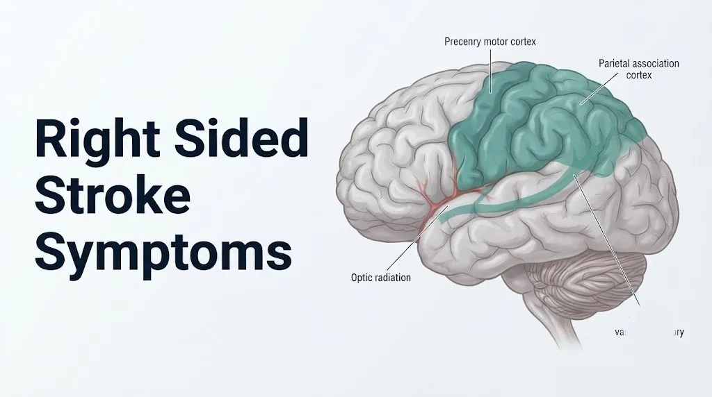
The right cerebral hemisphere is supplied by three major arterial territories. The right middle cerebral artery (MCA) is the largest and most clinically relevant. It supplies most of the right lateral hemisphere surface, including the right primary motor cortex (which controls left limb movement), the right somatosensory cortex (which processes left-sided touch and proprioception), the right parietal association cortex (which mediates spatial attention and self-awareness), and the right temporal lobe (which contributes to emotional processing and memory). The right anterior cerebral artery (ACA) supplies the medial surface of the right frontal and parietal lobes, which control left leg and foot movement. The right posterior cerebral artery (PCA) supplies the right occipital lobe (containing the primary visual cortex) and the posterior right thalamus.
Subcortical structures are equally important. The right internal capsule, a dense white matter structure passing through the center of the hemisphere, carries the right corticospinal tract (from the motor cortex to the spinal cord) and the right thalamocortical sensory fibers. A small lacunar infarction (a tiny deep infarct from occlusion of a perforating artery) within the posterior limb of the right internal capsule can produce pure left-sided motor weakness that is as severe as a large cortical infarct, because all the descending motor fibers are packed tightly into this small region.
Think of the right hemisphere like a central command station for the left side of the body and for the brain’s spatial and attentional awareness system. When the power goes out to any part of that station, the functions it controls go offline instantly.
The American Heart Association identifies the right MCA territory as the most common site of ischemic stroke overall, making right MCA syndrome the most frequently encountered right-sided stroke presentation in emergency settings.
Why Right Sided Stroke Causes Left Body Weakness: The Neuroanatomy Explained
Right sided stroke causes weakness on the left side of the body because the motor control pathway from the right hemisphere crosses to the opposite side of the body at the medullary pyramids, the paired ventral projections at the base of the brainstem where approximately 80 to 90% of the descending corticospinal tract fibers decussate.
The motor pathway begins in the right primary motor cortex at the precentral gyrus (Brodmann area 4). Upper motor neurons in layer V of this cortex send their axons downward through the right internal capsule (posterior limb), through the right cerebral peduncle in the midbrain, through the right pontine base, and into the right medullary pyramid. At the caudal medulla, approximately 85% of these fibers cross to the left side, forming the left lateral corticospinal tract. These crossed fibers descend through the left lateral funiculus of the spinal cord and synapse on left anterior horn cells, which send their axons out through the left spinal nerve roots to innervate left-sided skeletal muscles.
This crossing point is the anatomical reason that right brain injury produces left body weakness. It is not an accident of labeling. It is the fixed anatomical architecture of the human motor system, unchanged from individual to individual (with minor variations in the percentage of fibers that cross).
Facial weakness in right-sided stroke is produced by disruption of the right corticobulbar tract, which descends from the right motor cortex through the right internal capsule genu (the bend between the anterior and posterior limbs) to synapse on the facial nerve nucleus in the pons. Because of the bilateral cortical representation of the upper face (forehead and eyes), right-sided stroke spares the upper facial muscles in most cases. The lower face (cheek, lip, angle of mouth) is represented almost exclusively by the contralateral hemisphere, so right-sided stroke typically produces left lower facial drooping while the forehead wrinkles normally, a pattern that distinguishes central (cortical) from peripheral (cranial nerve) facial weakness.
According to the National Institute of Neurological Disorders and Stroke (NINDS), the corticospinal tract and corticobulbar tract together form the pyramidal motor system, and their disruption at any point from cortex to brainstem produces the upper motor neuron syndrome: weakness, spasticity, hyperreflexia, and the Babinski sign (upgoing plantar reflex).
Left-Sided Weakness and Paralysis From a Right Sided Stroke
Left-sided weakness and paralysis are the most visually apparent right sided stroke symptoms and represent the functional expression of right motor cortex and right corticospinal tract infarction, producing a clinical picture that ranges from mild arm drift to complete left hemiplegia.
The degree of weakness reflects both the extent and the location of the infarct. A large right MCA territory infarct involving the right precentral gyrus and internal capsule produces dense left hemiplegia: the arm is flaccid and falls when raised, the leg drags or cannot support weight, and the left corner of the mouth droops. An isolated infarct of the right ACA territory (the medial surface of the right frontal lobe) produces predominantly left leg weakness with relative sparing of the left arm, because the leg area occupies the medial wall of the precentral gyrus while the arm and face areas are on the lateral convexity supplied by the MCA.
The arm weakness pattern in right MCA stroke is characteristically asymmetric: the arm is more severely affected than the leg, and within the arm, the hand and fingers are more severely affected than the shoulder and elbow. This reflects the disproportionate cortical representation of the hand in the primary motor cortex, where the hand area (supplying fine motor control) occupies a larger surface area than any other body part.
The National Institutes of Health Stroke Scale (NIHSS), the standard clinical tool for quantifying stroke severity, assesses motor function on a scale from 0 (no drift) to 4 (no movement) for both the arm and leg separately. A right-sided stroke patient with left arm drift scoring 2 (arm falls before 10 seconds but does not hit the bed) and left leg scoring 3 (falls immediately) has a measurable, graded deficit that the NIHSS captures and that guides treatment decisions.
People with prior right hemisphere TIA or a history of small right hemisphere lacunar strokes may have pre-existing subtle left arm weakness that makes recognizing new additional weakness more difficult. Family members and caregivers of such individuals should establish a baseline arm strength assessment so that any new decline is recognizable against that baseline, rather than attributed to the pre-existing deficit.
Key Takeaway: Left-sided arm weakness is more severe than leg weakness in most right MCA strokes because the hand and finger area of the right primary motor cortex is large and located on the lateral convexity squarely within the MCA territory, making fine motor loss of the left hand an early and prominent sign.
Sensory Loss and Touch Disturbance From a Right Sided Stroke
Sensory loss from a right sided stroke reflects infarction of the right somatosensory cortex at the postcentral gyrus (Brodmann areas 3, 1, and 2) or disruption of the right thalamocortical sensory projections from the right ventral posterolateral (VPL) thalamic nucleus, producing left-sided loss of touch, pain, temperature, vibration, and proprioception.
The somatosensory pathway from the left body to the right cortex follows a two-neuron route. Primary sensory neurons (first-order neurons in dorsal root ganglia) send their axons up the left dorsal column of the spinal cord to synapse in the left cuneate and gracile nuclei in the medulla. Second-order neurons then cross the midline in the medial lemniscus and ascend to the right VPL thalamus. Third-order thalamocortical neurons project from the right VPL through the right internal capsule to the right postcentral gyrus. Infarction at any point in the right thalamocortical limb of this pathway produces left-sided sensory loss.
The sensory deficit is often incomplete and may be subtle enough that the patient does not spontaneously report it. A clinician must specifically test light touch, pinprick (pain), temperature discrimination, vibration sense (using a 128-Hz tuning fork), and joint position sense (proprioception) to characterize the deficit fully. Proprioceptive loss is particularly disabling: when the patient does not know where their left limb is in space, voluntary motor control of that limb is severely impaired even if motor strength is partially preserved.
The thalamic pain syndrome (also called Dejerine-Roussy syndrome) is a specific late complication of right thalamic or thalamocortical pathway infarction, in which the initial sensory loss resolves partially over weeks to months, only to be replaced by severe, burning, or aching pain on the left side of the body, often triggered by light touch. This delayed central pain state is produced by reorganized and dysregulated sensory processing in the recovering thalamocortical circuit.
Older adults with diabetic peripheral neuropathy who sustain a right-sided stroke have a particularly difficult clinical picture because baseline sensory impairment from peripheral neuropathy and new central sensory loss from the stroke superimpose, making it harder to identify what deficit is new and what represents pre-existing disease. Any older adult with diabetes who develops new asymmetric sensory changes should be evaluated for central nervous system pathology rather than attributed solely to worsening peripheral neuropathy.
Hemispatial Neglect: The Right Stroke Symptom Most People Miss
Hemispatial neglect is the failure to attend to, perceive, or respond to stimuli on the left side of the environment, produced by infarction of the right parietal association cortex and its connections with the right frontal eye fields and right thalamus, and it is the right-sided stroke symptom most likely to be missed by both patients and observers.
The right parietal lobe, specifically the right inferior parietal lobule (which includes the angular gyrus and supramarginal gyrus) and the right superior parietal lobule, serves as the brain’s dominant spatial attention system. Unlike the left hemisphere, which allocates attention primarily to the right side of space, the right hemisphere allocates attention to both sides of space. When the right parietal attention system is damaged by stroke, the left hemisphere continues to direct attention only rightward, and there is no competing signal directing attention leftward. The result is not blindness on the left (the visual system may be intact) but an active failure of the brain’s attentional spotlight to illuminate the left side of the environment.
Neglect manifests in striking and observable ways. The patient eats only the food on the right side of the plate and leaves the left side untouched. They shave only the right side of the face. They read only the right half of a sentence. They collide with objects on their left while walking. When asked to draw a clock, they cram all 12 numbers into the right half of the circle.
The clinical distinction between neglect and visual field loss (homonymous hemianopia) matters: a patient with hemianopia but intact parietal attention will compensate by turning their head or eyes to the left to scan the blind field. A patient with neglect will not, because the attentional failure is not in the eyes but in the brain’s spatial processing system.
Neglect is reported in approximately 30 to 80% of acute right hemisphere stroke patients depending on when and how it is assessed, according to research published in the journal Neuropsychological Rehabilitation. It is more prevalent and typically more severe after right than left hemisphere stroke, because the left hemisphere has no equivalent bilateral spatial attention function to compensate when the right hemisphere is damaged.
Anosognosia After Right Sided Stroke: When Patients Don’t Know They’re Impaired
Anosognosia is the unawareness or explicit denial of neurological disability following right-sided stroke, occurring in approximately 30 to 50% of patients with acute right hemisphere infarction, and it represents one of the most clinically dangerous features of right-sided stroke because it actively delays the patient from seeking help.
The neuroanatomical basis of anosognosia involves disruption of the right insular cortex and the right superior parietal lobule, regions that participate in self-monitoring, body ownership, and the integration of motor intention with sensory feedback. When these structures are damaged, the patient loses the neural machinery for detecting the mismatch between their intention to move the left arm and the arm’s failure to respond. The result is not confusion about symptoms: the patient genuinely perceives their left arm as functional even when it is paralyzed.
In practical terms, anosognosia means that a person having a right-sided stroke may calmly state that nothing is wrong, refuse to call for help, and resist assistance from concerned bystanders. They may explain away their arm’s immobility with confabulation (“I’m just tired” or “I always hold my arm that way”). They may become irritated or agitated when others express concern, because from their subjective experience, there is nothing to be concerned about.
This creates a specific emergency recognition responsibility for observers. When someone develops sudden left arm weakness, left facial drooping, or left-sided behavioral changes and simultaneously insists that they are fine and does not want help, the observer should not defer to the patient’s self-report. Anosognosia is a neurological symptom, not a considered judgment.
According to a review published in JAMA Neurology, anosognosia after right hemisphere stroke is independently associated with worse functional outcomes at 3 months, longer hospital stays, and higher rates of rehabilitation failure, because the patient’s inability to perceive deficits impairs their motivation and ability to engage with recovery exercises.
Pregnant women who sustain a right-sided stroke during pregnancy or the postpartum period (a period of elevated stroke risk due to hypercoagulability and potential amniotic fluid embolism) are particularly vulnerable if anosognosia delays their self-referral, because other pregnancy-related symptoms may be attributed to the pregnancy itself rather than recognized as stroke symptoms.
Key Takeaway: Anosognosia means the person having a right-sided stroke may sincerely believe and state that they are fine, so observers who see sudden left-sided weakness or behavioral change must call 911 regardless of the patient’s self-report.
Left Homonymous Hemianopia and Visual Symptoms of Right Sided Stroke
Left homonymous hemianopia is the loss of the left visual field in both eyes, produced by infarction of the right optic radiation (the geniculocalcarine tract running from the right lateral geniculate nucleus to the right primary visual cortex) or the right primary visual cortex itself in the right occipital lobe.
The visual pathway crosses at the optic chiasm, but the crossing is partial. The nasal (medial) retinal fibers from each eye cross to the opposite side, while the temporal (lateral) retinal fibers remain ipsilateral. After the chiasm, all fibers serving the left visual field (the nasal retinal fibers from the left eye and the temporal retinal fibers from the right eye) travel together in the right optic tract, synapse in the right lateral geniculate nucleus, and project via the right optic radiation to the right calcarine cortex. Infarction anywhere along the right optic radiation or right occipital cortex eliminates the left visual field in both eyes simultaneously.
The result is that the patient cannot see anything to the left of the vertical midline in either eye, a deficit that affects activities including driving, reading, and navigating around objects. Unlike hemispatial neglect, the patient with isolated hemianopia often recognizes that their left vision is absent and can learn to compensate by actively turning their head to scan the blind field.
Right PCA territory stroke is the most common cause of isolated left homonymous hemianopia because the PCA supplies the right occipital cortex. A right PCA stroke limited to the occipital cortex may produce pure hemianopia with no other neurological deficit, a presentation that can be mistaken for a retinal or optic nerve problem at initial evaluation. The key distinguishing feature is that homonymous hemianopia (involving the same visual field in both eyes) is always from a post-chiasmal (brain) lesion, while monocular visual loss (one eye only) suggests an anterior (eye or optic nerve) problem.
In people with pre-existing glaucoma, visual field testing for hemianopia is more complex, because glaucomatous field defects may overlap with stroke-related hemianopia and make both harder to detect and characterize accurately. Formal Goldmann or automated perimetry is the definitive test for mapping both deficits.
Speech and Communication Changes From a Right Sided Stroke
Speech changes from a right sided stroke differ fundamentally from those produced by left-sided stroke, and understanding the distinction matters because right-sided stroke speech changes are frequently minimized or missed in initial evaluation.
Left-sided stroke produces aphasia: the disruption of language comprehension and expression, from infarction of Broca’s area (left inferior frontal gyrus, Brodmann area 44 and 45) or Wernicke’s area (left posterior superior temporal gyrus, Brodmann area 22) in right-handed individuals. Right-sided stroke does not typically cause aphasia in right-handed people.
What right-sided stroke does produce is dysarthria and dysprosody. Dysarthria is the slurring or imprecision of speech from weakness of the left lower facial muscles and the articulatory muscles (lips, tongue, palate) controlled by the right corticobulbar tract. The words the patient chooses are correct and their comprehension is intact; the physical production of speech sounds is impaired. Speech sounds “thick” or “slurred” as if the speaker has had too much alcohol or has a numb mouth.
Dysprosody is the disruption of the emotional melody and intonation of speech. The right perisylvian cortex (right temporal and right inferior frontal regions homologous to Broca’s and Wernicke’s areas) is the primary neural substrate for prosody: the rises and falls in pitch, the stress patterns, and the emotional coloring that convey whether a statement is a question, a command, or an expression of sadness or joy. Right hemisphere stroke can disrupt both the production of prosody (the patient speaks in a flat, monotone voice) and the perception of prosody (the patient cannot reliably identify whether a speaker is happy, sad, or angry from their voice tone alone).
Dysprosody is frequently missed in emergency evaluation because the patient’s words are correct and comprehension appears intact. According to research published in the journal Brain and Language, right hemisphere lesions disrupt emotional prosody processing in a substantial proportion of right hemisphere stroke patients, but this deficit is rarely assessed systematically in the acute setting.
People who are professional performers, teachers, or anyone whose vocation depends on vocal expression may identify dysprosody earlier and more precisely than others, because they are attuned to subtle changes in vocal quality and emotional expressiveness that most people do not consciously notice.
Key Takeaway: Right-sided stroke does not cause aphasia in most right-handed people, but it does cause dysarthria (slurred articulation from facial weakness) and dysprosody (loss of emotional speech intonation), a distinction that changes both the diagnosis and the rehabilitation plan.
Emotional and Behavioral Symptoms of Right Hemisphere Stroke
Emotional and behavioral symptoms of right hemisphere stroke reflect infarction of the right orbitofrontal cortex, right anterior insula, right anterior cingulate cortex, and their connections with the limbic system, producing a clinical picture that can dominate the post-stroke presentation and is frequently mistaken for pre-existing psychiatric disease or personality change.
The most frequently observed behavioral changes following right hemisphere stroke include:
- Impulsivity and disinhibition: The patient acts without appropriate consideration of consequences, makes inappropriate comments, or disregards social norms. This reflects right orbitofrontal cortex dysfunction, which normally inhibits impulsive responses by integrating emotional signals with prefrontal decision-making circuits.
- Emotional lability: Rapid and exaggerated shifts in emotional expression (laughing or crying in response to mild stimuli) disproportionate to the patient’s subjective emotional state. This reflects disrupted control of emotional expression from right anterior cingulate and right corticobulbar pathways.
- Indifference or anosodiaphoria: The patient appears unconcerned about their deficits even when they have some awareness of them. Distinct from full anosognosia, this indifference reflects disrupted right insular connections to the limbic system.
- Poor judgment and risk estimation: The patient misjudges the severity of their condition, the safety of their actions, or the reliability of their own abilities. This is particularly dangerous when the patient insists on driving or returning to work prematurely after stroke.
- Depression post-stroke: Occurring in approximately 30% of stroke survivors within the first year, post-stroke depression has a specific neurobiological basis: disruption of right hemisphere frontal-subcortical circuits and reduced serotonergic input to the right prefrontal cortex from brainstem raphe nucleus projections, according to research published in the American Journal of Psychiatry.
If a stroke survivor in your household is experiencing thoughts of self-harm or hopelessness in the context of post-stroke depression, contact the 988 Suicide and Crisis Lifeline by calling or texting 988 at any time. This service is free, confidential, and available 24 hours a day, 7 days a week.
Behavioral changes are particularly difficult for family members to manage because they can look like willful personality change rather than neurological injury. Neuropsychological evaluation by a board-certified neuropsychologist is the appropriate clinical step for characterizing behavioral deficits and guiding behavioral management strategies in the rehabilitation phase.
Right Sided Stroke Symptoms in Women
Right sided stroke symptoms in women share the same core neurological profile as in men, but women are more likely than men to report atypical accompanying symptoms that can delay recognition and treatment.
The American Heart Association reports that approximately 55,000 more women than men have strokes each year in the United States, and that women are more likely to die from stroke than men, partly because they are older at the time of their first stroke (women live longer and stroke risk increases sharply with age) and partly because atypical symptom presentation delays their time-to-treatment.
Research published in Stroke journal has found that women with acute ischemic stroke are more likely than men to report symptoms including sudden severe headache, altered consciousness or confusion, generalized weakness (rather than clearly lateralized limb weakness), nausea and vomiting, and hiccups, in addition to or instead of the classic FAST signs. These atypical symptoms are more prevalent in posterior circulation strokes (basilar artery territory) and in strokes associated with atrial fibrillation, both of which are relatively more common in older women.
The result of atypical presentation is that women are less likely to be recognized as having a stroke by bystanders, less likely to call 911 as their first action, and more likely to arrive at the hospital outside the treatment window. A 2019 study published in the journal Neurology found that women were less likely than men to receive IV tPA within the treatment window after presenting to an emergency department with stroke symptoms.
Pregnancy and the postpartum period represent specific high-risk windows for women. The hypercoagulable state of pregnancy increases cerebral venous sinus thrombosis risk and arterial thromboembolism risk. Preeclampsia and eclampsia are associated with cerebral vasospasm and hemorrhagic stroke. Any woman in pregnancy or within 6 weeks postpartum who develops sudden neurological symptoms including left-sided weakness, visual changes, or severe headache should be evaluated by an emergency medicine physician immediately, because the obstetric context changes both the differential diagnosis and the treatment approach.
Right Sided Stroke Symptoms in Older Adults and High-Risk Populations
Right sided stroke symptoms in older adults are shaped by age-related changes in cerebrovascular physiology, reduced cognitive and neuroplastic reserve, and the higher prevalence of conditions that both cause stroke and complicate its presentation.
The most important individual risk factor for right-sided stroke in older adults is atrial fibrillation (AF), the most common sustained cardiac arrhythmia. In AF, the disorganized electrical activity of the atria causes blood to pool and form clots in the left atrial appendage. When these clots embolize, they travel through the systemic circulation and preferentially lodge in large cerebral arteries, causing large territory ischemic strokes. The right MCA is one of the most common embolization targets, explaining why AF-related strokes are frequently large territory right hemisphere events with dense left hemiplegia, neglect, and anosognosia combined.
According to the CDC, the prevalence of atrial fibrillation increases sharply with age, affecting approximately 2 to 3% of the general population but up to 9 to 10% of adults over age 80. An older adult without a known AF diagnosis who sustains a right hemisphere stroke should be evaluated with prolonged cardiac monitoring (at minimum a 24-hour Holter monitor, ideally a 30-day event monitor or implantable cardiac monitor) to detect paroxysmal AF that is present intermittently.
Hypertension, the leading modifiable stroke risk factor, is disproportionately prevalent in Black Americans, who have approximately twice the stroke incidence rate of white Americans, according to the CDC. Hypertension-related lacunar infarctions in the right internal capsule produce a specific clinical pattern: pure motor right-sided stroke with dense left hemiplegia and no cognitive, visual, or behavioral deficits, because the small infarct affects only the compact descending motor fibers without involving the cortex.
Older adults with pre-existing mild cognitive impairment or early dementia have a substantially more difficult post-stroke presentation because right hemisphere behavioral and cognitive deficits superimpose on baseline cognitive changes, making it harder to identify which deficits are new and which are pre-existing.
TIA Symptoms on the Right Side of the Brain: The Warning Stroke
A transient ischemic attack (TIA) affecting the right hemisphere produces the same symptom profile as a completed right-sided stroke but resolves completely within 24 hours (and typically within 60 minutes) because blood flow is restored before permanent infarction occurs.
TIA is the highest-risk period for completed stroke. A 2007 study published in Lancet Neurology found that approximately 10 to 15% of TIA patients have a completed stroke within 90 days, with the highest risk concentrated in the first 48 hours. The ABCD2 score (a clinical risk stratification tool incorporating Age, Blood pressure, Clinical features, Duration of symptoms, and Diabetes status) helps identify which TIA patients are at highest short-term stroke risk and require the most urgent workup.
Right-hemisphere TIA symptoms are identical to right-sided stroke symptoms but by definition resolve completely. The problem is that many people who experience these symptoms dismiss them precisely because they resolved: “It only lasted a few minutes, so it must not have been serious.” This is a clinically dangerous reasoning error. A TIA is a medical emergency. The transient nature of symptoms is not reassuring; it is a warning that a permanent stroke may be imminent.
TIA symptoms specific to right hemisphere involvement:
- Sudden left arm or leg weakness lasting minutes, then resolving completely
- Sudden left facial numbness or drooping that resolves within minutes to hours
- Brief episode of left visual field loss (amaurosis fugax if involving the right eye from right optic nerve ischemia, or left hemianopia if involving the right occipital cortex)
- Brief episode of confusion, behavioral change, or left-sided neglect that resolves
- Sudden slurred speech (dysarthria) that clears within minutes
- Sudden difficulty recognizing faces or voices (prosopagnosia or phonagnosia, from right temporal lobe TIA)
Any person who has symptoms consistent with a TIA, even if fully resolved, should be evaluated by a board-certified neurologist at an emergency department the same day, not at a scheduled outpatient appointment. The workup for TIA includes brain MRI with diffusion-weighted imaging (to identify any permanent infarction), CT angiography or MRA of the head and neck (to identify large vessel stenosis), cardiac monitoring for atrial fibrillation, and echocardiogram to assess for cardiac embolic sources.
Right Sided Stroke vs Left Sided Stroke: Key Symptom Differences
Right sided stroke and left sided stroke produce opposite lateralized motor and sensory deficits, but their most clinically distinguishing differences lie in the cognitive, behavioral, and communicative symptoms that reflect the distinct functional specializations of each hemisphere.
| Symptom Domain | Right Sided Stroke | Left Sided Stroke |
|---|---|---|
| Motor weakness | Left arm, leg, face (contralateral) | Right arm, leg, face (contralateral) |
| Sensory loss | Left body (contralateral) | Right body (contralateral) |
| Language (aphasia) | Rare in right-handers; possible in left-handers | Very common; Broca’s and Wernicke’s areas typically in left hemisphere |
| Speech production (dysarthria) | Present (left facial weakness) | Present (right facial weakness) |
| Emotional speech (prosody) | Frequently impaired (dysprosody) | Typically preserved |
| Spatial attention | Severely disrupted (hemispatial neglect, left-sided) | Mild right neglect possible, less severe |
| Self-awareness (anosognosia) | Frequent, can be severe | Uncommon |
| Visual field | Left homonymous hemianopia | Right homonymous hemianopia |
| Behavioral changes | Impulsivity, poor judgment, indifference, emotional lability | Catastrophic reaction (depression, anxiety about deficits) more typical |
| Awareness of deficits | Often markedly reduced | Usually preserved; patients often distressed about their deficits |
The behavioral contrast is diagnostically useful. A person with a right-sided stroke who is impulsive and apparently unconcerned about evident neurological deficits is showing a pattern consistent with right hemisphere damage. A person with a left-sided stroke who is distressed and anxious about their language difficulty is showing the more typical left hemisphere pattern where self-awareness is preserved.
This difference also has rehabilitation implications. Left-sided stroke patients often drive their own recovery through motivation and distress about their deficits. Right-sided stroke patients may require external motivational support from their rehabilitation team because their own insight into their deficits is impaired.
According to the American Heart Association, both right and left sided strokes carry substantial disability risk, but the specific deficits of right hemisphere stroke, particularly neglect and anosognosia, are independently associated with worse functional outcomes at one year because they interfere with the patient’s engagement in rehabilitation.
Emergency Symptoms: When to Call 911 for a Right Sided Stroke
Every right sided stroke symptom is a medical emergency. Do not wait to see if these symptoms resolve on their own.
Call 911 immediately if you observe or experience any of the following:
- Sudden weakness or inability to move the left arm, left leg, or left side of the face, even if partial or mild
- Sudden drooping of the left side of the face when the person tries to smile
- Left arm that drifts downward or falls when both arms are raised with eyes closed
- Sudden loss of vision or darkness in the left visual field of one or both eyes
- Sudden slurring of speech, even if the words themselves are correct
- A person who suddenly fails to notice or respond to stimuli on their left side
- A person who develops sudden confusion, behavioral change, or inappropriate emotional response
- A person who suddenly insists nothing is wrong despite observable left-sided weakness or facial drooping (anosognosia)
- A sudden severe headache described as “the worst headache of my life,” which can indicate subarachnoid hemorrhage from a ruptured cerebral aneurysm
- A person who loses consciousness or cannot be aroused following any of the above symptoms
Call 911 immediately. Do not drive the person to the hospital yourself. Paramedics can begin the pre-notification process that allows the receiving hospital’s stroke team to be ready before the patient arrives, reducing door-to-needle time for tPA administration to under 60 minutes.
The reason timing is so specific: intravenous tissue plasminogen activator (tPA) can be administered within 4.5 hours of symptom onset in eligible ischemic stroke patients, per AHA/ASA 2019 Stroke Guidelines. Mechanical thrombectomy for large vessel occlusion can extend the treatment window to up to 24 hours in carefully selected patients, per the 2018 DAWN and DEFUSE 3 trials published in the New England Journal of Medicine. But eligibility shrinks with every passing minute, because the ischemic core grows and the salvageable penumbra shrinks.
These presentations indicate acute right hemisphere stroke or hemorrhage, both of which require emergency neurological assessment and treatment at a stroke-certified hospital, not an urgent care appointment, a call to a primary care physician, or watchful waiting.
Right Sided Stroke Diagnosis and What to Expect in the ER
The emergency diagnosis of right-sided stroke begins the moment the patient or witness calls 911, and the ER process is designed to move from arrival to treatment decision within 60 minutes.
To help the ER team assess a right sided stroke most effectively:
- Note the exact time the first symptom appeared (or the last time the patient was known to be normal if they wake up with symptoms), because this determines treatment eligibility
- Tell the triage team immediately that you are concerned about stroke, using those words specifically, to trigger the stroke protocol and minimize wait time
- Bring a list of all current medications including blood thinners (warfarin, apixaban, rivaroxaban, dabigatran, clopidogrel) because anticoagulant use affects tPA eligibility
- Report any history of prior stroke, TIA, recent surgery, active bleeding, or recent trauma because these affect tPA and thrombectomy eligibility
- Report whether the patient is left-handed or right-handed because this informs the likelihood of right hemisphere language dominance
- Provide a clear description of which specific symptoms appeared, in what order, and how they have changed since onset
Imaging sequence in the acute stroke ER workup:
| Imaging Type | What It Detects | Time Frame |
|---|---|---|
| Non-contrast CT scan | Hemorrhagic stroke (excludes tPA contraindication); early ischemic changes | First 10 to 15 minutes after arrival |
| CT angiography (CTA) head and neck | Large vessel occlusion (thrombectomy candidate identification) | Immediately after non-contrast CT |
| CT perfusion (CTP) | Ischemic core vs. penumbra ratio (late window thrombectomy decision) | With CTA in selected patients |
| Diffusion-weighted MRI (DWI) | Most sensitive for acute ischemia (detects within minutes of onset) | After initial CT if time permits; primary in some centers |
The NIHSS (National Institutes of Health Stroke Scale) is administered by the stroke team to quantify deficits across 15 domains (consciousness, gaze, visual fields, facial palsy, motor arm, motor leg, ataxia, sensory, language, dysarthria, extinction and neglect). The total score guides treatment decisions and baseline prognosis. An NIHSS score of 6 or above in a right hemisphere stroke typically indicates a patient likely to benefit from thrombectomy if large vessel occlusion is confirmed.
According to the AHA/ASA 2019 Stroke Guidelines, the target door-to-needle time for IV tPA is under 60 minutes, and the target door-to-puncture time for mechanical thrombectomy is under 90 minutes. Arriving by ambulance rather than personal vehicle reduces door-to-treatment time by an average of 15 to 20 minutes at most stroke centers.
Frequently Asked Questions About Right Sided Stroke Symptoms
What are the first signs of a right sided stroke?
The first signs of a right sided stroke are typically sudden left-sided weakness or numbness of the face, arm, or leg, sudden left visual field loss, or sudden behavioral change such as confusion or neglect of the left side.
Because the right hemisphere is dominant for spatial awareness and self-monitoring, some patients experience anosognosia, meaning they do not recognize that anything is wrong despite visible left-sided weakness.
Call 911 immediately if any of these symptoms appear, because the treatment window for ischemic stroke begins closing from the moment of symptom onset.
Does a right sided stroke cause weakness on the left or right side of the body?
A right sided stroke causes weakness on the left side of the body, because the right corticospinal tract crosses to the opposite side at the medullary pyramids in the brainstem before descending to control left limb movement.
This contralateral relationship is a fixed anatomical feature of the human motor system and applies to facial weakness, arm weakness, and leg weakness in the same way.
The left lower face drops (not the upper face) because the lower face is controlled almost exclusively by the contralateral motor cortex, while the upper face has bilateral cortical representation.
What makes right sided stroke symptoms different from left sided stroke symptoms?
Right sided stroke produces contralateral left body deficits and distinctive right hemisphere cognitive symptoms including hemispatial neglect (ignoring the left side of the environment), anosognosia (unawareness of deficits), dysprosody (flat or monotone speech), and impulsivity.
Left sided stroke in right-handed individuals typically produces aphasia (language disruption), right-sided weakness, and right visual field loss, but patients with left hemisphere stroke usually retain awareness of their deficits.
The critical clinical difference is that right hemisphere stroke patients may deny or be unaware of their symptoms, making observer recognition and 911 activation by bystanders more important than in left hemisphere stroke.
Can a right sided stroke affect speech?
A right sided stroke can cause dysarthria (slurred or imprecise speech from left facial weakness) and dysprosody (loss of emotional intonation in speech), but it does not typically cause aphasia (language loss) in right-handed individuals.
In left-handed individuals who have right hemisphere language dominance (approximately 30% of left-handers), a right sided stroke can produce aphasia in addition to the typical right hemisphere deficits.
Dysprosody, the flattening of emotional speech tone, is frequently missed in initial emergency evaluation because the words are correct and comprehension appears intact.
What is hemispatial neglect and how does it happen after a right stroke?
Hemispatial neglect is the failure to attend to, perceive, or respond to stimuli on the left side of the environment, caused by infarction of the right parietal attention network that normally directs spatial awareness to both sides of space.
When the right parietal system is damaged, only the left hemisphere’s rightward-directed attention system remains active, and the left side of space drops out of awareness even though the eyes and visual pathways may be intact.
It is reported in approximately 30 to 80% of right hemisphere stroke patients in the acute setting and is associated with worse functional outcomes at one year than left hemisphere stroke without neglect.
How quickly do you need to get to the hospital for a right sided stroke?
You need to call 911 immediately at the first symptom, because intravenous tPA can only be given within 4.5 hours of symptom onset in eligible ischemic stroke patients, per AHA/ASA 2019 Stroke Guidelines.
Mechanical thrombectomy for large vessel occlusion can extend the treatment window up to 24 hours in carefully selected patients, based on the 2018 DAWN and DEFUSE 3 trials, but eligibility requires imaging confirmation and shrinks as the infarct core expands.
According to research published in the journal Stroke by Jeffrey Saver in 2006, approximately 1.9 million neurons die every minute during untreated ischemic stroke, making every minute from symptom onset to treatment directly measurable in neurological outcome.
What to Do With This Information Right Now
Right sided stroke produces a specific neurological signature that every adult should be able to recognize in themselves or in someone near them: sudden left-sided weakness, left facial drooping, loss of the left visual field, failure to notice the left side of the environment, slurred speech, or a sudden behavioral change that the person themselves may not recognize as abnormal.
The single most important clinical fact in this article is that calling 911 immediately is the only appropriate response to any of these symptoms. Do not drive to the hospital. Do not wait 20 minutes to see if symptoms improve. Do not defer to a patient who insists they are fine when you can see left-sided weakness, because anosognosia is a neurological symptom, not a reliable self-assessment.
If you or someone near you develops any symptom described in this article, the person observing those symptoms is responsible for making the call. Use the FAST framework as a quick check: Face drooping, Arm weakness, Speech changes, Time to call 911. But know that right-sided stroke can also present as left-side blindness, behavioral change, or denial of symptoms without any of the classic FAST signs, and those presentations are just as urgent. The treatment window is finite and measurable. Act on what you see