All Concussion Symptoms Eyes, Signs and Red Flags 2026
Concussion symptoms eyes include blurred vision, double vision, painful light sensitivity, difficulty tracking moving objects, and problems focusing at close range, all produced by the disruption of the brainstem oculomotor pathways, visual cortex circuits, and vestibular-ocular connections that a concussive force transmits through the brain. These are not surface-level eye injuries; they reflect disruption of the neural architecture that runs vision from the retina through the brainstem to the occipital cortex.
According to the Centers for Disease Control and Prevention (CDC), an estimated 1.6 to 3.8 million sports-related concussions occur annually in the United States, with research suggesting that visual and oculomotor symptoms are present in up to 90 percent of concussion patients, making them among the most prevalent and clinically telling features of this injury. Visual symptoms that worsen after initial improvement, or that appear suddenly hours after a head injury, can indicate intracranial hemorrhage rather than concussion and require immediate emergency evaluation.
This guide covers every documented concussion symptom involving the eyes, with the specific neural pathway disruption behind each one, explains which symptoms warrant monitoring at home versus same-day evaluation versus calling 911, distinguishes post-concussion vision syndrome from acute concussion symptoms, and addresses the populations for whom visual symptom assessment requires specific clinical modification.
How Concussion Affects the Eyes and Visual System
A concussion affects the eyes and visual system by disrupting the brainstem nuclei, white matter tracts, and cortical visual areas that coordinate eye movement, visual processing, light regulation, and gaze stabilization, not by injuring the eyes themselves.
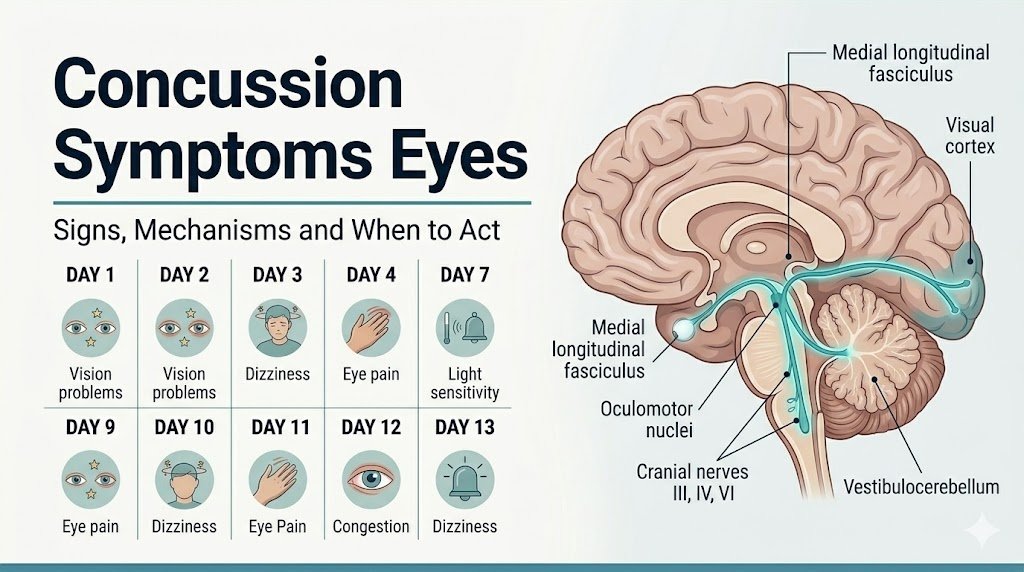
The visual system is not a single structure. It is a network extending from the retina through the optic nerve, the lateral geniculate nucleus of the thalamus, the primary visual cortex (V1) in the occipital lobe, and back through secondary visual areas (V2 through V5) that process motion, color, and spatial depth. Coordinating eye movement requires a separate oculomotor network: the cranial nerve III (oculomotor nerve), cranial nerve IV (trochlear nerve), and cranial nerve VI (abducens nerve), all connected through the medial longitudinal fasciculus (MLF), a white matter tract running vertically through the brainstem.
A concussive force produces diffuse axonal injury: the stretching and shearing of axons throughout white matter tracts. The MLF is particularly vulnerable because of its anatomical position in the dorsal pons and midbrain, a region exposed to the rotational acceleration forces that most concussions involve. When the MLF is disrupted, the three oculomotor cranial nuclei cannot communicate properly, producing the full range of gaze, tracking, and alignment problems that define concussion eye symptoms.
The energy crisis underlying these symptoms is established by prospective neurochemical research. The National Institute of Neurological Disorders and Stroke (NINDS) describes the acute concussion pathophysiology as a cascade of glutamate excitotoxicity, potassium ion efflux, calcium ion influx, and ATP depletion that impairs neural transmission across the affected circuits. Visual circuits, being among the most metabolically active in the brain, are disproportionately affected during this energy deficit period.
Quick Tip:
- The eyes themselves are almost always physically uninjured in a concussion; the problem is in the neural circuitry connecting brainstem to cortex
- Symptoms may be delayed by hours as the neurochemical cascade develops, meaning normal vision immediately post-impact does not guarantee the absence of a concussion
- People who wear corrective lenses or contact lenses should inform their examining clinician, because pre-existing refractive error can confound the assessment of post-concussion blurred vision
Concussion Symptoms Eyes: Full Overview
The full spectrum of concussion symptoms in the eyes encompasses seven distinct clinical categories, each produced by disruption of a different neural circuit, and each carrying its own level of urgency.
The following table presents the complete overview:
| Eye Symptom | Neural Pathway Disrupted | Anatomical Structure Involved | Urgency Level |
|---|---|---|---|
| Blurred vision | Visual cortex processing, accommodation | Occipital cortex V1, ciliary muscle via CN III | Moderate: evaluate within 24-48 hours |
| Double vision (diplopia) | Conjugate gaze coordination | MLF, CN III/IV/VI nuclei | High to Emergency: sudden onset = 911 |
| Light sensitivity (photophobia) | Trigeminovascular pathway, ipRGCs | Olivary pretectal nucleus, trigeminal nucleus | Moderate: monitor and limit exposure |
| Eye tracking difficulty | Smooth pursuit and saccadic pathways | Frontal eye fields, superior colliculus, PPRF | Moderate: evaluate within one week |
| Convergence insufficiency | Near-triad circuit | Edinger-Westphal nucleus, medial rectus | Moderate: neuro-optometry referral |
| VOR dysfunction | Vestibular-ocular reflex pathway | Flocculus, vestibular nuclei, abducens nucleus | Moderate to High: evaluate promptly |
| Unequal pupils (anisocoria) | Pupillary light reflex pathway | CN III, ciliary ganglion, Edinger-Westphal | Emergency: Call 911 immediately |
| Nystagmus (involuntary eye movement) | Cerebellar-vestibular pathway | Vestibulocerebellum, flocculus | High: same-day emergency evaluation |
People taking medications that affect pupillary function, including antihistamines, certain antidepressants, or opioid analgesics, should disclose these to their evaluating physician, because drug-induced pupillary changes can obscure the clinical significance of anisocoria after a head injury.
Blurred Vision After Concussion
Blurred vision after a concussion occurs when disruption of the primary visual cortex (V1 in the occipital lobe) or impaired function of the ciliary muscle via the Edinger-Westphal nucleus (which controls lens accommodation for near-focus) prevents the visual system from forming a sharp retinal image.
The distinction between cortical and accommodative causes of post-concussion blur matters clinically. Cortical blur affects vision at all distances and under all conditions: the image is simply not sharp regardless of distance. Accommodative blur is distance-dependent: vision is clearer at distance and deteriorates when focusing on near objects such as text or a phone screen. This is because accommodation, the process by which the ciliary muscle changes lens curvature to focus at close range, depends on the near-triad neural circuit through the Edinger-Westphal nucleus, which is highly vulnerable to concussive disruption.
According to a study published in JAMA Ophthalmology, accommodative insufficiency, the inability of the lens to change focus efficiently, is among the most frequently identified visual deficits in concussion patients in the subacute phase of recovery. The clinical presentation is a person who can drive and watch television without difficulty but who develops headaches, eye strain, and blurred vision within minutes of reading or screen use.
To describe blurred vision accurately to your physician or optometrist at an evaluation:
- Note whether blur is present at all distances or only at near range
- Record whether it is constant or intermittent and worsens with sustained visual tasks
- Rate severity on the Visual Analog Scale (0 = no blur, 10 = completely unable to see clearly)
- Note whether it was present immediately after the injury or developed over the subsequent hours to days
- Record whether previous corrective lenses adequately correct the blur or are no longer sufficient
Children and adolescents have a higher metabolic demand in developing visual cortex and accommodative circuits, making them more vulnerable to accommodative insufficiency after concussion than adults. A school-aged child with post-concussion blurred vision who returns to classroom reading without assessment will reliably report worsening symptoms and headaches during academic tasks.
Key Takeaway: Post-concussion blurred vision that is specifically worse at near range and worsens with sustained reading or screen use points to accommodative insufficiency from Edinger-Westphal nucleus disruption, not a primary eye injury, and warrants evaluation by a neuro-optometrist within one to two weeks of injury.
Double Vision After Concussion (Diplopia)
Diplopia (double vision) after a concussion is produced by misalignment of the two eyes’ visual axes, caused by disruption of the medial longitudinal fasciculus (MLF) or direct dysfunction of one or more of the oculomotor cranial nerves (CN III, IV, or VI) that move the extraocular muscles.
Think of the MLF as the brainstem’s telephone line between the three oculomotor offices. When both eyes are looking at the same object, CN III’s nucleus (which controls the medial rectus, pulling the eye inward) must perfectly coordinate with CN VI’s nucleus (which controls the lateral rectus, pulling the eye outward on the opposite side). The MLF connects these nuclei so they fire simultaneously. A concussion that shears or stretches the MLF produces a disconnect: one eye moves while the other lags, and the brain receives two non-identical images simultaneously.
The clinical urgency of diplopia after a head injury depends entirely on its onset pattern. Diplopia that develops immediately following the impact, persists consistently, and is associated with other mild concussion symptoms (headache, nausea, fatigue) in a context-appropriate injury is typically consistent with concussive brainstem disruption. Diplopia that appears suddenly hours to days after a head injury, particularly when the person had a brief period of normal vision (the “lucid interval”), is a red flag for an expanding intracranial hemorrhage compressing CN III or the brainstem and is a medical emergency.
The American Academy of Ophthalmology (AAO) states that new-onset diplopia after head trauma with any accompanying deterioration in alertness, increasing headache, or vomiting requires immediate emergency department evaluation with CT imaging to rule out an expanding subdural or epidural hematoma.
Adults over 60 taking anticoagulant medications (warfarin, apixaban, rivaroxaban) who sustain any head injury have a substantially elevated risk of intracranial hemorrhage and should proceed to emergency evaluation for any new diplopia, not wait to see if it resolves.
Light Sensitivity After Concussion (Photophobia)
Photophobia (light sensitivity) after a concussion is produced by hyperactivation of the trigeminovascular pathway and abnormal signaling from intrinsically photosensitive retinal ganglion cells (ipRGCs) that project to the olivary pretectal nucleus in the midbrain, creating a pain response to light intensity levels that would be normal before the injury.
The mechanism is neurological, not ophthalmological. The eyes themselves are not more sensitive to light. The brain’s pain-processing circuitry has become hyperresponsive to light signals because concussion disrupts the normal inhibitory regulation of the trigeminal nucleus caudalis, the same structure that mediates migraine headache. Light signals from ipRGCs (which express the photopigment melanopsin and project specifically to the olivary pretectal nucleus and suprachiasmatic nucleus) travel a dedicated non-image-forming visual pathway that is separate from the standard visual pathway. This pathway connects to the trigeminal pain system, which is why bright light after a concussion doesn’t just produce visual discomfort: it triggers genuine neurological pain.
Research published in the Journal of Neuro-Ophthalmology has documented that photophobia is one of the most persistent and functionally limiting symptoms in post-concussion syndrome, often lasting longer than other visual symptoms and correlating with delayed return to school and work. The mechanism shares features with migraine photophobia, which is why people with a pre-existing migraine history typically experience more severe and prolonged post-concussion photophobia than people without migraine: their trigeminovascular pathway is already sensitized.
Key management points for photophobia after concussion:
- Standard tinted sunglasses reduce brightness but do not address the trigeminovascular mechanism; FL-41 rose-tinted lenses, studied in the context of migraine and concussion photophobia, show some symptomatic benefit in emerging research
- Fluorescent lighting is particularly provocative because of its flickering frequency; LED lighting is typically better tolerated
- Dark rooms are not the recommended management approach; the CDC HEADS UP program guidance supports symptom-limited gradual light exposure rather than complete darkness, because prolonged light avoidance may prolong photosensitivity through decreased habituation
People with autism spectrum disorder (ASD) who sustain a concussion frequently have amplified photophobia because baseline sensory sensitivities interact with concussion-driven trigeminovascular hyperactivation, creating a particularly severe and prolonged photosensitivity presentation that requires modified assessment and management.
Eye Tracking Problems After Concussion
Eye tracking problems after a concussion occur when disruption of the smooth pursuit system (controlled by the medial temporal cortex and pons) or the saccadic system (controlled by the frontal eye fields and superior colliculus) prevents the eyes from following a moving target accurately or making precise rapid eye movements between fixed targets.
Normal vision requires two distinct types of eye movement. Smooth pursuits allow the eyes to follow a moving object, such as a ball in flight or text scrolling on a screen. Saccades are the rapid, ballistic eye movements used to jump between words while reading or to shift gaze from one object to another. Both systems are controlled by cortical and brainstem circuits that are highly vulnerable to the diffuse axonal injury of concussion.
Clinically, eye tracking deficits after concussion manifest as: difficulty keeping the eyes on a moving target (the eyes slip off and then jump back), difficulty reading because the eyes skip lines or lose place, and visual fatigue during sustained screen use that develops far faster than before the injury. These symptoms are frequently mistaken for reading comprehension problems or attention deficits, and in children they are sometimes inappropriately attributed to pre-existing learning disorders rather than to the concussion.
The Vestibular/Ocular Motor Screening (VOMS) tool, a validated clinical assessment developed and published in the context of sports concussion research in the British Journal of Sports Medicine, specifically evaluates smooth pursuit, horizontal and vertical saccades, near-point of convergence, and vestibulo-ocular reflex function. A study supporting its clinical use found that VOMS item scores above symptom provocation thresholds accurately predicted prolonged recovery in concussed athletes.
Adolescent baseball and softball players and tennis players, whose sports require precise saccadic and pursuit tracking, frequently report return-to-sport difficulty even after other symptoms resolve because their sport-specific visual demands expose residual tracking deficits that are not apparent during everyday activities.
Key Takeaway: Eye tracking deficits from concussion are frequently misattributed to attention or learning problems, particularly in children; a validated VOMS assessment by a sports medicine physician or neuro-optometrist specifically tests the saccadic and smooth pursuit systems and should be completed before any child returns to school or sport after a head injury.
Convergence Insufficiency After Concussion
Convergence insufficiency (CI) after a concussion is the inability of the eyes to maintain alignment while focusing on a near target, caused by disruption of the near-triad circuit that involves the Edinger-Westphal nucleus, the medial rectus muscles bilaterally, and the ciliary muscle, all coordinated through signals from the midbrain and frontal eye fields.
Convergence is the inward movement of both eyes simultaneously to focus on a close object, the same mechanism that makes text readable at arm’s length. The near-triad links three functions that always activate together: convergence (eyes moving inward), accommodation (lens thickening to increase refractive power), and pupillary constriction (the pupil narrowing to increase depth of focus). Concussion disrupts the neural coordination of this triad, so when the person tries to focus close, the eyes cannot maintain alignment, accommodation fails, and the image blurs or doubles.
The American Optometric Association (AOA) identifies convergence insufficiency as the most frequently diagnosed visual disorder following concussion, present in a clinically meaningful proportion of post-concussion patients who undergo comprehensive neuro-optometric evaluation. The defining clinical measurement is near-point of convergence (NPC): the closest distance at which both eyes can maintain alignment on a target. A normal NPC is approximately 6 centimeters or less from the nose. Post-concussion CI typically produces an NPC of 10 centimeters or greater, often accompanied by symptoms on the NPC testing task itself.
Patients with CI describe: words swimming or moving on the page during reading, eye fatigue developing within minutes of near work, headaches behind the eyes during sustained reading or computer use, and occasional diplopia when looking at near targets for extended periods.
People who had pre-existing, undiagnosed convergence insufficiency before their concussion will experience a dramatic worsening of pre-existing symptoms after the injury. A neuro-optometrist should document baseline NPC measurements and compare them to age-normalized reference values to determine how much of the deficit is new versus pre-existing.
Vestibulo-Ocular Reflex Dysfunction After Concussion
Vestibulo-ocular reflex (VOR) dysfunction after a concussion occurs when the neural pathway connecting the semicircular canals of the inner ear to the oculomotor system through the vestibular nuclei and vestibulocerebellum is disrupted, preventing the eyes from maintaining stable gaze during head movement.
The VOR is responsible for one of the most fundamental visual functions: keeping your eyes focused on a stationary point while your head moves. When you turn your head to the right while reading this text, your eyes automatically rotate left by an equal amount, maintaining the text in the center of your visual field. This reflex operates at speeds and latencies too fast for conscious control; it runs through the brainstem and cerebellum without cortical involvement. The pathway travels from the semicircular canals through CN VIII (the vestibulocochlear nerve), through the vestibular nuclei in the dorsal pons and medulla, to the flocculus and nodulus of the cerebellum (the vestibulocerebellum), and then to the oculomotor nuclei.
When this pathway is disrupted by concussion, the eyes no longer compensate accurately for head movement. The result is visual instability during motion: text appears to jump or blur when the person is walking, images appear to smear when the head moves, and visual scenes appear unstable during any physical activity. Patients often describe this as the world “moving” or “jiggling” during motion, and it is a major contributor to the exercise intolerance that many concussion patients report.
According to a prospective study published in the British Journal of Sports Medicine using the VOMS tool, VOR dysfunction is among the most common and functionally impactful findings in concussed athletes and is associated with prolonged recovery compared to athletes with normal VOR assessments.
Older adults with pre-existing vestibular disorders (benign paroxysmal positional vertigo, Meniere’s disease, vestibular neuritis) have baseline VOR dysfunction before any concussion occurs, making post-concussion assessment challenging. A vestibular audiologist or neuro-otologist should be part of the evaluation team for this population.
Key Takeaway: VOR dysfunction from concussion makes vision unstable during any head movement and is a primary reason concussed patients cannot return to exercise; if the world appears to move or jiggle during walking, this symptom requires VOMS assessment rather than being dismissed as dizziness.
Nystagmus and Pupil Changes After a Head Injury
Nystagmus (involuntary rhythmic eye movement) and abnormal pupil responses after a head injury indicate disruption of the cerebellar or brainstem circuits that stabilize gaze and regulate the pupillary light reflex, and both findings require same-day medical evaluation.
Nystagmus in the post-concussion context occurs when the cerebellar gaze-stabilization mechanisms, specifically the flocculus and the fastigial nucleus of the cerebellum, are unable to hold the eyes in a steady eccentric position of gaze. The eyes drift off-target (the slow phase) and then jump back (the fast phase), producing the characteristic saw-tooth oscillation. While some forms of nystagmus are benign and pre-existing, new-onset nystagmus after a head injury, particularly downbeat nystagmus (slow phase upward, fast phase downward) or gaze-evoked nystagmus (nystagmus appearing only in a specific direction of gaze), indicates cerebellar or posterior fossa pathology that must be distinguished from simple concussion by neuroimaging.
Pupil changes after head injury are among the most well-established emergency signs in neurology. Normal pupillary light reflex requires intact CN II (for the afferent, light-sensing limb) and CN III (for the efferent, constriction limb, through the Edinger-Westphal nucleus and ciliary ganglion). A dilated, unreactive pupil on one side following head trauma indicates compression of CN III by uncal herniation (the medial temporal lobe being pushed through the tentorium by rising intracranial pressure), which is a neurosurgical emergency.
The National Institute of Neurological Disorders and Stroke (NINDS) identifies unequal pupils (anisocoria) that are new after a head injury as one of the clearest clinical indicators of a life-threatening intracranial event, not a concussion, and states that immediate emergency evaluation is required without delay.
Children and adolescents with post-concussion nystagmus should be evaluated by a pediatric neurologist, because benign paroxysmal vertigo of childhood and other developmental vestibular conditions can produce nystagmus that superficially resembles post-concussion cerebellar nystagmus, and differentiating them requires a complete neurological examination.
Post-Concussion Vision Syndrome
Post-concussion vision syndrome (PCVS) is a persistent cluster of visual and oculomotor symptoms lasting beyond the expected acute recovery period (generally defined as beyond four weeks in adults and beyond four weeks in children, per the Concussion in Sport Group (CISG) Berlin Consensus Statement published in the British Journal of Sports Medicine in 2017), reflecting ongoing disruption of visual neural circuits rather than acute neurochemical dysfunction.
In the acute phase of concussion (zero to seventy-two hours), visual symptoms reflect the energy crisis: neurons with disrupted ion gradients cannot generate normal action potentials, and the affected visual circuits simply cannot function properly. Most acute visual symptoms resolve as the ionic crisis resolves and cerebral blood flow autoregulation normalizes over one to four weeks.
In PCVS, symptoms persist because of more lasting changes: incomplete axonal recovery in white matter tracts, maladaptive neural compensation strategies, and in some cases chronic neuroinflammation mediated by persistently activated microglia. Patients with PCVS typically report that visual symptoms improve with rest and worsen with any sustained visual demand, a pattern reflecting the inadequate metabolic reserve of partially recovered visual circuits.
The symptom profile of PCVS includes:
- Persistent accommodative insufficiency (difficulty with near focus that has not normalized)
- Persistent convergence insufficiency (near point of convergence remaining beyond 10 cm)
- Persistent photophobia not explained by migraine or other identified cause
- Persistent smooth pursuit and saccadic deficits identified on VOMS testing
- Visual motion sensitivity (discomfort or dizziness in environments with visual motion, such as scrolling screens, crowded spaces, or driving)
People with a prior history of concussion are at elevated risk for PCVS because their visual neural circuits may not have fully recovered from previous injuries before sustaining the current one. Research published in Pediatrics has documented that pediatric patients with repeated concussions have measurably prolonged visual symptom recovery compared to those experiencing a first concussion.
Concussion Eye Symptoms in Children and Adolescents
Concussion eye symptoms in children and adolescents follow a clinically distinct pattern from adult presentations because the pediatric visual cortex, oculomotor system, and cerebellum are still undergoing myelination and synaptic refinement, creating greater vulnerability to disruption and slower recovery.
Myelination of the visual association areas (V2 through V5) and the frontal eye fields continues into the mid-twenties. When these incompletely myelinated tracts are disrupted by a concussion in a 12-year-old, the recovery trajectory is fundamentally different from that in a 35-year-old with fully mature neural architecture. The pediatric brain has more plasticity (the ability to reorganize neural connections in response to injury) but also has less redundancy in visual processing pathways, making recovery less predictable.
Specific visual symptom features in pediatric concussion:
- Accommodative insufficiency is more prevalent in children than adults, likely reflecting the greater visual demands of school-related near work in this population
- Eye tracking deficits frequently manifest as reading difficulty, line-skipping, and loss of place in text, which are often misattributed to dyslexia or attention-deficit/hyperactivity disorder (ADHD)
- Photophobia is frequently more severe and longer-lasting in adolescents than in adults, particularly in teenagers who play sports on lit fields or courts
- Return-to-learn protocols require specific visual accommodation considerations: reduced near work demands, frequent breaks, and screen exposure limits before full academic reintegration
According to research published in Pediatrics, children who return to school too quickly after a concussion, before visual symptoms have adequately resolved, consistently report higher symptom burden and longer overall recovery times than those who follow structured return-to-learn protocols. Pediatric patients with persistent visual symptoms beyond four weeks should be referred to a neuro-optometrist with experience in pediatric concussion management, not dismissed as slow recoverers.
A pediatric sports medicine physician or pediatric neurologist should be the primary coordinating clinician for any child with visual symptoms persisting beyond two weeks post-concussion, given the academic, developmental, and return-to-play implications specific to this population.
Concussion Eye Symptoms in Female Athletes and Older Adults
Female athletes and older adults represent two populations in which concussion symptoms involving the eyes present with distinct clinical features that alter the assessment and management approach.
Female athletes have well-documented higher rates of concussion symptom burden compared to male athletes sustaining equivalent impacts, including higher rates of visual symptoms, longer symptom duration, and higher rates of meeting the threshold for prolonged recovery. Research published in the British Journal of Sports Medicine has found an association between concussion severity and hormonal cycle phase in female athletes, with preliminary evidence suggesting that concussions sustained during the luteal phase of the menstrual cycle (when progesterone is elevated) may be associated with more prolonged symptom courses, though this remains an area of active investigation rather than established clinical guideline. Female athletes should inform their evaluating sports medicine physician of their cycle phase at the time of injury as part of the clinical history.
Older adults present differently because the aging visual system has declining accommodation (presbyopia), reduced contrast sensitivity, slower saccadic latency, and increased susceptibility to VOR disruption from age-related vestibular changes. A 65-year-old who sustains a head injury may already have baseline accommodative insufficiency and reduced tracking speed, making it difficult to attribute new post-injury deficits to the concussion itself without pre-injury baseline measurements for comparison.
Additional considerations for older adults:
- Anticoagulant or antiplatelet therapy significantly elevates the risk of intracranial hemorrhage after head injury, lowering the threshold for emergency CT imaging
- Pre-existing macular degeneration or glaucoma can produce overlapping visual symptoms that complicate post-concussion visual assessment
- Age-related decline in cerebellar function amplifies VOR dysfunction symptoms, producing more severe gaze instability than would be expected in a younger patient with the same injury
A neuro-ophthalmologist should be involved in the evaluation of older adults with post-concussion visual symptoms, particularly when it is unclear how much of the deficit is attributable to aging versus the acute injury.
Key Takeaway: Female athletes should document their menstrual cycle phase at the time of concussion as part of the clinical history, and older adults on anticoagulants with any new visual symptom after head injury should proceed to emergency imaging rather than watchful waiting.
Concussion Eye Symptoms Versus Signs of a Brain Bleed
Distinguishing concussion eye symptoms from the eye findings of an intracranial hemorrhage (brain bleed) is the single most clinically important differentiation in post-head-injury assessment, because a brain bleed that is mistaken for a concussion can be fatal if it progresses without neurosurgical intervention.
Concussion produces diffuse, bilateral, and symmetrical visual disruption reflecting widespread neural dysfunction. Brain bleeds (epidural hematoma, subdural hematoma, subarachnoid hemorrhage, intracerebral hemorrhage) produce focal, asymmetric, and often progressive neurological findings that are fundamentally different in character.
| Feature | Concussion Eye Symptoms | Brain Bleed Eye Signs |
|---|---|---|
| Pupils | Equal, reactive to light | Unequal (anisocoria); one pupil fixed and dilated = CN III compression |
| Diplopia onset | Immediately post-impact, stable or improving | New onset hours after injury, or after a lucid interval |
| Visual field | No loss, or mild bilateral blur | Hemianopia (loss of half the visual field) if occipital hemorrhage |
| Symptom trajectory | Improves or stays stable over hours to days | Worsens progressively after initial stability |
| Consciousness level | Maintained or brief loss at impact; recovers | Deteriorating after a lucid interval (epidural pattern) |
| Nystagmus | Mild, if present; positional or gaze-evoked | Vertical, downbeat, or direction-changing; does not suppress with fixation |
| Accompanying signs | Headache, nausea, fatigue | Increasing headache, repeated vomiting, confusion, limb weakness |
The lucid interval deserves specific emphasis. In epidural hematoma (bleeding between the skull and the dura mater, usually from a torn middle meningeal artery), the patient may appear completely normal for minutes to hours after the injury before sudden rapid deterioration as the expanding blood clot compresses the brain. During this lucid interval, visual symptoms may be absent. New diplopia or unequal pupils appearing after a symptom-free period following head trauma is a neurosurgical emergency, not a delayed concussion symptom.
Emergency Eye Symptoms After a Head Injury
Certain concussion eye symptoms and post-head-injury visual findings require immediate emergency evaluation. Do not wait to see if these resolve on their own.
Call 911 or go to the nearest emergency room immediately if you observe or experience after a head injury:
- One pupil larger than the other (unequal pupils, anisocoria): indicates possible CN III compression from uncal herniation secondary to rising intracranial pressure, which is a neurosurgical emergency
- Sudden new double vision appearing hours after the injury, particularly after a period of normal vision (the lucid interval pattern of epidural hematoma)
- Complete loss of vision in one or both eyes following head trauma (indicates possible posterior cerebral artery territory involvement or occipital lobe hemorrhage)
- Loss of half the visual field (hemianopia): indicates a focal occipital or parietal hemorrhage requiring immediate imaging
- Rhythmic involuntary eye movement (nystagmus) that is vertical or direction-changing and does not suppress when the person fixates on a stationary target
- Any visual symptom that is rapidly worsening after an initial period of stability following a head injury
- Symptoms accompanied by deteriorating alertness, severe increasing headache, repeated vomiting, or confusion (signs of rising intracranial pressure)
- Loss of consciousness at any point following a head injury, even briefly
These presentations can indicate epidural hematoma, subdural hematoma, subarachnoid hemorrhage, or brainstem herniation, all of which are neurosurgical emergencies that require immediate CT imaging and cannot be assessed at an urgent care clinic or over the phone.
Neuro-Optometric Evaluation After Concussion
A neuro-optometric evaluation after concussion is a specialized clinical assessment performed by a neuro-optometrist or neuro-ophthalmologist that goes substantially beyond a standard vision check, specifically measuring the oculomotor functions most commonly disrupted by concussive brain injury.
Standard optometric examination (the routine annual eye exam) measures visual acuity, refractive error, and gross ocular health. It does not systematically assess smooth pursuit quality, saccadic accuracy, near-point of convergence under sustained demand, accommodative amplitude versus accommodative facility, or VOR gain. A person with significant post-concussion visual dysfunction can pass a standard vision check because their visual acuity at distance is unchanged, while their oculomotor system is substantially impaired.
A neuro-optometric evaluation typically includes:
- Near-point of convergence (NPC) testing: A target is moved toward the nose until the patient reports diplopia or the evaluator observes eye misalignment; repeated three times and averaged
- Smooth pursuit assessment: The patient follows a moving target in horizontal and vertical planes; deviation from smooth tracking is recorded
- Saccadic testing: The patient makes rapid eye movements between two fixed targets; latency, accuracy, and velocity are assessed
- Accommodative amplitude testing: The maximum near focus point is measured using a push-up or minus-lens method
- VOR gain testing: A simplified version can be performed clinically using head impulse testing
- Full VOMS assessment: Symptom provocation during each oculomotor subsystem test is rated on a 0-10 scale
The American Optometric Association (AOA) recommends that any athlete or patient with persistent visual symptoms after concussion, specifically symptoms lasting more than two weeks, receive a full neuro-optometric evaluation before returning to sport or full academic engagement. Findings guide the prescription of evidence-supported vision therapy interventions.
People with pre-existing convergence insufficiency, strabismus, or amblyopia should have pre-injury records available for their evaluating neuro-optometrist to establish what is attributable to the concussion versus pre-existing condition.
How Long Do Eye Symptoms Last After Concussion and Recovery
The duration of concussion eye symptoms varies substantially by symptom type, injury severity, patient age, and the presence of pre-existing visual conditions, ranging from days for acute blur and photophobia to weeks or months for convergence insufficiency and VOR dysfunction.
According to the Concussion in Sport Group (CISG) Berlin Consensus Statement, most adults recover from a concussion within 10 to 14 days, and most children and adolescents within four weeks, with visual symptoms typically resolving within this window in uncomplicated cases. However, research consistently documents that oculomotor deficits, particularly convergence insufficiency and smooth pursuit dysfunction, persist beyond the general symptom resolution timeline in a clinically meaningful proportion of patients, particularly those who had a high acute symptom burden or who returned to visually demanding activities too quickly.
The recovery timeline by visual symptom type:
| Symptom | Typical Resolution (Uncomplicated) | Prolonged Recovery Indicators |
|---|---|---|
| Acute blur (cortical) | 24-72 hours | Persisting beyond one week |
| Accommodative insufficiency | 1-4 weeks | Persisting beyond 4 weeks |
| Photophobia | 1-3 weeks | Persisting beyond 4 weeks; pre-existing migraine prolongs this |
| Eye tracking deficits | 2-4 weeks | Persisting beyond 4 weeks; high baseline visual demand prolongs this |
| Convergence insufficiency | 4-8 weeks with neuro-optometric management | Persisting beyond 8 weeks without treatment |
| VOR dysfunction | 2-6 weeks with vestibular rehabilitation | Persisting beyond 6 weeks |
| Diplopia from cranial nerve palsy | Weeks to months (CN IV most prolonged) | Persisting beyond 3 months warrants MRI |
Vision therapy for convergence insufficiency after concussion, specifically the program of in-office and home-based vergence and accommodative exercises studied in the Convergence Insufficiency Treatment Trial (CITT) research program, has the strongest evidence base for persistent post-concussion CI and is a structured rehabilitation program managed by a neuro-optometrist.
Pediatric patients and those with a history of multiple concussions should be followed with serial VOMS assessments rather than symptom report alone, because oculomotor deficits can persist in the absence of subjective visual complaints once the patient has adapted to degraded visual function.
Key Takeaway: Visual symptoms persisting beyond four weeks in any concussion patient, or beyond two weeks in a child, warrant formal neuro-optometric evaluation rather than continued watchful waiting, because evidence-supported vision rehabilitation exists for convergence insufficiency and tracking deficits and waiting delays treatment unnecessarily.
Frequently Asked Questions About Concussion Eye Symptoms
What eye symptoms are normal after a concussion?
Blurred vision, light sensitivity, difficulty tracking moving objects, and mild eye strain with near work are the most frequently observed visual symptoms in the first days after a concussion.
These symptoms occur because diffuse axonal injury and the acute neurochemical energy crisis disrupt the brainstem oculomotor pathways and visual cortex circuits, not because the eyes themselves are damaged.
Symptoms that are improving gradually over the first week are considered part of the expected recovery pattern; symptoms that are stable or worsening after the first 24 to 48 hours warrant clinical evaluation by an emergency medicine physician or primary care physician.
Is double vision after a concussion an emergency?
Double vision appearing immediately after a concussion and stabilizing or improving is generally consistent with concussive brainstem disruption and warrants same-day urgent evaluation rather than a 911 call, unless other emergency signs are present.
Double vision that appears for the first time hours after a head injury, especially after a symptom-free period, is a red flag for an expanding intracranial hemorrhage and requires immediate emergency room evaluation with CT imaging.
Call 911 immediately if new double vision is accompanied by a pupil that is larger on one side, deteriorating alertness, severe headache, repeated vomiting, or confusion.
How long does light sensitivity last after a concussion?
Light sensitivity (photophobia) after a concussion typically improves within one to three weeks in uncomplicated cases, driven by normalization of the trigeminovascular pathway hyperactivation as the acute neurochemical cascade resolves.
People with a pre-existing migraine history frequently experience more prolonged and severe photophobia because their trigeminovascular system was already sensitized before the injury.
Photophobia persisting beyond four weeks warrants evaluation by a neurologist or neuro-optometrist to assess for post-concussion vision syndrome and to consider whether migraine prevention strategies are appropriate.
Can a concussion cause permanent vision problems?
Most concussion-related visual symptoms resolve fully with time and, where indicated, neuro-optometric rehabilitation, and permanent vision loss from an uncomplicated concussion without intracranial hemorrhage is not a typical outcome.
Persistent convergence insufficiency, accommodative insufficiency, and smooth pursuit deficits that remain beyond six months post-injury represent the cases most likely to require ongoing vision therapy rather than eventual complete spontaneous resolution.
Any visual symptom that has not improved at all by six weeks post-concussion, particularly diplopia or a visual field deficit, should be evaluated by a neuro-ophthalmologist to rule out structural cranial nerve injury or occipital lobe pathology.
What is convergence insufficiency after a concussion?
Convergence insufficiency after a concussion is the inability of both eyes to maintain alignment on a near target simultaneously, produced by disruption of the near-triad neural circuit involving the Edinger-Westphal nucleus and the medial rectus muscles bilaterally.
The American Optometric Association identifies it as the most frequently diagnosed visual disorder following concussion, manifesting as blurred near vision, eye fatigue with reading, headaches behind the eyes during screen use, and occasional double vision at close range.
It is diagnosed by measuring near-point of convergence (a value greater than 10 centimeters is abnormal) and treated with a structured neuro-optometric vision therapy program under the care of a neuro-optometrist.
When should I go to the ER for eye symptoms after a head injury?
Go to the emergency room immediately for any of the following after a head injury: one pupil larger than the other, new double vision appearing hours after the injury (especially after an initially normal period), complete or partial vision loss, a visual symptom that is rapidly worsening, or any visual change accompanied by deteriorating alertness, severe headache, or confusion.
These findings can indicate intracranial hemorrhage with brainstem compression, which is a neurosurgical emergency requiring CT imaging within minutes, not hours.
Visual symptoms that are mild, present immediately after impact, and stable or improving can be evaluated by a primary care physician or sports medicine physician within 24 to 48 hours rather than at an emergency department.
Closing
The eye symptoms of a concussion are not simple vision problems. They are windows into the neural architecture of the brainstem, cerebellum, and visual cortex. Understanding which symptom reflects which disrupted pathway tells you something specific about severity, urgency, and what kind of evaluation you need.
The priority action depends on what you are experiencing. Sudden-onset double vision, a pupil that is larger on one side, or visual symptoms that appear after an initially normal period after a head injury: these are emergency room situations, not wait-and-see situations. Persistent blur with near work, eye tracking difficulty, or light sensitivity beyond two weeks: these warrant a formal neuro-optometric evaluation, not continued home management alone.
If you or someone you care for is experiencing visual symptoms after a head injury, document exactly what changed, when it changed, and whether it is improving, stable, or worsening. That timeline is the most important piece of clinical information you can bring to your evaluating physician