Early Glaucoma Symptoms: What Your Eyes Are Telling You
Early glaucoma symptoms are notoriously difficult to detect on your own, because the most common form of the disease destroys vision silently, without pain, without obvious visual disturbance, and often without any warning your eyes would recognize as a problem. What makes this particularly dangerous is that by the time most people notice something is wrong, they have already lost a substantial portion of their retinal ganglion cells permanently.
According to the National Eye Institute, glaucoma affects more than 3 million Americans and is the leading cause of irreversible blindness worldwide. Roughly half of those affected do not know they have it. That figure is not a failure of medicine. It is a direct consequence of how the disease works at the anatomical level.
This article covers every clinically recognized early symptom pattern across all major types of glaucoma, explains the specific mechanism behind each one, addresses who is at elevated risk and why, and tells you exactly when a symptom crosses into emergency territory. The physiology here is not simplified. You deserve to understand what is actually happening inside your eye.
What Is Glaucoma and How Does It Affect Vision
Glaucoma is a group of optic neuropathies characterized by progressive death of retinal ganglion cells (RGCs), whose axons form the optic nerve, ultimately producing irreversible loss of visual field.
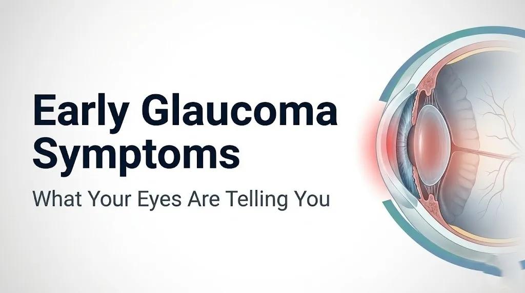
The most common mechanism involves elevated intraocular pressure (IOP). The ciliary body (a ring of tissue behind the iris) continuously produces aqueous humor, the clear fluid that fills the front compartment of the eye and maintains its structural shape. That fluid drains primarily through the trabecular meshwork, a sponge-like lattice at the angle where the iris meets the cornea, into Schlemm’s canal, and then through collector channels into the episcleral veins. When trabecular meshwork resistance increases, drainage slows while production continues. Pressure rises.
Think of the trabecular meshwork like a drain in a bathtub. When the drain gradually clogs with debris, the water level rises even though the tap rate stays exactly the same. In glaucoma, the “water level” is IOP. As IOP rises, it compresses and distorts the lamina cribrosa, a perforated connective tissue plate at the back of the eye through which RGC axons must travel to reach the brain. That compression impairs axoplasmic flow, the intracellular transport system that delivers survival signals from the brain back to the RGC cell bodies. Starved of these signals, RGCs undergo apoptosis. They die, and they do not regenerate.
Not every case of glaucoma involves elevated pressure, and not every elevated IOP causes glaucoma. That distinction matters enormously for understanding why some people with perfectly normal pressure readings still lose vision, which later sections address directly.
| Glaucoma Type | Primary Mechanism | Typical IOP | Symptom Pattern |
|---|---|---|---|
| Primary Open-Angle (POAG) | Trabecular meshwork outflow resistance | Elevated, often above 21 mmHg | Asymptomatic until late stage |
| Acute Angle-Closure | Complete iridotrabecular blockage | Acutely above 40 mmHg | Severe pain, red eye, halos, nausea |
| Normal-Tension (NTG) | Vascular insufficiency to optic nerve head | Within normal range (below 21 mmHg) | Asymptomatic or subtle |
| Ocular Hypertension | Elevated IOP, no optic nerve damage yet | Elevated | Often no symptoms at all |
| Pseudoexfoliation Glaucoma | Exfoliative material blocking trabecular meshwork | Often severely elevated | Asymptomatic or acute |
Older adults should be aware that the trabecular meshwork loses endothelial cells with age, which means outflow resistance gradually increases even without any identifiable systemic disease. The American Academy of Ophthalmology (AAO) Preferred Practice Pattern for Primary Open-Angle Glaucoma recommends baseline comprehensive eye examinations for all adults by age 40, regardless of symptoms.
Early Glaucoma Symptoms: The Clinical Picture in 2026
Early glaucoma symptoms in their most common form are the absence of detectable symptoms, which is what makes the disease so clinically treacherous.
In primary open-angle glaucoma, which accounts for approximately 74 percent of all glaucoma cases in the United States according to the American Glaucoma Society, a person can lose 30 to 40 percent of all their retinal ganglion cells before any subjective visual change is detectable under standard testing conditions. This is not a minor gap in awareness. It means the disease can be well into its damaging phase before you experience anything at all.
That said, there are early changes some people do notice, and recognizing their pattern matters:
- Subtle blurring at the outer edges of vision, noticeable only when fatigued or in specific lighting conditions
- Difficulty adjusting to low-light environments compared to previous years, related to early loss of peripheral rod photoreceptors that depend on functional RGC pathways
- Occasional headaches above or around the brow, more characteristic of mildly elevated IOP than of fully formed open-angle glaucoma but worth noting
- Needing more light to read or drive at night, a nonspecific but sometimes early functional indicator
- Slightly asymmetric vision between the two eyes that the person cannot pinpoint as a specific location of blur
The challenge with this list is that none of these symptoms are specific to glaucoma. Fatigue, refractive error changes, early cataracts, and dry eye disease produce similar functional complaints. That is precisely why clinical examination, not symptom self-assessment, is the only reliable early detection tool.
According to a 2021 analysis published in JAMA Ophthalmology, patients with confirmed glaucoma at their diagnosis had typically experienced detectable optic nerve changes on imaging for an average of four years before their first clinical visit for eye-related complaints. The disease was not silent to imaging equipment. It was silent to the patient.
Women and men present with open-angle glaucoma at similar frequencies, though women may delay seeking evaluation longer because they are less likely to have received prior IOP screening at a routine physical compared to men who receive occupational health screenings involving vision testing.
Why Early Glaucoma Symptoms Are Often Silent
Early glaucoma symptoms are silent primarily because peripheral vision, the first region of the visual field to be destroyed, is processed in a way the brain partially compensates for during the initial stages of loss.
The superior and inferior arcuate nerve fiber bundles are the first RGC axon pathways to deteriorate at the optic nerve head. These bundles carry visual signals from the mid-peripheral retina above and below the center of fixation. When arcuate fiber loss begins, the earliest visual field defects appear as small gaps or scotomas (blind spots) in the superior or inferior mid-peripheral field, outside the central 10 to 15 degrees of vision. The brain, which is extraordinarily good at pattern-completion, fills in these gaps using information from adjacent functional RGC pathways and from the fellow eye. You see an apparently complete image even when it has holes in it.
This neural compensation is not unique to glaucoma. It is the same mechanism that makes the physiological blind spot (the optic disc, where there are no photoreceptors) invisible to normal human perception. The brain silently interpolates the missing information.
The papillomacular bundle, which carries signals from the central 5 degrees of fixation and determines the visual acuity measured on a standard Snellen chart (the eye chart with the big E), is anatomically positioned to be damaged last at the superior temporal and inferior temporal optic nerve rim. This means a person can read 20/20 on an eye chart and still have advanced glaucoma with severe peripheral visual field loss.
According to the Early Manifest Glaucoma Trial (EMGT), published in the Archives of Ophthalmology, patients with established visual field defects at enrollment had lost a mean of approximately 40 percent of their optic nerve fibers by the time those defects were first detectable on standard automated perimetry. The damage always runs ahead of the detectable symptom.
For individuals with only one functioning eye due to amblyopia, prior eye injury, or other conditions, this compensatory mechanism between two eyes does not apply. Their single functional eye bears the full burden, and any early glaucomatous field loss is not offset by the fellow eye. Their risk from delayed diagnosis is correspondingly greater.
Key Takeaway: In primary open-angle glaucoma, your brain actively hides the early vision loss from your conscious perception by filling in missing peripheral information, which is why self-monitoring for early symptoms is clinically unreliable.
Peripheral Vision Loss in Glaucoma
Peripheral vision loss in glaucoma follows a predictable anatomical pattern determined by which retinal ganglion cell axon bundles are damaged first at the optic nerve head.
The earliest clinically detectable field defects appear as arcuate scotomas, crescent-shaped blind spots that curve through the superior or inferior mid-peripheral visual field, typically in the Bjerrum area (located roughly 10 to 20 degrees from the center of fixation). On a Humphrey Visual Field Analyzer, these appear as clusters of depressed test points in the arcuate zones corresponding to the superior and inferior optic nerve rim, where axons are most densely packed and most vulnerable to mechanical compression by the lamina cribrosa.
As glaucoma progresses without treatment, these arcuate defects enlarge and may merge across the horizontal midline, creating what is called a nasal step: a sharp horizontal boundary in the nasal visual field where damaged superior or inferior arcuate fibers meet the relatively preserved horizontal raphe RGC pathway. A nasal step on formal visual field testing is a hallmark of moderate glaucomatous damage.
In advanced disease, the expanding scotomas eventually leave only a small central island of vision, a condition often called “tunnel vision,” though this term is somewhat misleading. True tunnel vision implies a narrow cylindrical field. Glaucomatous end-stage vision loss more accurately resembles vision through a straw pointed at a specific spot, with surrounding complete darkness rather than blur.
The reason peripheral loss precedes central loss involves both the anatomy of the lamina cribrosa (superior and inferior poles are structurally weaker and have larger pores, making their axons more susceptible to IOP-driven distortion) and the ratio of axons to supporting blood vessels in these regions.
| Stage of Glaucoma | Visual Field Status | Patient-Perceived Symptom |
|---|---|---|
| Early (Stage 1-2) | Small arcuate scotoma, 10-20 degrees from fixation | Usually none; brain compensates |
| Moderate (Stage 3) | Expanded arcuate defect, nasal step present | May notice peripheral misses; bumping into objects |
| Advanced (Stage 4) | Severe superior and inferior field loss | Clear peripheral limitation; tunnel vision emerging |
| End-Stage (Stage 5) | Central island or temporal island only | Functional tunnel vision; major daily impairment |
Individuals with high myopia (nearsightedness above minus 6 diopters) are at increased risk for earlier and more severe peripheral field loss at any given IOP level, because the mechanically elongated myopic globe stretches and thins the lamina cribrosa, reducing its structural resistance to IOP-related deformation. A myopic individual with IOP of 18 mmHg may suffer the same degree of optic nerve damage as a non-myopic person with IOP of 25 mmHg, according to clinical observation published in the British Journal of Ophthalmology.
Glaucoma Symptoms Early in Open-Angle Glaucoma
Glaucoma symptoms early in the open-angle form are clinically distinguished from other glaucoma types by their slow, painless, and bilateral nature, though the degree of damage is almost always asymmetric between the two eyes.
Primary open-angle glaucoma (POAG) is bilateral in approximately 90 percent of cases, but it rarely progresses at exactly the same rate in both eyes. One eye typically sustains greater optic nerve damage and visual field loss than the other, which is why ophthalmologists pay close attention to interocular IOP asymmetry and cup-to-disc ratio differences.
The cup-to-disc ratio is a measure of the relative size of the central cup (the pale, excavated central zone of the optic disc) compared to the total disc diameter. A ratio above 0.5 in either eye, or a difference of 0.2 or more between the two eyes, is clinically suspicious for glaucoma and warrants formal evaluation with optical coherence tomography (OCT) and visual field testing. This asymmetry is detectable only with equipment, not with self-examination.
Here is what the clinical early symptom picture of open-angle glaucoma actually looks like in practice:
- No pain at any stage of early disease
- No redness during typical pressure elevations
- No perception of the arcuate scotomas forming in the mid-peripheral field
- Possible slight difficulty with contrast sensitivity in low light, noticed as needing more light than before
- Possible subtle difficulty with tasks requiring peripheral awareness, such as sports or driving in complex traffic
- No change in visual acuity on a standard eye chart until very late disease
The painless nature of POAG is directly related to the fact that IOP rises gradually, over years to decades. Gradual IOP elevation does not stretch the corneal endothelium fast enough to trigger the acute edema and pain response seen in angle-closure crisis. The trabecular meshwork and optic nerve experience slow, cumulative damage rather than acute traumatic deformation.
According to the Ocular Hypertension Treatment Study (OHTS), individuals with IOP between 24 and 32 mmHg (elevated but not acutely dangerous) and no optic nerve damage at enrollment had a five-year risk of developing definite POAG of approximately 9.5 percent in the untreated group, compared to 4.4 percent in those treated with pressure-lowering drops. The disease develops slowly enough to miss entirely without regular professional screening.
For women who are pregnant or using hormonal contraceptives, IOP can fluctuate due to progesterone-mediated changes in aqueous humor production and trabecular outflow facility. These fluctuations do not produce symptoms but can complicate the interpretation of IOP measurements taken during pregnancy.
High Pressure in Eyes Symptoms
High pressure in the eyes, clinically called ocular hypertension, most commonly produces no symptoms whatsoever, which is the single most important clinical fact about this condition.
IOP is measured in millimeters of mercury (mmHg). The statistically defined normal range is approximately 10 to 21 mmHg, based on population distribution data. Pressures above 21 mmHg are considered elevated. However, the relationship between IOP and symptoms is not linear, and it is not reliable as a self-detection tool.
Moderate IOP elevation (22 to 30 mmHg) is almost universally asymptomatic. The corneal endothelium and epithelium compensate adequately at these pressures, so no corneal edema, no halos, and no pain develop. The person feels nothing unusual. This is exactly the IOP range in which most people with POAG are diagnosed, and it is also the range in which the optic nerve is sustaining ongoing damage without any perceptible signal from the eye.
At higher IOP elevations (30 to 40 mmHg), some individuals may begin to experience:
- Mild aching or pressure sensation around or behind the eye, not sharp pain
- Mild frontal headache, sometimes attributed to eyestrain or tension
- Slightly increased sensitivity to bright light
- Subtle reduction in visual clarity, especially in low-contrast situations
These symptoms are intermittent and nonspecific. Most people attribute them to fatigue, screen use, sinusitis, or tension headache. They rarely trigger a visit to an ophthalmologist on their own.
The reason moderate IOP elevation does not produce dramatic symptoms is anatomical. The sclera (the white outer coat of the eye) is a tough, fibrous structure capable of accommodating moderate, chronic pressure increases without deforming rapidly. The gradual mechanical stress on the lamina cribrosa occurs internally, invisibly, without surface indicators until optic nerve damage is already substantial.
IOP measurement and interpretation are further complicated by central corneal thickness (CCT). Standard Goldmann applanation tonometry, the gold standard for clinical IOP measurement, can systematically underestimate true IOP in patients with thin corneas (below 520 micrometers) and overestimate it in those with thick corneas (above 580 micrometers). This means a person with thin corneas and a measured IOP of 18 mmHg may have a true IOP closer to 22 or 23 mmHg, placing them in a higher risk category than their raw measurement suggests. The OHTS identified thin central corneal thickness as one of the strongest independent predictors of glaucoma conversion in their cohort.
Key Takeaway: Ocular hypertension almost never produces pain or noticeable visual symptoms until IOP rises suddenly and acutely, which is why IOP measurement by a qualified eye care professional is the only reliable way to detect elevated eye pressure.
What Are the Symptoms of High Eye Pressure
The symptoms of high eye pressure depend almost entirely on the rate at which IOP rises, not just the absolute level it reaches.
Slow, chronic IOP elevation (the kind seen in most POAG) is asymptomatic regardless of the final pressure reached, unless corneal decompensation occurs. Rapid, acute IOP elevation (the kind seen in angle-closure crisis) produces a dramatic and unmistakable symptom cluster. Understanding this rate-dependency is the key to interpreting any visual or ocular sensation correctly.
For chronically elevated IOP (POAG range, 22 to 35 mmHg over months to years):
| Symptom | Mechanism | Clinical Significance |
|---|---|---|
| None (most common) | Gradual, compensated mechanical stress | Requires screening to detect |
| Mild brow ache | Moderate IOP elevation affecting ciliary body nerve endings | Nonspecific; often misattributed |
| Subtle low-light difficulty | Early peripheral RGC loss affecting rod photoreceptor input pathways | Suggests possible early field loss |
| Intermittent halos in dim light | Brief IOP spikes causing transient corneal edema | Warrants ophthalmology evaluation |
For acutely elevated IOP (angle-closure range, above 40 mmHg over minutes to hours):
- Severe, sudden eye pain (often described as the worst eye pain experienced)
- Frontal headache radiating to the brow and temple
- Nausea and vomiting (triggered by vagal response to severe pain and IOP spike)
- Blurred vision
- Halos around lights
- A visibly red eye
- The affected eye feels hard to the touch compared to the fellow eye
The phrase “feels hard to the touch” deserves explanation. In a normal eye, gentle digital palpation through the upper eyelid produces a slight give, like pressing a firm water balloon. In an eye with IOP above 40 to 50 mmHg, that give is largely absent. The globe feels tight, almost rigid. This is not a reliable self-test, but it is an observation that prompted many patients in emergency medicine case reports to describe the sensation of their eye “feeling different.”
Women are at higher risk for acute angle-closure than men, because women have anatomically shallower anterior chambers on average. This means there is less space between the iris and the cornea. In conditions that cause the pupil to dilate moderately (dim lighting, emotional stress, certain medications), the iris can bow forward enough to contact the trabecular meshwork and trigger an acute closure episode. Anticholinergic medications, antihistamines, some antidepressants, and decongestants can precipitate angle-closure in anatomically susceptible eyes by inducing mid-dilation.
Glaucoma Red Eye Symptoms and Acute Angle-Closure
Glaucoma red eye symptoms are the hallmark of acute angle-closure glaucoma, which is medically distinct from primary open-angle glaucoma and represents a genuine ocular emergency.
In acute angle-closure, the peripheral iris physically blocks the trabecular meshwork. Aqueous outflow stops completely. IOP rises from a normal baseline to 40, 50, or even 70 mmHg within minutes to hours. At these extreme pressures, the ciliary body becomes ischemic. Inflammation follows. The limbal and conjunctival blood vessels dilate dramatically, producing a deep, circumferential redness (called ciliary flush or circumlimbal injection) that is concentrated around the iris periphery rather than at the edges of the white sclera.
This pattern of redness differs from the diffuse redness of conjunctivitis (pink eye). In conjunctivitis, the redness is most intense at the periphery of the white of the eye and fades toward the iris. In acute angle-closure, the redness is deepest around the corneal edge, giving the eye a characteristic angry, violet-red color around the colored part of the eye.
The cornea in acute angle-closure also has a characteristic appearance. The extreme IOP overwhelms the corneal endothelial pump, which normally actively removes fluid from the corneal stroma to maintain its clarity. Fluid accumulates in the stroma and epithelium, giving the cornea a steamy, hazy appearance visible to an observer. The patient perceives this as blurred vision that does not clear with blinking.
Key symptoms of acute angle-closure glaucoma:
- Sudden, severe, unilateral eye pain (occasionally perceived as headache only)
- Deep circumferential redness concentrated around the corneal rim
- Steamy or hazy appearance of the cornea
- Fixed, mid-dilated pupil (approximately 4 to 6 mm, oval or irregular, not reactive to light)
- Halos around lights (secondary to corneal edema scattering light)
- Blurred vision that does not resolve with blinking
- Nausea and vomiting (not GI in origin; driven by pain-mediated vagal activation)
- Frontal headache that may be severe enough to suggest migraine
Individuals of Southeast Asian and East Asian descent have anatomically narrower anterior chamber angles on average compared to people of European descent, which gives them a substantially higher lifetime risk of acute angle-closure. The American Academy of Ophthalmology estimates that individuals of Asian descent represent approximately 87 percent of all acute angle-closure cases globally.
Key Takeaway: The redness in acute angle-closure glaucoma is concentrated around the edge of the cornea (circumlimbal), the pupil is mid-dilated and non-reactive, and the cornea looks hazy. This is not pink eye. This is an emergency.
Halos Around Lights and Other Visual Changes in Glaucoma
Halos around lights are a specific visual symptom in glaucoma caused by corneal edema disrupting the uniform refractive properties of the cornea, and their significance depends entirely on whether they are acute or intermittent in onset.
The cornea is transparent because its collagen fibrils are arranged in a highly regular lattice, and because its relative dehydration (maintained actively by the endothelial pump) keeps the spacing between those fibrils uniform. When fluid accumulates in the corneal stroma and epithelium under elevated IOP, the regular lattice becomes irregular. Incoming light rays from a point source (a streetlight, a headlight) are scattered at slightly different angles by these irregularly spaced fibrils, producing a diffraction ring around the light source that appears as a rainbow-colored halo.
Halos in glaucoma can appear in two distinct clinical scenarios:
In acute angle-closure crisis, halos appear suddenly alongside severe pain, redness, and blurred vision. This combination is an emergency. The corneal edema is the result of IOP exceeding 40 to 50 mmHg overwhelming the endothelial pump.
In chronic open-angle glaucoma or ocular hypertension, halos may appear transiently, typically in the morning (when IOP is naturally at its diurnal peak for many individuals) or after prolonged darkness (when the pupil is dilated, altering anterior chamber dynamics). These intermittent halos without pain are a warning sign that warrants prompt ophthalmology evaluation, but they are not an emergency in the same urgent sense as the angle-closure presentation.
Other visual changes associated with glaucoma at various stages:
- Difficulty with dark adaptation: Peripheral rod photoreceptors lose their functional RGC conduit early in glaucoma, producing specific difficulty transitioning from bright to dim environments.
- Reduced contrast sensitivity: The ability to distinguish objects from similarly colored backgrounds decreases before any measurable acuity loss on a Snellen chart, because contrast sensitivity testing engages spatial frequency channels processed by mid-peripheral RGC pathways.
- Visual flickering or gray-out in one eye: Brief episodes of visual obscuration reported by some patients with intermittent IOP spikes or compromised optic nerve head perfusion.
According to a 2019 study published in JAMA Ophthalmology, contrast sensitivity reduction detectable on formal testing preceded visual field defects measurable on standard automated perimetry in a subset of patients with confirmed early POAG. This finding supports the clinical view that functional visual changes precede the conventional symptom threshold.
People with pre-existing cataracts may experience halos around lights for reasons unrelated to IOP, because lens opacities also scatter light. Distinguishing cataract-related halos from IOP-related halos requires clinical examination. A board-certified ophthalmologist can differentiate these sources through slit-lamp biomicroscopy, corneal clarity assessment, and IOP measurement at the time of the symptom.
Normal Tension Glaucoma Symptoms
Normal tension glaucoma symptoms are clinically identical to those of high-pressure open-angle glaucoma at the perceptible level, which is to say, they are largely absent until the disease is moderately advanced.
Normal-tension glaucoma (NTG) is defined as progressive optic nerve damage and visual field loss occurring in the presence of IOP consistently at or below 21 mmHg. It accounts for approximately 30 to 40 percent of all primary open-angle glaucoma cases in the United States, according to epidemiological data from the Baltimore Eye Survey published in the Archives of Ophthalmology. In populations of Japanese descent, NTG may represent the majority of all glaucoma cases.
The mechanism behind NTG differs from conventional pressure-driven POAG. In NTG, the primary driver of optic nerve damage appears to be vascular insufficiency to the optic nerve head, rather than mechanical IOP compression. The optic nerve head is supplied by short posterior ciliary arteries and the peripapillary choroid. When ocular perfusion pressure falls (either because systemic blood pressure is low or because the blood vessels supplying the optic nerve are dysregulated), the lamina cribrosa and RGC axons become ischemic.
Several systemic conditions are associated with NTG and point toward this vascular mechanism:
- Migraine with aura: Associated with cortical spreading depression and vasospasm, which may also affect posterior ciliary circulation
- Raynaud phenomenon: Episodic vasospasm of digital and peripheral vessels, suggesting a systemic propensity for vasospasm including at the optic nerve head
- Nocturnal arterial hypotension: Blood pressure that drops excessively during sleep (confirmed by 24-hour ambulatory blood pressure monitoring) reduces nocturnal ocular perfusion pressure during the hours when IOP may be at its diurnal peak
- Sleep apnea: Associated with intermittent hypoxia and nocturnal IOP spikes; epidemiological data published in the American Journal of Ophthalmology found a 1.67-fold higher glaucoma risk in patients with obstructive sleep apnea compared to matched controls
The visual field defect pattern in NTG may differ subtly from pressure-driven POAG. NTG scotomas tend to be closer to fixation and denser at their edges, reflecting the pattern expected from vascular damage to a specific sector of the optic nerve head capillary bed rather than diffuse mechanical compression.
Because IOP is normal in NTG, standard community screening that relies only on IOP measurement will fail to detect it. NTG requires optic nerve structural assessment by OCT and formal visual field testing for diagnosis.
Glaucoma Early Symptoms in High-Risk Populations
Glaucoma early symptoms carry different clinical weights depending on your demographic background, because the disease does not distribute itself evenly across the population.
African American individuals face a glaucoma risk approximately four times higher than non-Hispanic white individuals of the same age, according to data from the Barbados Eye Study and the Baltimore Eye Survey. POAG typically presents at younger ages (the risk substantially increases after age 40, compared to after age 60 in non-Hispanic white populations) and progresses more aggressively. The reasons include genetic factors involving variants in the MYOC and OPTN genes, a higher prevalence of thin central corneal thickness (which underestimates IOP on standard tonometry), and differences in optic nerve head morphology that may reduce structural tolerance to IOP.
Hispanic individuals over 60 have glaucoma prevalence rates approaching those of African American populations, according to the Los Angeles Latino Eye Study published in Ophthalmology. Despite this, screening rates in this population remain lower than in non-Hispanic white populations of comparable age.
Individuals with pseudoexfoliation syndrome deserve particular attention. In pseudoexfoliation, abnormal fibrillar material produced by abnormal basement membrane metabolism deposits throughout the eye, including in the trabecular meshwork. This material dramatically increases outflow resistance, producing some of the highest IOP elevations seen in any open-angle glaucoma type. IOP in pseudoexfoliation glaucoma can reach 35 to 50 mmHg. Despite these extreme pressures, the onset is often still relatively asymptomatic because the elevation is chronic rather than sudden.
People with pigment dispersion syndrome are predominantly young, myopic men between 20 and 40 years of age. In this condition, friction between the iris and the lens disperses melanin granules from the iris pigment epithelium into the aqueous humor. These granules clog the trabecular meshwork, elevating IOP. Exercise and pupil dilation can acutely worsen IOP by increasing pigment release. A young patient complaining of blurred vision or halos for one to two hours after intense aerobic exercise deserves a glaucoma evaluation, not a reassurance that they are “too young” for glaucoma.
To describe your visual symptoms accurately to an ophthalmologist:
- Note whether the symptom occurs in one eye or both eyes, and if you can tell.
- Record what time of day the symptom is most noticeable.
- Note whether specific activities (exercise, dark environments, medication use) precede or worsen it.
- Describe the symptom without simplifying: “halo around lights in dim rooms but not in daylight” is far more diagnostically useful than “my vision is blurry.”
- Bring a list of all medications including inhaled steroids, topical steroid creams, eye drops, antihistamines, and antidepressants to your appointment.
Key Takeaway: African American adults over 40 and Hispanic adults over 60 should not wait for symptoms to appear before seeking glaucoma screening. At these ages and for these groups, routine screening is the standard of care, not a precaution.
Corticosteroid Use and High Eye Pressure Symptoms
Corticosteroid use is a medically significant and frequently underrecognized cause of elevated IOP and secondary glaucoma, producing high eye pressure symptoms that mirror those of primary glaucoma because the mechanism of trabecular meshwork damage is functionally similar.
Corticosteroid-induced ocular hypertension occurs when corticosteroid medications, regardless of their route of administration, increase the expression of fibronectin, laminin, and other extracellular matrix proteins within the trabecular meshwork endothelial cells. This accumulation stiffens and clogs the meshwork, reducing aqueous outflow facility and driving IOP upward. The same drainage pathway affected in POAG is affected here, but the trigger is pharmacological rather than degenerative.
This is not limited to eye drops. The following routes of corticosteroid administration can all produce clinically significant IOP elevation:
- Topical ophthalmic corticosteroids (strongest IOP effect; prednisolone, dexamethasone, fluorometholone)
- Inhaled corticosteroids (budesonide, fluticasone; moderate IOP risk with long-term use)
- Nasal corticosteroid sprays (lower but measurable IOP risk with chronic use)
- Oral corticosteroids (prednisone, methylprednisolone; significant IOP risk with doses above 10 mg/day for more than a few weeks)
- Intraarticular and epidural corticosteroid injections (systemic absorption can elevate IOP in susceptible individuals)
- Topical skin corticosteroids applied near the eye (perorbital application; some systemic and local absorption occurs)
Approximately 30 to 40 percent of the general population is classified as a “steroid responder,” meaning their trabecular meshwork endothelial cells are genetically more sensitive to corticosteroid-induced fibronectin accumulation. Within this group, IOP can rise 10 to 15 mmHg above baseline within two to four weeks of starting potent topical ophthalmic steroids. In a subset of individuals with an underlying glaucoma diagnosis or a first-degree family history of glaucoma, the response can be even more pronounced.
Symptoms of corticosteroid-induced IOP elevation follow the same pattern as POAG: almost universally asymptomatic until IOP is extremely elevated or optic nerve damage has occurred. A person on chronic inhaled fluticasone for asthma who has never had an eye examination may be accumulating optic nerve damage from elevated IOP without any eye symptoms.
Patients currently taking any form of corticosteroid for any medical condition should specifically inform their ophthalmologist or optometrist of the full medication list and the duration of use. Any patient starting a prolonged course of systemic corticosteroids who has a personal or family history of glaucoma should request baseline IOP measurement and optic nerve assessment before starting and at regular intervals during treatment.
Glaucoma Symptoms in Adults Over 60
Glaucoma symptoms in adults over 60 remain predominantly asymptomatic in the early and moderate stages, but this age group faces specific clinical complexities that make accurate symptom interpretation more difficult and professional evaluation more time-sensitive.
The prevalence of glaucoma in adults over 60 is substantially higher than in younger populations. According to the National Eye Institute, approximately 1 in 40 Americans over 60 has some form of glaucoma, and that prevalence increases further with each decade. In adults over 80, the prevalence of POAG alone exceeds 10 percent in some population studies.
Several factors unique to older adults change the clinical picture:
The trabecular meshwork loses approximately 0.6 percent of its endothelial cells per year after age 20, according to histopathological studies published in Investigative Ophthalmology and Visual Science. By age 70, cumulative cell loss from decades of natural aging, combined with any pathological process, substantially reduces outflow reserve. Even IOP in the high-normal range (17 to 20 mmHg) may exceed the aging trabecular meshwork’s capacity to compensate.
Older adults also frequently take medications that can mask or complicate glaucoma symptoms:
- Oral beta-blockers (metoprolol, atenolol) taken for cardiac or hypertensive conditions reduce systemic blood pressure, which can also reduce ocular perfusion pressure. This may accelerate NTG progression while also potentially lowering IOP slightly. The net effect on optic nerve health is complex.
- Antihistamines and bladder anticholinergic agents (oxybutynin, solifenacin) commonly used in older adults can dilate the pupil sufficiently to precipitate acute angle-closure in anatomically susceptible eyes.
- Topical nasal decongestants and systemic pseudoephedrine can alter pupillary dynamics in ways that increase angle-closure risk.
The coexistence of cataracts, age-related macular degeneration, and dry eye disease in older adults can obscure early glaucomatous visual field changes on both subjective report and formal testing. A cataract that reduces visual clarity across the entire visual field can mask early arcuate scotomas on automated perimetry by reducing the overall reliability of the test.
Adults over 60 should schedule comprehensive glaucoma evaluations at least every one to two years, regardless of symptom status. For those with established glaucoma, IOP management targets may need to be adjusted more aggressively than in younger patients because the aging optic nerve has less structural reserve to tolerate pressure fluctuations.
How Glaucoma Is Diagnosed from Early Symptoms
Glaucoma is diagnosed through a combination of structural and functional tests because no single measurement is sufficient to confirm or exclude the condition, and symptoms alone cannot direct the diagnostic process.
The standard diagnostic workup for glaucoma or suspected glaucoma includes:
- IOP measurement by Goldmann applanation tonometry: The clinical gold standard. The tonometer probe gently contacts the anesthetized cornea and measures the force required to flatten a standard area. Adjusted for central corneal thickness (pachymetry), this provides the most accurate available IOP estimate.
- Central corneal thickness (pachymetry): An ultrasound or optical measurement of corneal thickness. Thin corneas (below 520 micrometers) suggest that true IOP is higher than the tonometry reading indicates and independently predict faster glaucoma progression.
- Gonioscopy: A specialized contact lens is placed on the anesthetized eye to directly visualize the anterior chamber angle. This is the only way to definitively distinguish open-angle from closed-angle anatomy and to assess the degree of trabecular meshwork pigmentation, exfoliative material deposition, or angle narrowing.
- Optical coherence tomography (OCT) of the optic nerve head and retinal nerve fiber layer: A non-contact imaging technique using near-infrared light to generate precise, cross-sectional maps of the RNFL thickness around the optic disc. RNFL thinning below age-adjusted normative values, particularly in the superior and inferior sectors, is a structural marker of glaucomatous damage that appears before measurable visual field loss on standard perimetry.
- Humphrey Visual Field Analyzer (automated static perimetry): Tests sensitivity at a grid of points in the visual field. Early glaucomatous defects produce characteristic patterns (arcuate scotomas, nasal step) that are graded by indices including Mean Deviation (MD) and Pattern Standard Deviation (PSD).
- Optic disc photography: High-resolution fundus photographs document the cup-to-disc ratio, neuroretinal rim configuration, disc hemorrhages, and baseline appearance for longitudinal comparison.
According to the AAO Preferred Practice Pattern for Primary Open-Angle Glaucoma, a definitive diagnosis requires at least two reliable, reproducible abnormal visual field tests or confirmed structural RNFL thinning on OCT combined with corresponding field loss. A single abnormal test is insufficient because test variability is significant, particularly in early disease where defects are small.
Adults with a first-degree family history of glaucoma, IOP above 21 mmHg on any measurement, a cup-to-disc ratio above 0.6 in either eye, or thin corneas should be evaluated by a board-certified ophthalmologist or a fellowship-trained glaucoma specialist for a full diagnostic workup.
Visual Field Testing and Optic Nerve Imaging in Glaucoma
Visual field testing and optic nerve imaging are the two pillars of glaucoma monitoring, and understanding what each measures helps you interpret what your eye doctor is telling you.
Optical coherence tomography (OCT) of the RNFL is the most sensitive structural test currently in clinical use for glaucoma. OCT generates a 360-degree map of RNFL thickness in micrometers around the optic disc. The superior and inferior sectors of the RNFL are the first to thin in POAG, corresponding to the arcuate nerve fiber bundles most vulnerable at the optic nerve head. Modern OCT instruments compare each individual measurement to a large normative database of age-matched controls. Results are color-coded: green indicates normal, yellow indicates borderline, red indicates outside normal limits.
The critical clinical concept with OCT is that structural damage detected on OCT typically precedes visual field defects on perimetry by three to five years in early POAG, based on longitudinal data from the Advanced Imaging for Glaucoma Study published in Ophthalmology. This means a patient can have OCT findings that are unambiguously abnormal while their Humphrey visual field is still statistically within normal limits. This is the structural-functional gap, and it is the reason why glaucoma specialists follow both tests rather than relying on either one alone.
Humphrey automated perimetry tests 54 to 76 points in the central 24 to 30 degrees of the visual field, presenting light stimuli of varying intensity at each location. The patient presses a button when they detect a stimulus. Glaucomatous field defects appear as clusters of depressed points in the arcuate zones. The Mean Deviation (MD) index summarizes the overall field loss (0 dB is normal; values below -6 dB suggest moderate, -12 dB severe, -20 dB very severe loss). The Pattern Standard Deviation (PSD) identifies localized clusters of depressed points that are more specific for glaucomatous damage than diffuse depression from cataracts or media opacity.
| Test | What It Measures | When It Becomes Abnormal in POAG | Limitation |
|---|---|---|---|
| IOP (tonometry) | Aqueous pressure in mmHg | At any stage; not definitive | Normal IOP does not exclude glaucoma |
| OCT (RNFL) | Nerve fiber layer thickness | Before visual field loss (structural-functional gap) | Age-dependent normative database; media opacity degrades image |
| Humphrey VF (perimetry) | Sensitivity at 54-76 visual field points | After approximately 40% RGC loss | High test-retest variability in early disease |
| Optic disc photos | Cup-to-disc ratio, rim anatomy | When cupping is progressive | Requires serial photography; subjective interpretation |
| Pachymetry | Central corneal thickness | Baseline only; adjusts IOP interpretation | Not a glaucoma test; modifies IOP data |
For adults who have been told they are a “glaucoma suspect” (elevated IOP, suspicious disc, or thin RNFL without definitive diagnosis), a board-certified ophthalmologist or glaucoma specialist typically schedules follow-up at six-month intervals, alternating OCT and visual field testing, until the pattern clarifies or treatment indication is established.
When High Eye Pressure Requires Urgent Evaluation
High eye pressure requires urgent professional evaluation under specific clinical circumstances, and distinguishing urgent from elective referral is clinically meaningful.
Elective evaluation (within four to eight weeks) is appropriate when:
- IOP is found to be above 21 mmHg on routine screening by an optometrist, with no symptoms, no optic disc changes, and no visual field complaints
- A person has a first-degree family member newly diagnosed with glaucoma and has not had a baseline eye examination in the past two years
- Halos around lights are noticed occasionally and only in dim conditions, without any pain, redness, or blurred vision
- A person of African American or Hispanic descent over 40 has never had a comprehensive eye examination
Urgent evaluation (within one to two weeks) is appropriate when:
- IOP above 30 mmHg is found on any measurement
- Optic disc asymmetry or a large cup-to-disc ratio (above 0.7) is noted on any eye examination
- Halos around lights are newly appearing and recurrent, even without pain
- A person with known ocular hypertension or glaucoma suspect status notices any new visual disturbance not previously present
Same-day emergency evaluation is required when:
- Any combination of sudden eye pain, redness, blurred vision, halos, nausea, or vomiting occurs together
- Any sudden unilateral vision loss occurs, regardless of whether pain is present
- A known glaucoma patient notices a dramatic, sudden change in vision quality that differs from their established pattern
According to the European Glaucoma Society Guidelines, acute angle-closure with IOP above 30 to 35 mmHg documented at an eye care visit should trigger same-day treatment, including topical IOP-lowering agents, systemic acetazolamide, and urgent laser peripheral iridotomy or surgical intervention. Delays beyond a few hours in confirmed acute angle-closure increase the risk of permanent, irreversible optic nerve damage.
For adults on blood pressure medications, particularly those whose physician has recently increased or changed their antihypertensive regimen, a drop in systemic blood pressure can worsen optic nerve perfusion in NTG. A new visual change that begins shortly after a medication change warrants prompt ophthalmology evaluation, not just a call to the prescribing physician.
Emergency Symptoms of Acute Angle-Closure Glaucoma
Certain symptoms associated with glaucoma require immediate emergency evaluation. Do not wait to see if these resolve on their own.
Emergency Symptoms: When to Call 911 or Go to the ER
Call 911 or go to the nearest emergency room immediately if you experience:
- Sudden severe pain in or around one eye, especially if it is worse than any eye pain you have previously experienced
- Sudden blurred vision in one eye that does not clear with blinking, combined with any other symptom on this list
- A visibly red eye with a hazy or steamy-looking cornea and a pupil that appears fixed and does not respond to light
- Halos around lights combined with severe eye pain, even if the halos appeared before without pain
- Nausea and vomiting accompanied by any eye pain or visual change, particularly if you have no gastrointestinal complaint to explain them
- Frontal headache severe enough to be considered among the worst headaches of your life, combined with any eye symptom
- Any sudden unilateral vision loss, with or without pain
These presentations can indicate acute angle-closure glaucoma with IOP above 40 to 60 mmHg or an associated ocular vascular emergency such as central retinal artery occlusion, and they require emergency medical assessment, not an urgent care appointment or a phone call. Irreversible optic nerve damage from acute angle-closure can occur within hours of symptom onset. Permanent vision loss from central retinal artery occlusion can occur within 90 minutes. Emergency assessment is the only appropriate response.
Frequently Asked Questions About Early Glaucoma Symptoms
What are the very first signs of glaucoma that most people miss?
The very first signs of glaucoma are almost always undetectable by the affected person, because open-angle glaucoma causes peripheral visual field loss that the brain compensates for silently.
Subtle functional changes such as difficulty adjusting to dim light, slightly reduced peripheral awareness during driving or sports, and occasional halos around lights in the morning may appear before formal field defects are detectable on standard testing.
Because these changes are nonspecific and the brain fills in missing peripheral information, a comprehensive eye examination with optic nerve imaging and visual field testing is the only reliable way to detect the disease early.
Can you feel high eye pressure as a symptom?
Most people cannot feel chronically elevated eye pressure at all, because the gradual IOP rise in primary open-angle glaucoma does not trigger the corneal edema or ciliary body pain that produces perceptible symptoms.
Moderate IOP elevations of 22 to 30 mmHg may occasionally produce a mild aching sensation around the brow or a vague sense of pressure behind the eye, but these are nonspecific and frequently attributed to other causes.
Only acute, rapid IOP elevation above 40 mmHg, as in acute angle-closure crisis, reliably produces severe, unmistakable pain.
Can you have glaucoma with normal eye pressure?
Yes. Normal-tension glaucoma, in which progressive optic nerve damage and visual field loss occur at IOP consistently below 21 mmHg, accounts for approximately 30 to 40 percent of all open-angle glaucoma cases in the United States.
In normal-tension glaucoma, the primary mechanism is vascular insufficiency to the optic nerve head rather than mechanical pressure, and it is associated with conditions such as migraine, Raynaud phenomenon, nocturnal hypotension, and sleep apnea.
This means a normal IOP reading does not exclude glaucoma, and diagnosis requires optic nerve structural imaging and visual field testing, not IOP measurement alone.
How quickly does glaucoma cause blindness if untreated?
The rate of progression varies widely. Primary open-angle glaucoma is typically a slowly progressive disease measured in years to decades without treatment, but approximately 10 to 15 percent of patients progress rapidly regardless of IOP control, according to data from the Early Manifest Glaucoma Trial.
Acute angle-closure glaucoma can cause permanent optic nerve damage within hours if IOP remains above 50 mmHg without treatment, which is why it represents a true ocular emergency.
Pseudoexfoliation glaucoma progresses faster than POAG on average, with higher rates of conversion to functional blindness within a 10-year follow-up period compared to matched POAG controls in population studies.
What does glaucoma vision loss actually look like?
Glaucoma vision loss appears as missing patches in the peripheral visual field that the affected person typically does not perceive because the brain compensates by filling in the gaps.
On formal visual field testing, early glaucoma produces crescent-shaped blind spots (arcuate scotomas) in the superior or inferior mid-peripheral field, and these progressively enlarge and merge toward the center of vision over time.
Central visual acuity, measured by a standard eye chart, remains intact until very late-stage disease, which is why many patients are genuinely surprised by an advanced glaucoma diagnosis despite reading 20/20 at their last vision check.
At what age should you get tested for glaucoma?
The American Academy of Ophthalmology recommends a baseline comprehensive eye examination, including glaucoma screening, for all adults by age 40, and every one to two years from age 65 onward.
African American adults should begin routine glaucoma screening at age 40 and follow up every one to two years, given their substantially higher risk and earlier typical age of onset.
Adults with a first-degree family member diagnosed with glaucoma, a history of elevated IOP, thin central corneas, high myopia, pseudoexfoliation syndrome, or long-term corticosteroid use should begin screening earlier and follow up more frequently regardless of age, as recommended by a board-certified ophthalmologist.
Closing
Early glaucoma symptoms demand a specific response: not symptom-watching, but proactive screening. Because the disease destroys peripheral vision silently while leaving central acuity intact until late-stage disease, the absence of symptoms is not evidence of healthy optic nerves. It is simply a consequence of how glaucomatous damage works at the retinal ganglion cell level.
The practical action here is straightforward. If you are over 40, have a family history of glaucoma, are of African American or Hispanic descent, use corticosteroids in any form, or have been told your eye pressure is elevated, schedule a comprehensive eye examination with a board-certified ophthalmologist now. Ask specifically about optic nerve OCT imaging and visual field testing, not just a standard vision prescription check.
The one thing you can take from everything covered here is this: the window for effective glaucoma treatment is real, it is open right now for most people reading this, and the only way to know whether your optic nerve is healthy is to have it assessed professionally. Regular, scheduled examination is what turns early detection from a possibility into a reality.