Low Estrogen Symptoms: The Complete Body Guide for 2026
Symptoms of low estrogen can appear across almost every organ system in the body, from the brain and bones to the skin and bladder, because estradiol (E2), the most potent form of estrogen, acts as a regulatory signal in tissues far beyond the ovaries. A woman does not need to be approaching menopause to experience these symptoms; low estrogen affects women at virtually every adult age, and recognizing its full clinical picture is the first step toward understanding what your body is doing and why.
According to the National Institutes of Health (NIH) National Institute on Aging, estrogen plays a role in bone density maintenance, cardiovascular health, brain function, skin integrity, and urogenital tissue health throughout a woman’s adult life. This is not a hormone that only matters in the decades surrounding menopause. Its effects are measurable in women as young as their early 20s when production is disrupted.
This article covers the complete symptom picture of low estrogen, including the physiological mechanisms driving each symptom, how presentation changes by age and individual circumstance, which symptoms signal a need for urgent medical evaluation, and what the difference is between low estrogen production and impaired estrogen metabolism. It is written for the reader who wants real answers, not a generic list.
Symptoms of Low Estrogen: What Is Actually Happening in Your Body
Symptoms of low estrogen occur because estradiol acts as a biological signaling molecule that binds to estrogen receptor alpha (ER-alpha) and estrogen receptor beta (ER-beta) in tissues throughout the entire body, and when those receptors go underactivated, multiple physiological processes slow down, become dysregulated, or stop functioning correctly.
Think of estradiol like a system-wide broadcast signal. When the signal is strong, tissues from the bone to the brain receive instructions to maintain collagen, regulate temperature, keep mucous membranes moist, maintain serotonin tone, and build bone matrix. When the signal weakens, all those downstream processes begin to falter simultaneously, which is why low estrogen produces symptoms that seem to have nothing to do with each other.
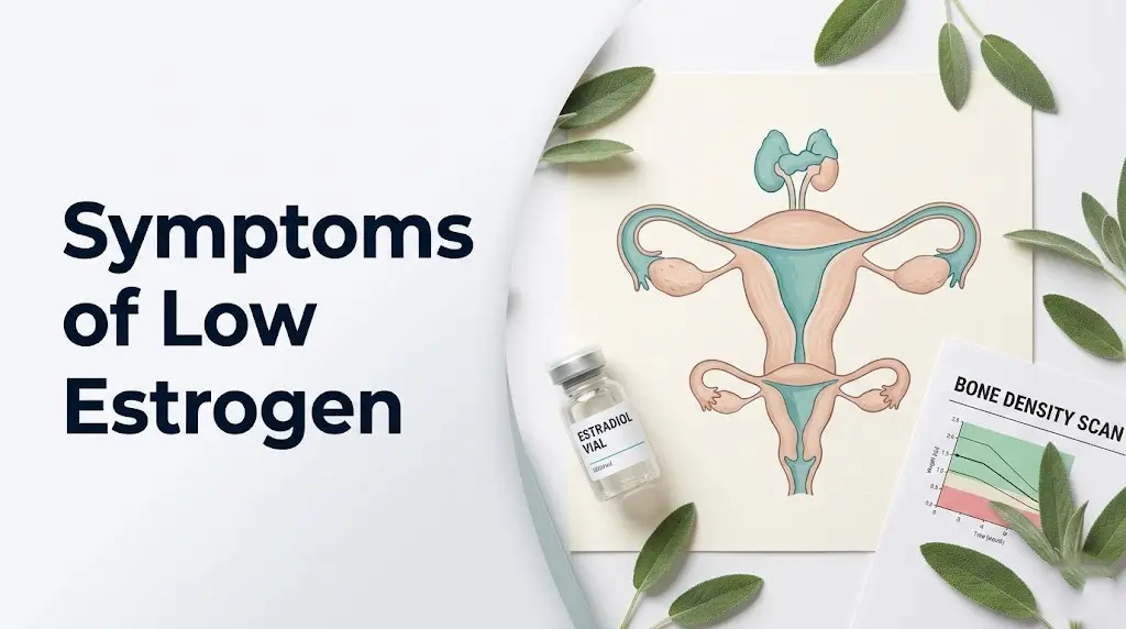
The primary source of estradiol in premenopausal women is the ovarian follicle, specifically the granulosa cells surrounding the developing egg. When follicle numbers decline, as they do in natural perimenopause, or when the hypothalamic-pituitary-ovarian (HPO) axis is suppressed by extreme caloric restriction, chronic stress, or illness, estradiol production falls. The anterior pituitary responds by secreting rising levels of follicle-stimulating hormone (FSH), which is why elevated FSH is the primary blood marker used to confirm low estrogen states clinically.
What makes low estrogen particularly difficult to recognize is that no two women experience exactly the same symptom combination. A 2022 analysis published in the Journal of Clinical Endocrinology and Metabolism found that symptom burden in women with documented low estradiol levels varied substantially based on ER-alpha receptor density in individual tissues, meaning two women with identical blood estradiol levels can have very different symptom presentations.
Key symptoms across the most commonly affected body systems:
- Vasomotor symptoms: hot flashes and night sweats
- Urogenital symptoms: vaginal dryness, painful intercourse, recurrent urinary tract infections
- Skeletal symptoms: accelerating bone density loss, joint pain
- Neurological symptoms: mood dysregulation, anxiety, brain fog, poor sleep
- Integumentary symptoms: dry skin, hair thinning, collagen loss
- Metabolic symptoms: weight redistribution, fatigue, insulin sensitivity changes
Women who have undergone surgical removal of both ovaries (bilateral oophorectomy) experience an abrupt drop in estradiol rather than the gradual decline of natural menopause. Their symptom onset is typically faster and more severe than that of women who enter perimenopause gradually.
Signs and Symptoms of Low Estrogen You Might Not Recognize
The signs and symptoms of low estrogen that most people recognize are hot flashes and irregular periods. The ones that most frequently go unrecognized include heart palpitations, recurrent urinary tract infections, joint stiffness, worsening dental health, and subtle cognitive changes that are often mistaken for stress or aging.
Heart palpitations in low estrogen states occur because estradiol regulates the cardiac autonomic balance. Specifically, estradiol enhances parasympathetic tone through nitric oxide (NO) signaling in the vascular endothelium and modulates cardiac beta-adrenergic receptor sensitivity. When estradiol declines, sympathetic nervous system activity becomes relatively more dominant, producing sensations of rapid or irregular heartbeat. A 2021 study published in Menopause (the peer-reviewed journal of The Menopause Society) found that up to 43% of perimenopausal women report heart palpitations as a primary symptom, yet fewer than a quarter had discussed it with their provider in the context of hormonal change.
Dental health deterioration is another commonly overlooked sign. The periodontal ligament and alveolar bone both contain estrogen receptors, and declining estradiol accelerates alveolar bone resorption. This makes gum recession, increased tooth sensitivity, and tooth mobility early physical markers of systemic estrogen decline in some women.
| Sign or Symptom | Why It Occurs | Commonly Recognized? |
|---|---|---|
| Hot flashes | KNDy neuronal dysregulation narrows thermoneutral zone | Yes |
| Heart palpitations | Sympathetic dominance from reduced NO-mediated cardiac regulation | Rarely |
| Recurrent UTIs | Urethral and vaginal epithelial thinning reduces barrier function | Rarely |
| Joint stiffness | Reduced synovial fluid production and cartilage collagen loss | Sometimes |
| Worsening gum recession | Alveolar bone resorption from ER-alpha receptor underactivation | No |
| Dry eyes | Lacrimal gland ER-beta receptor underactivation reduces tear film | No |
| Increased allergy sensitivity | Mast cell degranulation threshold lowered in low-estrogen states | No |
Women taking selective serotonin reuptake inhibitors (SSRIs) or antipsychotic medications may find some low-estrogen symptoms are masked or confused with medication side effects, particularly mood changes, fatigue, and sexual dysfunction. This overlap makes clinical review by both a psychiatrist and an obstetrician-gynecologist appropriate when symptom presentation is complex.
What Are the Symptoms of Low Estrogen at Each Stage
What are the symptoms of low estrogen depends significantly on which stage of estrogen decline a woman is in, because symptom pattern, severity, and affected organ systems shift as estradiol levels fall from mildly reduced to severely depleted.
Early-stage estrogen decline, which often begins in perimenopause anywhere from 2 to 10 years before the final menstrual period, produces intermittent rather than constant symptoms. Irregular menstrual cycles are frequently the first clinical sign, caused by erratic FSH and LH surges that fail to produce consistent ovulation. Sleep disruption, particularly difficulty maintaining sleep in the early morning hours, appears early because GABA-A receptor sensitivity in the brain is partly dependent on estradiol, and its decline disrupts the architecture of slow-wave and REM sleep phases before vasomotor symptoms become prominent.
As estradiol continues to fall, vasomotor symptoms intensify. The thermoneutral zone, the temperature range within which the body does not need to sweat or shiver, narrows from approximately 0.4 degrees Celsius in premenopausal women to a much narrower band in estrogen-deficient states. Research from JAMA Internal Medicine has described the narrowing of this thermoregulatory window as the primary driver of hot flashes and night sweats, mediated by the KNDy neuronal system in the hypothalamic arcuate nucleus releasing unchecked neurokinin B signals.
| Stage | Estradiol Level Range | Primary Symptoms | Secondary Symptoms |
|---|---|---|---|
| Early perimenopause | 30 to 100 pg/mL (declining from 200+) | Cycle irregularity, mild sleep disruption, mood changes | Breast tenderness, PMS intensification |
| Late perimenopause | 15 to 40 pg/mL | Frequent hot flashes, night sweats, vaginal dryness onset | Cognitive changes, joint symptoms beginning |
| Postmenopause / severe deficiency | Below 15 pg/mL | Persistent vaginal atrophy, bone loss acceleration, cognitive symptoms | Urinary incontinence, cardiovascular risk rise |
| Premature ovarian insufficiency (under 40) | Below 20 pg/mL sustained | All of the above at younger age | Fertility loss, greater cardiovascular and bone risk from longer deficiency duration |
Adolescent girls and young adult women with hypothalamic amenorrhea, a condition caused by insufficient caloric intake relative to energy expenditure, can have estradiol levels below 20 pg/mL with a symptom picture that resembles early postmenopause, including stress fractures from bone density loss and severe mood dysregulation. Their HPO axis suppression is potentially reversible with nutritional rehabilitation, unlike the follicular depletion of natural menopause.
Key Takeaway: Early-stage low estrogen often announces itself through sleep disruption and cycle irregularity long before hot flashes develop, and recognizing those early signals is the single most useful clinical insight from this section.
Low Estrogen Levels Symptoms: The Complete Clinical Picture
Low estrogen levels symptoms span at least seven distinct organ systems, which is why estrogen deficiency is often initially misattributed to thyroid disease, depression, chronic fatigue syndrome, or autoimmune conditions before a hormonal cause is identified.
The musculoskeletal system is among the most consequentially affected. Osteoblasts (bone-building cells) require ER-alpha receptor activation to maintain their activity. When estradiol declines, osteoblast activity slows while osteoclast (bone-resorbing cell) activity, which is upregulated by RANKL, IL-1, IL-6, and TNF-alpha in low-estrogen states, continues at a higher relative rate. According to the American College of Obstetricians and Gynecologists (ACOG), women can lose up to 20% of their bone density in the five to seven years following menopause if estrogen deficiency is untreated, with the steepest loss occurring in the first one to two years.
Cardiovascular symptoms, while often attributed to aging rather than hormonal change, have a direct estrogen-dependent mechanism. Estradiol promotes endothelial nitric oxide synthase (eNOS) expression in arterial walls, which keeps blood vessels dilated and flexible. As estradiol falls, eNOS activity decreases, arterial stiffness increases, LDL cholesterol tends to rise, and HDL cholesterol tends to fall. These shifts are measurable within the first one to two years of significant estrogen decline.
The integumentary system responds visibly because the dermal fibroblasts responsible for producing collagen type I and type III both require estradiol signaling. A study published in the British Journal of Dermatology found that women lose approximately 30% of their skin collagen in the first five years after menopause, with the rate slowing to approximately 2% per year thereafter. This produces the characteristic skin thinning, reduced elasticity, and increased dryness that many women notice in perimenopause and postmenopause.
The full low-estrogen symptom picture across systems:
- Reproductive: irregular or absent periods, painful intercourse, vaginal dryness
- Skeletal: accelerating bone loss, fracture risk, joint inflammation
- Cardiovascular: rising LDL, arterial stiffness, palpitations
- Neurological: mood changes, anxiety, brain fog, memory lapses
- Integumentary: skin thinning, hair loss, dry or brittle nails
- Urinary: urgency, frequency, recurrent infections
- Metabolic: abdominal weight redistribution, insulin sensitivity reduction, fatigue
Women with hypothyroidism or autoimmune thyroid disease (Hashimoto’s thyroiditis) frequently experience symptom overlap with low estrogen. Both conditions produce fatigue, hair loss, dry skin, and cognitive symptoms. Evaluating thyroid-stimulating hormone (TSH), free T4, and serum estradiol simultaneously is often clinically necessary to distinguish the contribution of each hormonal axis.
Low Estradiol Symptoms and What Your Lab Results Mean
Low estradiol symptoms reflect the specific deficiency of estradiol (E2), the most biologically active of the three estrogen types, rather than of estrone (E1) or estriol (E3). This distinction matters clinically because estradiol is the form most tightly coupled to ER-alpha receptor signaling in bone, brain, and cardiovascular tissue.
A serum estradiol test measures circulating E2 in picograms per milliliter (pg/mL). According to Mayo Clinic laboratory reference ranges, premenopausal women in the follicular phase typically have estradiol levels between 30 and 120 pg/mL, with midcycle ovulatory peaks reaching 200 to 400 pg/mL. Women with levels consistently below 30 pg/mL and elevated FSH above 40 mIU/mL meet the laboratory criteria used to support a clinical diagnosis of premature ovarian insufficiency (POI) when under age 40, or natural menopause when above.
One key point that most symptom guides omit: normal-range estradiol does not exclude symptoms. Women in the lower portion of the normal range (30 to 50 pg/mL in the follicular phase) can experience significant symptoms if their tissue ER-alpha receptor density is relatively high. The symptom threshold is partly determined by an individual’s receptor sensitivity, not just by the absolute circulating estradiol level. This is why some women feel pronounced low-estrogen symptoms with levels that a laboratory report labels as technically normal.
Quick Tip:
- When requesting a serum estradiol test, ask for FSH to be measured simultaneously, as the FSH-to-estradiol ratio provides more clinical information than either value alone.
- Request the test in the early follicular phase (days 2 to 4 of the menstrual cycle) for the most reproducible baseline comparison.
- Women on hormonal contraception (combined pill or hormonal IUD) will have artificially suppressed FSH and estradiol, making interpretation complex. Inform your provider if you are using any form of hormonal contraception.
Women taking aromatase inhibitors for breast cancer treatment will have profoundly suppressed estradiol levels (often below 5 pg/mL), producing a rapid and severe low-estrogen symptom picture that requires specialized management by both an oncologist and a gynecological specialist experienced in hormonal health in cancer survivors.
Key Takeaway: A technically normal serum estradiol result does not rule out low-estrogen symptoms in a woman whose individual receptor sensitivity is high, which is why clinical symptom assessment must always accompany laboratory interpretation.
Estrogen Drop Symptoms: What Changes First When Levels Fall
Estrogen drop symptoms follow a recognizable sequence, though the precise order varies by how rapidly the drop occurs, whether it is gradual (as in natural perimenopause) or abrupt (as after surgical removal of the ovaries).
The first system to signal a problem is almost always the central nervous system. Before hot flashes become prominent, many women notice changes in sleep quality, particularly early morning awakening between 3 a.m. and 5 a.m., and increased emotional reactivity. Both symptoms reflect estradiol’s role in regulating GABA-A receptor sensitivity in the brain’s limbic system and in supporting serotonin transporter (SERT) expression. As estradiol declines, SERT expression decreases, which paradoxically raises synaptic serotonin briefly before downstream regulatory changes reduce serotonin signaling efficiency.
Menstrual irregularity typically emerges next. Cycles may shorten first (from 28 days to 24 or 25 days) due to shortened follicular phases, then become unpredictable in length, and eventually stop. This pattern is the clinical hallmark of perimenopause, though women with hypothalamic amenorrhea from undereating or overtraining may experience abrupt cessation of periods without the irregular-cycle transition phase.
Vasomotor symptoms follow. The rapid or sudden drop that occurs after surgical oophorectomy can produce severe hot flashes within days of the procedure because the thermoregulatory system has had no time to adapt. Gradual perimenopause typically produces milder initial vasomotor symptoms that intensify over months to years as estradiol falls further.
To describe estrogen drop symptoms accurately to your provider:
- Note the exact timing of each symptom relative to your menstrual cycle if still cycling.
- Track hot flash frequency and duration over two weeks using a written log or phone app.
- Rate sleep disruption on a 1 to 10 scale each morning and note which symptom woke you.
- Record any mood changes with specific triggers or patterns noted.
- Bring this log to your appointment rather than relying on memory, as symptom frequency and pattern directly inform the clinical assessment.
Low Estrogen Symptoms in Women Across Different Life Stages
Low estrogen symptoms in women look different at 25 than at 45 than at 65, because the cause of the deficiency, the body’s baseline hormonal context, and the affected organ systems’ prior exposure to estrogen all change the clinical picture.
In women of reproductive age (18 to 40), low estrogen is almost always caused by HPO axis suppression rather than follicular depletion. The most common causes in this age group include hypothalamic amenorrhea (from caloric restriction, excessive exercise, or chronic psychological stress), hyperprolactinemia (from a pituitary adenoma or from dopamine-blocking medications), and premature ovarian insufficiency. Symptoms in this group frequently include absent or irregular periods, fertility difficulties, low libido, vaginal dryness, and mood dysregulation.
In perimenopausal women (typically 40 to 52), the pattern is more variable. According to The Menopause Society, the average duration of the perimenopausal transition is four years, though it ranges from one year to a decade. During this window, estradiol levels fluctuate widely, which means symptoms can be intermittent and confusing. Women may have hot flashes for three months, then feel entirely normal, then have them return more intensely.
In postmenopausal women (12 months or more after the final menstrual period), symptoms stabilize but do not disappear. The genitourinary syndrome of menopause (GSM), which encompasses vaginal dryness, vulvar atrophy, urinary urgency, and recurrent UTIs, affects an estimated 50 to 70% of postmenopausal women according to ACOG, yet fewer than 25% discuss it with a healthcare provider due to embarrassment or the mistaken belief that it is a normal part of aging rather than a treatable hormonal condition.
Women who experience menopause before age 40 due to surgery, chemotherapy, or premature ovarian insufficiency face a longer period of estrogen deficiency than women who enter natural menopause at the population average of 51. This extends their cumulative exposure risk for bone density loss, cardiovascular disease, and cognitive decline, making early evaluation by a reproductive endocrinologist or gynecologist with expertise in early menopause especially important.
Key Takeaway: Low estrogen in women under 40 is almost always caused by HPO axis suppression rather than ovarian aging, making the cause, treatment approach, and specialist referral entirely different from perimenopause management.
Symptoms of Decreased Estrogen: Bone, Heart, and Metabolic Effects
Symptoms of decreased estrogen in the skeletal, cardiovascular, and metabolic systems develop more slowly than vasomotor or neurological symptoms, but they carry greater long-term health consequences because their effects are cumulative and partly irreversible without intervention.
Bone density loss accelerates measurably in the first one to two years of significant estrogen decline. Estradiol suppresses the expression of RANKL (receptor activator of nuclear factor kappa-B ligand), a cytokine that activates osteoclasts. When estradiol falls, RANKL expression rises, osteoclast activity accelerates, and bone mineral density decreases at a rate that can be two to three times the premenopausal baseline. Women may not feel bone density loss, which is why the National Osteoporosis Foundation recommends a DEXA scan (dual-energy X-ray absorptiometry) within one to two years of menopause or at diagnosis of POI, regardless of whether fracture symptoms are present.
Cardiovascular effects of decreased estrogen include measurable increases in LDL cholesterol and triglycerides, measurable decreases in HDL cholesterol, increasing arterial stiffness, and declining eNOS activity in the vascular endothelium. A large analysis published in JAMA Internal Medicine in 2022 found that postmenopausal women who experienced menopause before age 45 had a cardiovascular risk profile more consistent with women 10 years older, underscoring the cardiovascular significance of prolonged estrogen deficiency.
Metabolically, declining estradiol shifts fat storage patterns. Estradiol promotes peripheral (gluteal and femoral) fat storage through ER-alpha receptor action on adipose tissue. As it declines, fat redistribution toward the visceral abdominal compartment occurs, which is metabolically more active in producing inflammatory cytokines and is associated with increased insulin resistance. This metabolic shift can occur even in women whose total body weight does not change.
Women with pre-existing type 2 diabetes or metabolic syndrome who enter perimenopause experience a compounding effect: declining estradiol worsens insulin sensitivity at the same time that glucocorticoid patterns and activity levels often change. An endocrinologist review is appropriate for these women alongside gynecological management.
Very Low Estrogen Symptoms: When the Deficiency Is Severe
Very low estrogen symptoms occur when estradiol levels fall below approximately 10 to 15 pg/mL, a level seen in postmenopausal women who are not receiving hormone therapy, in women with bilateral oophorectomy, in women undergoing chemotherapy, and in women with severe long-standing hypothalamic amenorrhea.
At this level of deficiency, vaginal atrophy becomes clinically significant. The vaginal epithelium transitions from its premenopausal thick, rugated, glycogen-rich state to a thin, pale, fragile state that is prone to tearing, bleeding, and secondary infection. The vulvar vestibule and clitoris also undergo structural changes, with clitoral retraction and labia minora thinning occurring in severe and prolonged estrogen deficiency. These changes are classified under the clinical term genitourinary syndrome of menopause (GSM) when they produce symptoms including dryness, burning, dyspareunia (pain with intercourse), urinary urgency, or recurrent infections.
Bone loss at very low estrogen levels accelerates beyond the perimenopausal rate. Women with estradiol below 10 pg/mL who do not receive treatment can lose 3 to 5% of bone mineral density per year in the lumbar spine and femoral neck. This rate is associated with a fracture risk that doubles within five years of severe deficiency onset, according to data reviewed by the National Osteoporosis Foundation.
Cognitively, very low estrogen is associated with verbal memory decline, increased word-finding difficulty, and reduced processing speed. These symptoms are distinct from dementia but can significantly affect daily function. Research from the NICHD has found that the hippocampus, which is central to verbal memory, contains a high density of ER-alpha receptors and is particularly sensitive to estradiol withdrawal.
Women who have undergone total hysterectomy with bilateral salpingo-oophorectomy and who are not receiving hormone therapy face the most rapid onset of very-low-estrogen symptoms. For these women, the standard recommendation from ACOG is to initiate estrogen therapy unless there is a specific contraindication, as the benefits of preventing accelerated bone loss, cardiovascular risk, and GSM outweigh the risks for most women without a history of estrogen-receptor-positive breast cancer.
Key Takeaway: Genitourinary syndrome of menopause (GSM) is a treatable clinical condition produced by very low estrogen, not an inevitable part of aging, and the majority of women with GSM symptoms who discuss them with their gynecologist have effective treatment options available.
Loss of Estrogen Symptoms: Skin, Hair, and Body Composition Changes
Loss of estrogen symptoms in the skin, hair, and body are among the most visible and immediately noticed, yet they are also among the most frequently dismissed as “just getting older” rather than recognized as clinically meaningful signs of hormonal change.
The dermis loses both collagen production rate and collagen cross-linking density as estradiol falls. Collagen type I fibers, which give skin its tensile strength, and type III fibers, which provide elasticity, are both produced less efficiently when ER-alpha receptor activity in dermal fibroblasts decreases. The practical result is skin that appears thinner, crepier, and more prone to bruising. Women who apply pressure to their skin may notice it takes longer to return to its normal contour, a clinical sign of reduced dermal elasticity.
Hair changes reflect estradiol’s role in regulating the hair follicle cycle. In adequate estrogen states, follicles spend a longer proportion of their cycle in the anagen (active growth) phase and a shorter time in the telogen (resting and shedding) phase. When estradiol declines, the anagen phase shortens, more follicles shift to telogen simultaneously, and diffuse hair thinning occurs across the scalp rather than the patterned loss seen in androgenetic alopecia. The temporal and frontal hairlines typically remain intact in estrogen-related hair loss, which distinguishes it visually from male-pattern baldness.
Body composition changes extend beyond skin and hair. Lean muscle mass is partly maintained by estrogen’s anabolic signaling in skeletal muscle satellite cells. As estradiol falls, muscle protein synthesis declines, contributing to reduced strength, slower recovery from physical activity, and the gradual increase in fat-to-muscle ratio that many women notice in their 40s even without changes in diet or exercise.
Women who are very lean or who have low body fat percentage (under approximately 18 to 20%) experience more pronounced low-estrogen skin and hair symptoms because peripheral estrogen conversion from androgens in adipose tissue is a secondary estrogen source that thinner women have less access to. For women in this category, the symptom picture can be more pronounced despite other health-promoting behaviors.
Symptoms of Poor Estrogen Metabolism
Symptoms of poor estrogen metabolism are distinct from symptoms of absolute low estrogen production, and this distinction is one that virtually no mainstream health content addresses. A woman can have adequate ovarian estradiol production yet still experience estrogen-related symptoms if her liver is not clearing estrogen metabolites efficiently.
The liver metabolizes estrogen through a two-phase process. Phase I metabolism involves hydroxylation by cytochrome P450 enzymes, primarily CYP1A2 and CYP3A4, converting estradiol into hydroxylated metabolites. The two most clinically significant are 2-hydroxyestrone (considered protective and weakly estrogenic) and 16-alpha-hydroxyestrone (more strongly estrogenic and less easily cleared). Phase II metabolism then conjugates these metabolites for excretion, primarily through catechol-O-methyltransferase (COMT) and sulfotransferase (SULT1A1) enzymes.
When either phase is impaired, estrogen metabolites accumulate. Women with a COMT gene polymorphism that reduces COMT enzyme activity clear catechol estrogens more slowly, which has been associated in observational research with increased estrogen-sensitive tissue exposure, more pronounced PMS symptoms, heavier menstrual bleeding, and increased breast tissue density. This is not the same as classical low estrogen; it is more accurately described as poor estrogen clearance with resulting metabolite accumulation.
Symptoms that may reflect poor estrogen metabolism rather than low production include:
- Worsening PMS symptoms despite apparently normal cycle regularity
- Heavy menstrual bleeding with breast tenderness and bloating
- Headaches specifically in the late luteal phase (days 21 to 28)
- Estrogen-sensitive fibrocystic breast changes worsening over time
- Persistent fatigue despite normal serum estradiol and FSH levels
Evaluating estrogen metabolism requires more than a standard serum estradiol test. A dried urine test for comprehensive hormones (DUTCH test) or a urinary estrogen metabolite panel measures estrogen metabolite ratios, including the 2:16 hydroxyestrone ratio, and can identify phase I or phase II impairment. This testing is not universally offered in primary care and may require referral to a gynecologist or reproductive endocrinologist with a clinical interest in hormonal health.
Chronic alcohol use impairs hepatic phase II estrogen metabolism directly, as does non-alcoholic fatty liver disease (NAFLD). Women with either condition who experience worsening hormonal symptoms should have their liver function assessed as part of a comprehensive hormonal workup.
Key Takeaway: Poor estrogen metabolism can produce estrogen-related symptoms even when circulating estradiol levels appear normal on a standard blood test, which is why urinary metabolite testing provides clinical information that serum testing alone cannot.
Low Estrogen Symptoms in Your 20s
Low estrogen symptoms in your 20s are frequently misdiagnosed or attributed to stress, depression, or poor sleep rather than recognized as signs of a hormonal deficiency, because both clinicians and patients tend to assume that low estrogen is primarily a concern for women over 40.
The three most common causes of low estrogen in women in their 20s are hypothalamic amenorrhea (HA), premature ovarian insufficiency (POI), and hyperprolactinemia. HA, which occurs when the HPO axis is suppressed by caloric restriction, excessive exercise, or psychological stress, is particularly prevalent among collegiate athletes, women with restrictive eating disorders, and women in high-stress professional environments. The National Institute of Child Health and Human Development (NICHD) estimates that HA accounts for approximately 20 to 35% of amenorrhea cases in women of reproductive age, with many cases going undiagnosed for years.
Symptoms of low estrogen in this age group present differently from perimenopausal symptoms because the neurological and cardiovascular systems have not had decades of estrogen priming. Hot flashes may be mild or absent even with clinically low estradiol. Instead, the predominant symptoms in younger women include:
- Absent or irregular menstrual periods (the most consistent clinical sign)
- Low libido and vaginal dryness despite young age
- Stress fractures or unexpectedly poor bone density on DEXA scan
- Mood dysregulation, anxiety, or depressive symptoms without a primary psychiatric cause
- Persistent fatigue disproportionate to sleep quality and activity level
- Difficulty conceiving
POI in women under 40 is associated with significant long-term health implications because of the extended duration of estrogen deficiency. According to ACOG, women with POI have a higher lifetime risk of cardiovascular disease, osteoporosis, and all-cause mortality compared with women who enter natural menopause at the population average. Early diagnosis and appropriate management, which often includes hormone therapy until the typical age of natural menopause (approximately 51), significantly reduces these risks.
A young woman with absent periods for three months or more, especially combined with any of the symptoms above, should be evaluated by a primary care physician or obstetrician-gynecologist. The evaluation typically includes serum estradiol, FSH, LH, prolactin, TSH, and a pregnancy test as an initial panel.
Low Estrogen and Brain Fog: The Cognitive Connection
Low estrogen and brain fog are linked through multiple neurological pathways, and the cognitive symptoms of estrogen deficiency are among the most distressing and least discussed aspects of hormonal change.
The hippocampus, the brain region most involved in verbal memory, word retrieval, and new learning, contains a high density of ER-alpha receptors. Estradiol activates these receptors to promote dendritic spine density (the structural basis for synaptic connections) and to enhance acetylcholine release in hippocampal circuits. When estradiol falls, synaptic density in the hippocampus decreases measurably, and acetylcholine signaling efficiency drops. The practical experience of this is difficulty remembering names, losing words mid-sentence, and finding it harder to hold multiple pieces of information in working memory simultaneously.
The prefrontal cortex, which governs executive function, planning, and sustained attention, is also estrogen-sensitive. Research from the University of Illinois published in the Menopause journal found that perimenopausal women showed measurable reductions in verbal memory and processing speed during periods of documented low estradiol, with partial reversal when estradiol levels rose again during the fluctuating hormonal pattern of early perimenopause. This fluctuation-linked cognitive change is one reason some women notice that their cognitive symptoms come and go rather than being constant.
Low estrogen and brain fog should be distinguished from the cognitive symptoms of primary depression or anxiety, with which they frequently co-occur. The key clinical distinction is timing relative to the menstrual cycle (or its cessation), sleep quality (estrogen-related cognitive symptoms often worsen directly after nights of poor sleep from night sweats), and whether cognitive symptoms resolve with adequate sleep and stress reduction or persist regardless.
According to the National Institutes of Health, brain fog and cognitive difficulty during perimenopause typically do not progress to dementia or clinically significant cognitive impairment for most women. However, women with early perimenopause, POI, or surgical menopause who are not receiving hormone therapy may face a modestly higher long-term risk of cognitive decline, which is an area of active research.
Women with ADHD diagnoses should be aware that estradiol decline can worsen ADHD symptom burden through its effects on dopamine and norepinephrine signaling in the prefrontal cortex. This interaction often requires collaborative management between a psychiatrist and a gynecologist with hormonal expertise.
Key Takeaway: The brain fog of low estrogen is a real, measurable neurophysiological event rooted in reduced hippocampal dendritic density and declining acetylcholine signaling, not a vague complaint or a side effect of aging.
Low Estrogen and Joint Pain: Why Your Joints Are Affected
Low estrogen and joint pain are connected through the estrogen receptor system in synovial tissue, articular cartilage, and subchondral bone, making joint symptoms one of the more surprising but well-documented manifestations of estrogen deficiency.
Synovial membranes, which line the joint capsule and produce synovial fluid for lubrication and nutrient delivery to cartilage, contain both ER-alpha and ER-beta receptors. Estradiol stimulates the production of hyaluronic acid, a primary component of synovial fluid that gives it its viscous, lubricating properties. When estradiol falls, hyaluronic acid production decreases, synovial fluid volume and viscosity drop, and joints that previously moved smoothly begin to feel stiff and uncomfortable, particularly after periods of rest such as morning awakening.
Articular cartilage itself contains chondrocytes (cartilage cells) with estrogen receptors. Estradiol promotes chondrocyte collagen synthesis and inhibits the production of matrix metalloproteinases (MMPs), enzymes that degrade cartilage matrix. In low-estrogen states, MMP activity increases and cartilage collagen synthesis decreases, accelerating the cartilage thinning that underlies osteoarthritis. Research published in Arthritis and Rheumatism found that postmenopausal women have a higher incidence and faster progression rate of knee and hip osteoarthritis than age-matched men, with estrogen deficiency identified as a primary contributing factor.
The most commonly affected joints in low-estrogen states are:
- Knees (highest synovial tissue estrogen receptor density)
- Hips (high load-bearing with cartilage thinning risk)
- Small hand joints (often presenting as morning stiffness that resembles early rheumatoid arthritis)
- Lumbar facet joints (contributing to low back stiffness)
- Temporomandibular joint (jaw clicking or stiffness, often overlooked as hormonal)
Women with pre-existing autoimmune arthritis such as rheumatoid arthritis (RA) or psoriatic arthritis may notice their disease activity worsens during perimenopause. Estradiol has immunomodulatory effects that can suppress some inflammatory pathways relevant to autoimmune joint disease, and its decline can trigger flares. A rheumatologist and gynecologist working collaboratively is appropriate for these patients.
Joint stiffness that is symmetric, accompanies systemic fatigue and morning stiffness lasting more than 45 minutes, or is accompanied by measurable joint swelling should be evaluated by a rheumatologist to distinguish estrogen-related joint changes from an emerging inflammatory arthritis.
Low Estrogen Urinary Symptoms
Low estrogen urinary symptoms are caused by atrophic changes in the urethral mucosa and bladder trigone, both of which contain estrogen receptors and rely on adequate estradiol signaling to maintain their structural integrity and normal function.
The urethra and bladder trigone (the triangular region at the base of the bladder near the urethral opening) are embryologically derived from the same tissue as the vaginal epithelium, the urogenital sinus. Both share high ER-alpha receptor density. As estradiol declines, the urethral epithelium thins, loses glycogen-rich cells, and becomes more susceptible to bacterial adhesion. Simultaneously, urethral closure pressure decreases, which reduces the mechanical barrier that normally prevents urine leakage. The result is a cluster of urinary symptoms that includes:
- Urinary urgency: a sudden, compelling need to urinate that is difficult to defer
- Urinary frequency: urinating more than eight times in 24 hours or more than once at night
- Stress urinary incontinence: leakage with coughing, sneezing, or physical exertion
- Recurrent urinary tract infections (UTIs): two or more per year, caused by thinned epithelium and reduced protective Lactobacillus flora in the vaginal environment
- Dysuria: burning or discomfort with urination in the absence of active infection
According to ACOG, genitourinary syndrome of menopause (GSM), which encompasses these urinary and vaginal symptoms, affects between 50 and 70% of postmenopausal women. Unlike vasomotor symptoms (hot flashes), which often improve spontaneously over time, GSM symptoms do not improve without treatment. They typically worsen progressively with continued estrogen deficiency.
Recurrent UTIs in a postmenopausal woman with no other identified risk factors should prompt evaluation for GSM by a gynecologist. Local vaginal estrogen therapy (vaginal estrogen cream, ring, or tablet) is the evidence-based first-line treatment for GSM-related urinary symptoms and has minimal systemic absorption, making it appropriate for most women including many with a history of hormone-sensitive conditions. The Menopause Society and ACOG both endorse local vaginal estrogen as safe and effective for long-term use in appropriate patients.
Younger women with hypothalamic amenorrhea or POI can also develop early urogenital atrophy symptoms, though typically milder than in postmenopausal women. A gynecologist should assess any woman under 40 with recurrent UTIs and no anatomical or behavioral risk factors for the possibility of an underlying hormonal contribution.
Emergency Symptoms: When to Call 911 or Go to the ER
Certain symptoms associated with low estrogen and its associated conditions require immediate emergency evaluation. Do not wait to see if these resolve on their own.
Call 911 or go to the nearest emergency room immediately if you experience:
- Chest pain, pressure, or tightness, particularly if combined with shortness of breath, jaw pain, arm pain, or sweating. Women in perimenopause and postmenopause have an increased cardiovascular risk profile, and these symptoms can indicate an acute myocardial infarction. Women’s cardiac presentations often differ from the classic male presentation and may involve more subtle symptoms.
- Sudden severe headache unlike any previous headache, especially if accompanied by vision changes or neck stiffness. This presentation can indicate subarachnoid hemorrhage or hypertensive emergency, both of which require immediate imaging.
- Heart palpitations accompanied by loss of consciousness, near-fainting, or collapse. While palpitations alone in low-estrogen states are common and typically not dangerous, those accompanied by syncope or pre-syncope require cardiac evaluation to exclude arrhythmia.
- Sudden weakness, numbness, or paralysis on one side of the body, facial drooping, sudden severe difficulty speaking, or sudden loss of vision in one or both eyes. These are stroke warning signs. Women in low-estrogen postmenopausal states have rising cardiovascular and cerebrovascular risk.
- A bone fracture occurring from minimal trauma, particularly of the hip, wrist, or spine. This requires emergency orthopedic evaluation and indicates severely compromised bone density that needs immediate comprehensive assessment.
These presentations can indicate acute myocardial infarction, stroke, hypertensive emergency, serious cardiac arrhythmia, or osteoporotic fracture requiring emergency orthopedic and medical management. None of these warrant a wait-and-see approach or an urgent care appointment.
Signs of Low Estrogen and When to See a Doctor
Signs of low estrogen that warrant prompt medical evaluation include menstrual cycle changes lasting more than three months, vasomotor symptoms that significantly disrupt sleep or daily function, urogenital symptoms producing pain or recurrent infections, any cognitive change that appears disproportionate to sleep quality or stress, and bone fracture from minimal trauma.
The clinical pathway for evaluation typically begins with a primary care physician or obstetrician-gynecologist. Initial testing generally includes serum estradiol, FSH, LH, TSH, prolactin, and a complete blood count. Specific additional tests are ordered based on initial results and clinical picture.
To prepare for your first appointment regarding possible low estrogen symptoms:
- Track your menstrual cycle dates for the past three to six months.
- Write down every symptom you have noticed with the approximate date of onset.
- Note the timing of symptoms relative to your cycle (if still cycling) or relative to the past year.
- List all medications, supplements, and hormonal contraceptives you currently use.
- Record your family history of premature menopause, osteoporosis, or premature cardiovascular disease.
| Symptom Pattern | Appropriate Provider | Urgency Level |
|---|---|---|
| Irregular cycles + hot flashes in 40s | Primary care or OB-GYN | Routine appointment within 4 to 6 weeks |
| Absent periods under 40 for 3+ months | OB-GYN or reproductive endocrinologist | Appointment within 2 to 4 weeks |
| Recurrent UTIs in postmenopausal woman | OB-GYN | Within 2 to 4 weeks |
| Severe cognitive symptoms | OB-GYN plus neurologist if needed | Within 2 to 4 weeks |
| Hot flashes plus heart palpitations only | OB-GYN plus cardiologist if palpitations are severe | OB-GYN within 2 to 4 weeks; cardiac evaluation if palpitations cause lightheadedness |
| Suspected fracture or bone pain | Emergency medicine, then bone health specialist | Same day or emergency evaluation |
| Severe mood symptoms, tearfulness, hopelessness | OB-GYN plus primary care or mental health provider | Within 1 to 2 weeks |
Women with suspected POI (absent periods under 40 with elevated FSH on two separate tests at least four weeks apart) should be referred to a reproductive endocrinologist who specializes in ovarian insufficiency. This condition has implications for fertility, bone health, cardiovascular risk, and long-term hormonal management that go beyond standard menopause care.
If you are experiencing suicidal ideation, hopelessness, or thoughts of self-harm alongside severe hormonal symptoms, contact the 988 Suicide and Crisis Lifeline by calling or texting 988 immediately. This service is free, confidential, and available 24 hours a day, 7 days a week. Severe mood symptoms in the context of low estrogen can be both physiologically driven and clinically serious, and they deserve urgent attention from both a mental health provider and a clinician who can address the hormonal component.
Frequently Asked Questions About Symptoms of Low Estrogen
What does low estrogen feel like physically?
Low estrogen physically feels like a combination of heat surges that rise from the chest to the face (hot flashes), dry or uncomfortable vaginal tissue, joints that feel stiff after rest, skin that feels thinner or drier than before, and fatigue that is not fully explained by sleep quality.
Some women describe it as a body that suddenly feels less resilient, where recovery from exercise takes longer, sleep is lighter, and temperature regulation feels unreliable.
The specific symptom combination varies by individual, as estrogen receptor density differs between women, meaning two people with identical estradiol levels can feel quite different physically.
Can low estrogen cause anxiety and mood changes?
Yes, low estrogen directly causes anxiety and mood changes through its effects on serotonin transporter expression, GABA-A receptor sensitivity, and dopamine signaling in the limbic system and prefrontal cortex.
When estradiol declines, serotonin signaling efficiency decreases and GABA-A receptor sensitivity in the amygdala is altered, reducing the brain’s natural capacity to regulate stress responses and emotional reactivity.
A 2020 study published in the journal Menopause found that perimenopausal women with documented low estradiol had significantly higher rates of new-onset anxiety than age-matched premenopausal women, independent of other psychosocial factors.
How do you know if your estrogen is low without a blood test?
Without a blood test, the most reliable clinical indicators of low estrogen are menstrual cycle changes (shortening, lengthening, or stopping) in a woman under 51, combined with two or more of the following: hot flashes, night sweats, vaginal dryness, new-onset sleep disruption, or joint stiffness.
No symptom-based assessment can confirm or rule out low estrogen with clinical accuracy, and symptom patterns overlap with thyroid dysfunction, autoimmune conditions, and primary mood disorders.
A serum estradiol and FSH test remains the only reliable way to confirm estrogen deficiency, and is available through any primary care physician or obstetrician-gynecologist.
What are the symptoms of low estrogen in your 20s?
Low estrogen in your 20s most commonly presents as absent or irregular menstrual periods, low libido, vaginal dryness, fatigue, mood dysregulation, and, in women with prolonged deficiency, unexplained stress fractures or poor bone density.
Unlike perimenopausal women, younger women with low estrogen often do not experience prominent hot flashes, which is one reason the diagnosis is frequently delayed.
Any woman under 40 with absent periods for three or more months should be evaluated by a primary care physician or obstetrician-gynecologist, with serum estradiol, FSH, prolactin, and TSH as part of the initial workup.
Can low estrogen cause weight gain even with a healthy diet?
Yes, low estrogen contributes to weight gain and body composition changes even without changes in caloric intake, primarily through shifting fat storage patterns from peripheral (hip and thigh) to visceral (abdominal) distribution.
Estradiol promotes peripheral fat storage through ER-alpha receptor action on adipose tissue, and when estradiol falls, the hormonal signal directing fat to peripheral depots weakens, favoring visceral fat accumulation that is metabolically more inflammatory.
Additionally, estradiol supports lean muscle mass maintenance; as it declines, muscle protein synthesis decreases and fat-to-muscle ratio rises, which can change body weight distribution and resting metabolic rate even in women following a consistent diet and exercise routine.
What is the difference between low estrogen and poor estrogen metabolism?
Low estrogen refers to insufficient estradiol production, primarily from declining ovarian function or HPO axis suppression, producing low circulating estradiol levels on a serum blood test.
Poor estrogen metabolism refers to impaired hepatic clearance of estrogen metabolites through phase I (CYP1A2, CYP3A4 enzymes) and phase II (COMT, SULT1A1 enzymes) detoxification pathways, which can produce estrogen-related symptoms even when serum estradiol levels appear normal.
Distinguishing between the two requires a urinary estrogen metabolite panel or DUTCH test in addition to standard serum estradiol and FSH testing, and should be evaluated by a gynecologist or reproductive endocrinologist with expertise in hormonal assessment.
Closing
Recognizing the symptoms of low estrogen as a whole-body hormonal event, rather than a vague collection of complaints that might be stress or aging, is the clinical shift that leads women to the right evaluation and the right provider. The physiology is real, the mechanisms are well-documented, and the symptoms are treatable in most cases.
The most practically important step you can take is to track your symptoms with dates and describe them specifically to a primary care physician or obstetrician-gynecologist, rather than minimizing them or waiting to see if they resolve. Ask specifically for serum estradiol, FSH, and TSH to be tested together, and ask what your results mean relative to your age and reproductive status.
You understand your body better than any symptom list. What this article gives you is the physiological framework to communicate what you are experiencing with clinical precision, get the right testing, and advocate for evaluation that takes your hormonal health seriously at every age.