Symptoms of Pulled Chest Muscle in Women: Full Guide
The symptoms of a pulled chest muscle in women include sharp, localized anterior chest pain that worsens with specific movements, tenderness when you press directly on the sore area, and sometimes a deep aching that radiates toward the shoulder or arm, but the most clinically important fact is that these same symptoms can also signal a cardiac event, and in women, distinguishing between the two requires more clinical precision than a general symptom list provides.
The American Heart Association (AHA) has established that women are more likely than men to experience myocardial infarction without classic substernal chest pressure, presenting instead with fatigue, jaw pain, nausea, or arm aching that can be indistinguishable from musculoskeletal chest wall pain without a clinical evaluation. According to the National Heart Lung and Blood Institute (NHLBI), cardiovascular disease is the leading cause of death in women in the United States, which makes accurate triage of chest pain in women a genuinely high-stakes clinical problem.
This article covers the specific anatomy of the chest wall structures most commonly strained in women, how each symptom is produced mechanically, the female-specific hormonal and anatomical factors that affect both injury risk and presentation, and the precise clinical threshold at which chest pain in a woman warrants emergency evaluation rather than home monitoring.
Symptoms of Pulled Chest Muscle Female: What You Need to Know First
The symptoms of a pulled chest muscle in a female patient are primarily localized, movement-dependent chest wall pain with direct tenderness at the site of injury, combined with tightness or restricted range of motion in the shoulder and arm on the affected side.
What separates a musculoskeletal chest wall injury from a cardiac one is the behavior of the pain. Musculoskeletal pain from a strained pectoralis major or intercostal muscle is typically reproducible: pressing directly on the sore spot makes the pain sharper. The pain also changes predictably with arm movement, deep breathing, or twisting. Cardiac pain does not follow this pattern because it originates from visceral nociceptors in the myocardium, which send signals via the cardiac sympathetic plexus (T1-T5 afferents) to the same dorsal horn segments as the chest wall, producing referred pain that the brain cannot accurately localize to surface pressure.
The core symptom set for a pulled chest muscle in women includes:
- Sharp or stabbing anterior chest pain at the site of the strained muscle, worsening with pushing, pulling, or reaching overhead
- Tenderness that is precisely reproducible when a finger is pressed directly on the injured area
- Tightness or a sense of restricted expansion when breathing in deeply, produced by the resistance of the strained muscle to stretch
- Mild swelling or bruising over the affected muscle in Grade 2 or 3 tears (partial or complete disruption)
- Aching that persists at rest and intensifies with coughing or sneezing
- Limited range of motion of the ipsilateral shoulder and arm, particularly with adduction and internal rotation against resistance
Women with fibromyalgia should be aware that widespread central sensitization can amplify the pain of even a minor chest wall strain far beyond what the tissue damage alone would predict, making severity assessment based on pain intensity alone unreliable in this population.
What Is a Pulled Chest Muscle in Women and How Does It Occur
A pulled chest muscle in women is a strain of the pectoralis major, pectoralis minor, serratus anterior, or intercostal muscles of the chest wall, occurring when the contractile or tensile force applied to the muscle tissue exceeds the structural tolerance of the muscle fibers or their attachment at the musculotendinous junction.
The musculotendinous junction is the most mechanically vulnerable point in any skeletal muscle because it is where the force-generating contractile fibers transition to the inextensible tendinous tissue. Think of this junction like the connection between a bungee cord and its hook: when the cord is stretched beyond its elastic limit, the failure almost always happens at that attachment point rather than in the middle of the cord. In the pectoralis major, the most common injury site is the musculotendinous junction at the intertubercular sulcus of the humerus, where the bilaminar pectoral tendon inserts.
Three mechanisms produce chest muscle strains in women most often:
- Eccentric overload: the muscle is lengthening while simultaneously contracting against a load (as in lowering a heavy object or performing the descending phase of a push-up or chest press)
- Sudden unplanned stretch: catching a fall, a sudden reaching movement, or absorbing an unexpected impact
- Repetitive low-load strain: repeated pushing, carrying, or overhead work that accumulates microtrauma faster than the muscle can repair
According to the American Academy of Orthopaedic Surgeons (AAOS), pectoralis major rupture occurs far more frequently in males than females, likely because male pectoral muscle mass and the training loads applied to it in weightlifting contexts are substantially higher. However, lower-grade strains (Grade 1 and Grade 2) of the chest wall in women are seen regularly in clinical practice among recreational athletes, swimmers, climbers, and those performing manual labor.
Women over 40 who have not maintained regular upper body strengthening may have a higher strain risk during sudden or unfamiliar physical demands because muscular endurance and musculotendinous junction integrity both decline with disuse.
Pectoralis Major and Chest Wall Anatomy in Women
The pectoralis major is a large, fan-shaped muscle covering the anterior chest wall, and its anatomy in women differs from men in two clinically meaningful ways: breast tissue alters the surface anatomy and load distribution, and the ratio of muscle mass to total chest wall mass is proportionally lower, affecting the mechanical advantage of the muscle during pushing and pressing movements.
The pectoralis major has two distinct heads. The clavicular head originates from the medial half of the clavicle and produces shoulder flexion and internal rotation. The sternocostal head originates from the anterior surface of the sternum and the costal cartilages of ribs 1 through 6, and it is the larger and more powerful of the two, producing adduction and internal rotation of the humerus. Both heads merge into a bilaminar tendon that inserts at the intertubercular sulcus on the anterior humerus. Understanding this anatomy matters because the specific head injured determines both the pain location and the movement that aggravates it: clavicular head strains produce pain at the upper chest and clavicular region, while sternocostal head strains produce pain at the midsternal and lateral chest wall.
Directly beneath the pectoralis major lies the pectoralis minor, which runs from the anterior surfaces of ribs 3 through 5 to the coracoid process of the scapula. Its function is to stabilize the scapula and draw it anteriorly and inferiorly. Pectoralis minor strain produces a different symptom pattern: deep anterior chest pain near the coracoid process (just below the clavicle, lateral to the shoulder), worsening with scapular retraction or overhead reaching.
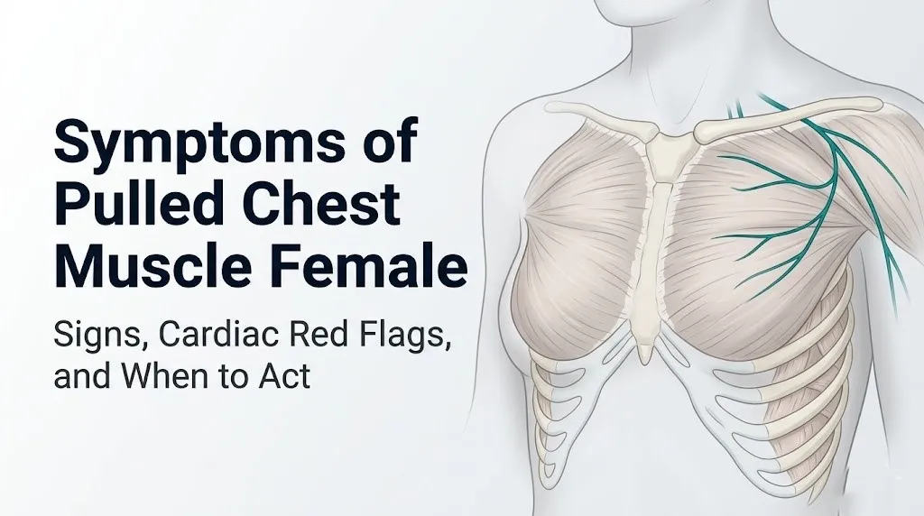
The motor and sensory supply to the pectoralis major travels via the lateral pectoral nerve (C5-C7, from the lateral cord of the brachial plexus) and the medial pectoral nerve (C8-T1, from the medial cord of the brachial plexus). Sensory fibers from the anterior chest wall also travel via the anterior cutaneous branches of intercostal nerves T1 through T6. This shared nerve supply is the anatomical basis for the arm and shoulder pain that can accompany pectoral muscle injury.
According to the National Institute of Arthritis and Musculoskeletal and Skin Diseases (NIAMS), skeletal muscle in women has a higher proportion of Type I (slow-twitch, fatigue-resistant) fibers compared to men, which provides some protection against strain during sustained low-load activity but may reduce the rapid force generation capacity needed to protect the musculotendinous junction during sudden eccentric loading.
Key Takeaway: Pressing directly on the sore spot in the anterior chest and reproducing your exact pain is the single most reliable indicator that the source is musculoskeletal rather than cardiac, because visceral cardiac pain does not map to specific surface pressure points the way somatic chest wall pain does.
Pulled Chest Muscle Pain Symptoms in Women
The pain of a pulled chest muscle in women is characteristically sharp, well-localized to a specific area of the anterior chest wall, and reproducible by palpation or by the specific movement that caused the injury.
The sharpness of the pain reflects activation of type A-delta nociceptive fibers in the strained muscle, which respond to mechanical disruption with rapid, precisely-localized pain signals. As inflammation develops over the first 24 to 48 hours, type C (slow, diffuse, burning) fibers become increasingly sensitized by prostaglandin E2 and bradykinin, adding a deeper aching quality to the initial sharp pain. This is why chest muscle strain pain often evolves from a sharp catching sensation at the moment of injury to a persistent, broader aching in the hours and days that follow.
The pain location gives a clinical clue about which structure is involved:
- Upper anterior chest, below the clavicle: clavicular head of pectoralis major or pectoralis minor
- Mid to lateral anterior chest: sternocostal head of pectoralis major
- Along the lateral rib margin: serratus anterior or external intercostal muscles
- Deep chest wall pain, worsened by twisting: internal intercostal muscle involvement
A 2021 study published in the British Journal of Sports Medicine noted that chest wall musculoskeletal pain that is reproducible by palpation with the patient in a relaxed supine position has a high positive predictive value for a musculoskeletal rather than cardiac source, though the study authors explicitly noted this finding does not eliminate the need for cardiac evaluation in patients with risk factors.
Women taking oral contraceptive pills or using hormonal contraception should note that their slightly elevated venous thromboembolism risk makes pulmonary embolism a relevant alternative cause if their chest pain is accompanied by any shortness of breath, particularly if leg swelling or recent travel is also present.
Chest Muscle Strain Symptoms When Breathing
Chest muscle strain that worsens with breathing is one of the most frequently reported and most clinically useful symptom patterns because it reflects the mechanical coupling between respiratory movement and the strained chest wall tissue.
Every breath requires the rib cage to expand, which lengthens the intercostal muscles, stretches the costal attachments of the pectoralis major’s sternocostal head, and increases tension across the entire anterior chest wall. When any of these tissues is strained, that unavoidable respiratory stretch activates the sensitized nociceptors in the injured tissue, producing a characteristic sharpening of pain at peak inhalation. Patients often describe this as a catching or stabbing sensation that forces them to take shallow breaths to avoid triggering it.
The important distinction is between pain that worsens at peak inhalation and eases immediately at the end of the breath (musculoskeletal origin) versus pain that worsens with breathing and is also associated with pleuritic features like a persistent dry cough or low-grade fever (suggesting pleuritis from viral infection or, more urgently, from pulmonary embolism).
To describe breathing-related chest pain accurately to a provider:
- Note whether the pain sharpens at the start, middle, or peak of inhalation
- Note whether the pain improves immediately when you stop inhaling (musculoskeletal) or persists throughout the breath cycle (more likely visceral or pleural)
- Record whether coughing triggers the same pain and whether it is identical in character to the breathing pain
- Note whether taking a breath in a specific position (leaning forward or to one side) reduces the pain
- State whether the pain has any component that is present regardless of breathing, even at complete rest
The Mayo Clinic notes that pleuritis produces breathing-related chest pain that is typically described as sharp and stabbing, precisely worsened at peak inhalation, and may be partially eased by lying on the affected side (which limits that side’s respiratory expansion). This pattern overlaps closely with musculoskeletal chest wall strain, making clinical examination and imaging necessary to differentiate the two.
Adolescent female athletes who develop breathing-related chest wall pain after contact sports or gymnastics should be evaluated for rib stress fractures, which produce a breathing-related pain pattern essentially identical to intercostal muscle strain but require different management and a longer return-to-sport timeline.
Pulled Chest Muscle Symptoms Left Side and Right Side Differences in Women
The side on which a pulled chest muscle occurs changes the clinical picture in two ways: it determines the likely mechanism of injury and the specific muscles affected, and left-sided chest pain carries a higher urgency for cardiac evaluation than right-sided pain in female patients.
Left-sided anterior chest pain in a woman always requires higher clinical caution. The left ventricle, left atrium, and the left anterior descending coronary artery (the vessel most often associated with anterior myocardial infarction) all refer ischemic pain to the left anterior chest, left shoulder, and left arm via the T1-T5 cardiac sympathetic afferents. A strained left pectoralis major produces pain in exactly the same anatomical territory. The American Heart Association states that women experiencing a heart attack are more likely than men to describe their pain as pressure, aching, or fullness rather than crushing, and may also have left jaw pain, left arm heaviness, or profound fatigue accompanying the chest discomfort.
Right-sided chest pain from a pulled muscle is still clinically significant but the cardiac differential is narrower, because right-sided chest pain is less commonly the primary presentation of left-sided cardiac ischemia. Right-sided chest wall pain in women raises its own differential:
- Right pectoralis major strain from asymmetric loading (right-hand dominant work, pushing heavy objects)
- Costochondritis at the right sternocostal junctions
- Right-sided pleuritis from pneumonia or pulmonary embolism
- Referred pain from the right hemidiaphragm via the phrenic nerve (C3-C5) due to hepatic or biliary pathology
Women with scoliosis affecting thoracic curvature have asymmetric loading across the chest wall musculature by definition, making side-specific strain patterns more complex and potentially requiring evaluation by a board-certified sports medicine physician familiar with scoliosis-related musculoskeletal asymmetry.
Key Takeaway: Left-sided chest pain in any woman requires cardiac evaluation before accepting a musculoskeletal explanation, regardless of how well the pain is reproduced by palpation, because the anatomical territory of cardiac referred pain and pectoral muscle strain pain is identical.
Referred Arm and Shoulder Pain from Chest Muscle Strain in Women
A pulled chest muscle can cause pain that travels down the arm or into the shoulder in women, and this occurs through the shared nerve supply of the pectoralis major and the upper extremity.
The pectoralis major is innervated by the lateral pectoral nerve (C5-C7) and the medial pectoral nerve (C8-T1), both of which are branches of the brachial plexus. The brachial plexus also supplies the arm and hand via the median, ulnar, and radial nerves. When significant inflammation or mechanical tension develops in the pectoral muscle, the sensitization of the pectoral nerve branches can produce referred pain along the C8-T1 dermatomal distribution: the medial forearm, the medial hand, and the fourth and fifth fingers. This referred pain pattern is called a C8-T1 dermatomal referral and it is anatomically distinct from radiculopathy (nerve root compression at the spine), though clinically they can feel similar.
The shoulder pain associated with pectoral muscle strain reflects the mechanical consequence of the muscle’s function: because the pectoralis major produces adduction and internal rotation of the humerus, when it is strained and in spasm, it creates a pulling sensation at its humeral insertion (the intertubercular sulcus), which patients feel as anterior shoulder pain or a sense of tightness deep in the shoulder joint.
What makes this clinically important in women: the arm and shoulder pain of a pectoral muscle strain via the medial pectoral nerve (C8-T1 distribution) can be virtually indistinguishable in location from the left arm aching that the American Heart Association identifies as an atypical myocardial infarction symptom in women. This is the most dangerous diagnostic overlap in this topic, because a woman may attribute her left arm aching to a known recent chest workout when it is actually a cardiac event.
Women with thoracic outlet syndrome (compression of the brachial plexus or subclavian vessels at the thoracic outlet between the clavicle and first rib) may already experience baseline arm tingling or weakness, which can make it harder to recognize when new arm symptoms are arising from a pectoral strain or from a cardiac source rather than their underlying thoracic outlet condition.
Chest Muscle Strain vs. Heart Attack Symptoms in Women
Distinguishing a pulled chest muscle from a heart attack in women is one of the most clinically consequential differentiations in emergency medicine because the two conditions can produce nearly identical symptom patterns in female patients, and the consequences of misidentifying a cardiac event as musculoskeletal are potentially fatal.
The American Heart Association has published extensive guidance noting that women having a heart attack are more likely than men to present without the classic crushing substernal pressure. Instead, they more commonly experience fatigue, shortness of breath, nausea, jaw pain, upper back pain, and arm aching, often without any sensation they would describe as chest pain at all. When women do have chest pain during a heart attack, it may be sharp or burning rather than pressure-like, closely mimicking the character of musculoskeletal chest wall pain.
| Feature | Pulled Chest Muscle | Heart Attack in Women |
|---|---|---|
| Pain reproducibility | Reproduced precisely by pressing on the sore spot | Not reproduced by surface palpation |
| Relationship to movement | Consistently worsens with specific arm/trunk movements | Variable; may not clearly worsen with chest wall movement |
| Onset | Usually related to a specific activity or exertion | May occur at rest, during emotional stress, or during light activity |
| Associated symptoms | Muscle tightness, limited arm range of motion | Fatigue, nausea, jaw pain, arm aching, shortness of breath, sweating |
| Response to rest | Pain typically eases with stillness | Pain often persists at rest or worsens with lying flat |
| Fever or systemic symptoms | Absent (unless concurrent illness) | Absent in MI; diaphoresis and pallor possible |
| History | Typically follows identifiable physical demand | May occur without preceding physical exertion |
| Duration | Persists days to weeks with gradual improvement | Does not improve without medical intervention |
Two female-predominant cardiac conditions deserve specific mention. Takotsubo cardiomyopathy (stress cardiomyopathy, ICD-10: I51.81), which the Journal of the American College of Cardiology has documented as occurring in women at approximately nine times the rate of men, produces acute chest pain and shortness of breath triggered by emotional or physical stress, with a near-normal initial electrocardiogram in many cases. Microvascular coronary artery disease, affecting the small coronary vessels rather than the large epicardial arteries, produces exertional or emotional chest pain in women without the classic ST-elevation changes that ECG monitoring typically catches.
Women with any of the following should not attempt to self-differentiate chest pain as musculoskeletal without medical evaluation: age over 55, hypertension, diabetes mellitus, smoking history, family history of premature coronary artery disease, prior cardiac events, or current use of stimulant medications.
Hormonal and Biological Factors Affecting Chest Muscle Strain in Women
The hormonal environment of a woman’s body at any given life stage directly affects how susceptible her chest wall musculature is to strain, how intensely she experiences the associated pain, and how efficiently the tissue repairs itself after injury.
Relaxin, a peptide hormone produced by the corpus luteum during the luteal phase of the menstrual cycle and in far higher concentrations by the placenta during pregnancy, acts on relaxin receptors in connective tissue fibroblasts to upregulate matrix metalloproteinase activity and reduce collagen cross-linking. This makes ligaments and the musculotendinous junction more extensible but also more susceptible to strain beyond their elastic limit. Research published in the Journal of Orthopaedic and Sports Physical Therapy has documented that female athletes have higher musculotendinous injury rates during the luteal phase of the menstrual cycle, when relaxin levels are at their monthly peak, though studies specific to pectoral injury remain limited.
Estrogen has direct anabolic effects on skeletal muscle through estrogen receptor-alpha (ERα), which is expressed in satellite cells (muscle stem cells responsible for repair). Estrogen promotes satellite cell activation after injury and supports Type I muscle fiber maintenance. After menopause, declining estrogen reduces satellite cell responsiveness and decreases collagen synthesis capacity in the musculotendinous junction, slowing repair and increasing re-injury risk.
Quick Tip:
- Women who train intensely in the days before menstruation, when relaxin is at its monthly peak, may benefit from reducing maximal eccentric chest loading during that window to reduce musculotendinous junction strain risk
- Women in the perimenopausal transition who notice that chest wall strains take longer to resolve than they did a decade ago are experiencing a real biological change in tissue repair capacity, not a perception error
- Women with hypothyroidism should inform their primary care physician if they develop a chest wall strain, as thyroid hormone plays a role in skeletal muscle protein synthesis and a low thyroid state can impair recovery
Key Takeaway: Relaxin peaks during the luteal phase of the menstrual cycle and during pregnancy, making the musculotendinous junction of the pectoral muscles more susceptible to strain at those times, not because women are less physically capable, but because the connective tissue biology is in a different mechanical state.
Chest Muscle Strain During Pregnancy Symptoms
Chest muscle strain during pregnancy is a distinct clinical presentation because pregnancy changes the anatomy, biomechanics, and hormonal environment of the chest wall simultaneously, making strains more likely and their symptoms harder to interpret.
By the second trimester, the growing uterus displaces the diaphragm superiorly by up to four centimeters, which widens the costal angle, increases rib cage circumference, and shifts the effective attachment geometry of the sternocostal head of the pectoralis major. This altered geometry means that movements that previously caused no chest wall stress may now apply unexpected eccentric load to the lower fibers of the pectoralis major and to the serratus anterior along the lateral rib margin.
Symptoms of pectoral or intercostal muscle strain during pregnancy:
- A dull to sharp aching at the lower rib margins or anterior chest, worsening with lifting, reaching overhead, or rolling in bed
- A catching sensation with deep inhalation, distinct from the respiratory restriction caused by uterine elevation of the diaphragm alone
- Tenderness when pressing along the lower costal margin or along the lateral sternum where the sternocostal head attaches
- Aching that is positional and worsens lying on the affected side
Chest pain during pregnancy that is not clearly positional and not reproducible by palpation should be evaluated promptly. The American College of Obstetricians and Gynecologists (ACOG) identifies cardiac conditions, pulmonary embolism (for which pregnancy increases risk significantly due to hypercoagulable state and venous stasis), and HELLP syndrome as causes of chest and upper abdominal pain in pregnancy that require immediate evaluation.
Imaging choices are modified in pregnancy. Chest X-ray with appropriate abdominal shielding is generally considered acceptable when clinically indicated. MRI without contrast is preferred over CT for musculoskeletal evaluation when possible. Any imaging decision during pregnancy should involve the treating obstetrician-gynecologist.
Chest Muscle Strain in Postmenopausal and Older Women
Postmenopausal women represent a distinct clinical group for chest muscle strain because declining estrogen, reduced skeletal muscle mass (sarcopenia), and elevated cardiovascular risk all converge to make both the injury and its differential diagnosis more complex.
After menopause, the loss of estrogen-mediated anabolic support for skeletal muscle (via ERα in satellite cells) contributes to sarcopenia: a progressive loss of muscle mass and strength that, according to the National Institute of Arthritis and Musculoskeletal and Skin Diseases (NIAMS), can begin as early as the fifth decade and accelerates in the years immediately following menopause. Reduced pectoral muscle mass means less structural protection for the musculotendinous junction during loaded movements, and reduced collagen synthesis capacity means that microtears accumulate faster than they are repaired when eccentric training loads are applied without adequate progressive loading.
The cardiovascular risk context changes the clinical urgency of chest pain evaluation entirely. The NHLBI reports that after age 55, women’s cardiovascular disease risk rises sharply toward the male risk level as the cardioprotective effects of estrogen decline. A postmenopausal woman with new chest pain cannot be assumed to have a musculoskeletal cause without cardiac evaluation, regardless of how closely her symptoms resemble a strain pattern.
Specific considerations for older women with suspected chest muscle strain:
- Rib stress fractures become more likely with osteoporosis, producing a breathing-related chest wall pain pattern indistinguishable from intercostal muscle strain on clinical examination alone
- Vertebral compression fractures at the thoracic level (T4-T12) can produce anterior chest wall referred pain that mimics anterior muscle strain
- Women on long-term aromatase inhibitor therapy (used in breast cancer management) have accelerated bone loss and elevated musculoskeletal pain risk, altering both the injury pattern and the pain threshold
Any postmenopausal woman with new chest wall pain requires evaluation by a primary care physician within 24 hours, and if any cardiac features are present, evaluation by an emergency medicine physician the same day.
Early vs. Severe Pulled Chest Muscle Symptoms in Women
The clinical presentation of a pulled chest muscle in women changes predictably as the severity of the injury increases, and the American Academy of Orthopaedic Surgeons (AAOS) muscle strain grading system provides a useful framework for understanding this progression.
| Strain Grade | Tissue Involved | Pain Quality | Functional Impact | Typical Timeline |
|---|---|---|---|---|
| Grade 1 (mild) | Microscopic myofibril tears; structural integrity intact | Mild to moderate, localized aching; some tenderness to palpation | Minimal; full movement possible, slightly painful | 1 to 3 weeks |
| Grade 2 (moderate) | Partial macroscopic tear; some fibers disrupted | Moderate to severe; tenderness, possible visible bruising; pain on active contraction | Reduced strength and range of motion; visible guarding | 4 to 8 weeks |
| Grade 3 (severe) | Complete muscle or tendinous disruption | Severe at injury; may improve paradoxically as fibers lose tension; palpable defect possible | Significant weakness; inability to perform resisted adduction | Weeks to months; may require surgical evaluation |
Grade 1 strains produce diffuse tenderness over the muscle without clear bruising. Grade 2 strains may produce ecchymosis (bruising) over the chest wall within 24 to 72 hours as blood tracks from the torn fibers into the subcutaneous tissue. Grade 3 tears can produce a visible or palpable deformity: the muscle bunches up toward its origin as the tendon retracts, creating an asymmetric chest wall contour.
Women who train with weights or perform gymnastics and develop sudden severe chest pain with a feeling of a “pop” followed by immediate loss of strength during a pushing or pressing movement should be evaluated for a Grade 2 or Grade 3 pectoral tear by a primary care physician or board-certified sports medicine physician within 24 to 48 hours, and should ask specifically about musculoskeletal ultrasound or MRI to assess the extent of fiber disruption.
Causes and Risk Factors for Chest Muscle Strain in Women
Chest muscle strain in women arises from a defined set of physical mechanisms, and several risk factors specific to female anatomy, hormonal status, and training patterns increase the probability of injury in particular subgroups.
Primary causes of chest muscle strain in women:
- Bench press or chest press with excessive load or inadequate warm-up (the eccentric descent phase places the greatest force on the sternocostal head of the pectoralis major)
- Sudden pushing movement against a resisting force: pushing a stalled vehicle, opening a heavy door unexpectedly, or catching a fall
- Swimming (particularly freestyle and butterfly strokes, which place high cyclic loads on the pectoralis major through shoulder adduction)
- Gymnastic rings, dips, and wide-grip push-up variations that place the pectoral fibers at maximum length under load
- Overhead throwing or reaching with sudden onset
- Repetitive carrying or manual labor involving sustained arm adduction against load
Risk factors that amplify chest muscle strain likelihood in women:
- Inadequate progressive loading when beginning or returning to upper body strength training
- Hormonal contraceptive use with luteal-phase relaxin peaks (though evidence on hormonal contraceptive effects on injury risk is mixed in the current literature)
- Pregnancy (elevated relaxin, altered biomechanics)
- Postmenopausal status (reduced estrogen and sarcopenia)
- Hypothyroidism (impaired muscle protein synthesis and repair)
- Prior ipsilateral pectoral injury (scar tissue at the musculotendinous junction reduces elasticity)
- Insufficient warm-up before high-intensity upper body activity
- Rapid increase in training volume or load (greater than 10 percent weekly increase in load is associated with elevated musculotendinous injury risk, according to guidelines cited by the British Journal of Sports Medicine)
Women with systemic lupus erythematosus (SLE) taking long-term corticosteroids are at elevated risk for musculotendinous junction weakness due to corticosteroid-mediated inhibition of collagen synthesis, making even moderate physical demands capable of producing Grade 2 or Grade 3 strain.
Key Takeaway: The single highest-risk scenario for a Grade 2 or Grade 3 pectoral tear in women is performing heavy eccentric chest press or dip exercises with insufficient progressive preparation, particularly during the luteal phase of the menstrual cycle when relaxin-driven connective tissue laxity is at its monthly peak.
How a Pulled Chest Muscle Is Diagnosed in Women
A pulled chest muscle in women is diagnosed through a structured clinical history, physical examination including direct palpation and resisted muscle testing, and targeted imaging when the degree of injury or the differential diagnosis makes it necessary.
The diagnostic process a primary care physician or sports medicine physician typically follows:
- Clinical history: mechanism of injury (was there a specific movement or load?), exact pain location, onset and progression, aggravating and relieving factors, cardiac risk factor review (hypertension, diabetes, smoking, family history, prior cardiac events, menopausal status), medication history including hormonal contraceptives and corticosteroids
- Palpation examination: the physician systematically presses along the pectoralis major (from its clavicular and sternocostal origins to its humeral insertion), the pectoralis minor (at the coracoid process), and the intercostal spaces to identify the precise point of maximal tenderness. Reproducibility of the patient’s exact pain by palpation is the most clinically useful finding supporting a musculoskeletal cause
- Resisted muscle testing: the patient attempts to perform shoulder adduction (pressing the arm inward against the examiner’s resistance) with the elbow at 90 degrees. Pain and weakness during this maneuver specifically implicates the pectoralis major
- Range of motion assessment: the physician assesses shoulder flexion, abduction, and horizontal adduction to identify which movements are restricted
- Cardiac risk stratification: in any woman with chest pain, the physician assesses the probability of a cardiac cause using established cardiovascular risk factors, symptom character, and vital signs. If any cardiac concern exists, an ECG and troponin measurement are ordered before attributing the pain to musculoskeletal causes
Imaging options for confirmed or suspected chest wall strain:
| Test | What It Evaluates | When It Is Ordered |
|---|---|---|
| Musculoskeletal ultrasound | Real-time visualization of pectoral muscle fiber integrity, partial or complete tears, hematoma | Suspected Grade 2 or Grade 3 tear; preferred first-line imaging |
| MRI of the chest wall | Detailed assessment of muscle fiber disruption, edema pattern, extent of tear | When ultrasound is inconclusive or surgical planning is needed |
| Chest X-ray | Rules out pneumothorax, rib fractures, pleural effusion | When breathing-related pain raises concern for rib fracture or pleural pathology |
| ECG and serial troponin | Rules out myocardial infarction | Any woman with cardiac risk factors or atypical symptom features |
| CT pulmonary angiography | Rules out pulmonary embolism | Dyspnea with chest pain; particularly in pregnancy or women on hormonal contraceptives |
Women should bring to their appointment a clear account of the specific activity that preceded the pain, whether they felt a pop or sudden increase in pain at a specific moment, and a complete list of their medications including hormonal contraceptives, as these affect both the injury context and imaging decisions.
Emergency Symptoms: When to Call 911 or Go to the ER
Certain chest pain presentations in women require immediate emergency evaluation regardless of how closely the symptoms resemble a musculoskeletal injury. Do not wait to see if these resolve on their own.
Call 911 or go to the nearest emergency room immediately if you experience:
- Chest pain with shortness of breath, even mild, that was not present before the pain started (potential pulmonary embolism, NSTEMI, or Takotsubo cardiomyopathy)
- Chest pain with sweating, nausea, or vomiting (potential myocardial infarction; these are identified by the American Heart Association as common atypical MI presentations in women)
- Chest pain with jaw pain, left arm aching, or upper back pain occurring together (potential myocardial infarction with atypical radiation pattern)
- Sudden severe chest pain that is the worst pain you have ever felt (potential aortic dissection)
- Chest pain with lightheadedness, dizziness, or near-fainting (potential hemodynamic compromise from cardiac or vascular cause)
- Chest pain with a rapid or irregular heartbeat that you can feel (potential arrhythmia associated with cardiac ischemia or Takotsubo cardiomyopathy)
- Chest pain in a woman who is pregnant, accompanied by any shortness of breath or leg swelling (potential pulmonary embolism, for which pregnancy is a well-established risk factor)
- Chest pain that is not reproducible by pressing on the sore area and does not change with arm or trunk movement (this absence of musculoskeletal behavior is a red flag for visceral cardiac or pulmonary cause)
- Chest pain in any woman with known heart disease, diabetes, hypertension, or prior cardiac events, regardless of how mild the pain seems
These presentations can indicate myocardial infarction, pulmonary embolism, aortic dissection, or Takotsubo cardiomyopathy and require emergency medical assessment and intervention, not an urgent care appointment or a phone call.
Recovery and What to Expect from Chest Muscle Strain in Women
Recovery from a pulled chest muscle in women follows a predictable biological timeline based on the severity of the injury, though hormonal, age-related, and comorbidity factors can extend or complicate this timeline in ways that general recovery guides do not acknowledge.
Grade 1 chest muscle strains in healthy premenopausal women without complicating conditions typically resolve within one to three weeks with activity modification, rest from aggravating movements, and a structured return to activity that reintroduces loading gradually. The healing process follows three phases: the inflammatory phase (days 1 to 5, characterized by pain, swelling, and tenderness as neutrophils and macrophages clear damaged tissue), the proliferative phase (days 5 to 21, during which satellite cells proliferate and begin depositing new myofibrils), and the remodeling phase (weeks 3 to 12, during which new muscle fibers mature and the musculotendinous junction regains tensile strength).
Grade 2 strains require a longer proliferative and remodeling phase, typically four to eight weeks, and benefit from supervised rehabilitation with a physical therapist to restore full range of motion and rebuild the neuromuscular control of the pectoral muscles before returning to loaded activity. Returning to heavy pressing or throwing movements before the remodeling phase is complete significantly elevates re-injury risk.
Grade 3 tears (complete rupture) in women are rare but require evaluation by an orthopedic surgeon for assessment of whether surgical repair is appropriate. Non-operative management is sometimes chosen for lower-demand individuals, but surgical reattachment is associated with better long-term strength outcomes in women who wish to maintain an active lifestyle, according to clinical case series published in the Journal of Bone and Joint Surgery.
Postmenopausal women should expect recovery timelines at the longer end of these ranges due to the reduced satellite cell response and collagen synthesis capacity that accompany declining estrogen. A board-certified sports medicine physician can guide a return-to-activity plan that accounts for this biological reality while maintaining the long-term musculoskeletal health benefits of progressive upper body loading.
Frequently Asked Questions About Symptoms of Pulled Chest Muscle in Women
What does a pulled chest muscle feel like in a woman?
A pulled chest muscle in a woman typically feels like a sharp or aching pain at a specific point on the anterior chest wall that is precisely reproduced when you press directly on the sore area.
The pain usually worsens with arm movements that activate the pectoral muscles, particularly pushing, reaching overhead, or crossing the arm across the body, and may intensify with deep breathing if the strain involves the intercostal or sternocostal attachments.
Pain that is not reproducible by pressing on the chest surface, or that is accompanied by sweating, nausea, or shortness of breath, requires immediate evaluation by an emergency medicine physician to rule out a cardiac cause.
How do I know if my chest pain is a pulled muscle or my heart?
Chest muscle pain can typically be reproduced by pressing directly on the sore spot, worsens with specific arm movements, and is usually related to a recent physical activity.
Cardiac chest pain in women is often not reproducible by surface palpation, may be accompanied by fatigue, jaw pain, arm aching, or shortness of breath, and can occur without any physical exertion, particularly in conditions like Takotsubo cardiomyopathy and microvascular coronary artery disease, which are more prevalent in women.
Any woman with cardiac risk factors (hypertension, diabetes, smoking, family history of premature heart disease, or menopause) who develops chest pain should be evaluated by a primary care physician or emergency medicine physician before attributing the pain to a musculoskeletal cause.
Can a pulled chest muscle cause pain that goes down the arm in women?
A pulled chest muscle can cause arm pain in women through the medial pectoral nerve (C8-T1) and lateral pectoral nerve (C5-C7), which connect the pectoralis major to the brachial plexus supplying the arm.
This referred arm pain typically follows a C8-T1 dermatomal distribution, producing aching or tingling along the medial forearm and into the fourth and fifth fingers on the affected side.
Because this pain pattern overlaps closely with the atypical left arm aching of a heart attack in women, any new left arm pain accompanying left chest pain should be evaluated by an emergency medicine physician the same day it occurs.
How long does a pulled chest muscle take to heal in women?
A Grade 1 chest muscle strain in a healthy woman typically heals within one to three weeks with activity modification and a gradual return to loading.
Grade 2 partial tears take four to eight weeks, and postmenopausal women or those with conditions affecting muscle repair (hypothyroidism, diabetes, long-term corticosteroid use) should expect recovery at the longer end of these ranges due to reduced satellite cell responsiveness and collagen synthesis capacity.
Returning to full loading activities before the remodeling phase is complete, typically before four weeks even for mild strains, measurably increases the risk of re-injury at the same musculotendinous junction site.
What are the signs that chest muscle pain in a woman is serious?
Chest muscle pain in a woman is serious when it is not reproducible by pressing on the chest, is accompanied by shortness of breath, sweating, nausea, jaw pain, arm aching, or lightheadedness, or occurs in a woman with cardiovascular risk factors.
Chest pain that is described as pressure, fullness, or squeezing rather than sharp and positional is more likely to have a cardiac origin and warrants emergency evaluation regardless of the woman’s age.
Call 911 immediately if any of these features are present alongside chest pain, because myocardial infarction, pulmonary embolism, and Takotsubo cardiomyopathy can all produce chest pain in women that initially resembles a musculoskeletal injury.
Can a pulled chest muscle get worse with deep breathing?
A pulled chest muscle, particularly of the intercostal muscles or the sternocostal head of the pectoralis major, consistently worsens with deep breathing because inhalation stretches the strained muscle fibers and intercostal attachments as the rib cage expands.
The pain typically sharpens at peak inhalation and eases immediately when the breath is released, producing a characteristic “catching” or stabbing quality that forces shallow breathing.
Breathing-related chest pain that does not ease at the end of exhalation, or that is accompanied by cough, fever, or any shortness of breath, may indicate pleuritis or pulmonary embolism rather than a musculoskeletal strain, and warrants evaluation by a primary care physician or emergency medicine physician the same day.
Taking the Right Next Step With Your Chest Pain
A pulled chest muscle in a woman is a real, common, and manageable musculoskeletal injury. It has a specific anatomical source, a predictable symptom pattern, a reliable clinical test (direct palpation reproducing the pain), and a well-understood recovery timeline. What makes this different from most musculoskeletal injuries is the diagnostic context: chest pain in a woman never gets the benefit of the doubt that identical pain in other body regions would receive.
If your symptoms clearly match the musculoskeletal pattern described here, specifically pain that you can reproduce by pressing on the sore spot, that worsens with specific movements, and that followed an identifiable physical demand, contact a primary care physician or board-certified sports medicine physician within 24 to 48 hours to confirm the diagnosis and establish a safe return-to-activity plan.
If there is any uncertainty, any cardiac symptom alongside the chest pain, or any cardiovascular risk factor in your history, do not wait. Call 911 or go to the nearest emergency room immediately. That decision takes less than a minute and it is always the right one when you are not sure.