Early Psoriasis Symptoms: What Your Skin Is Telling You
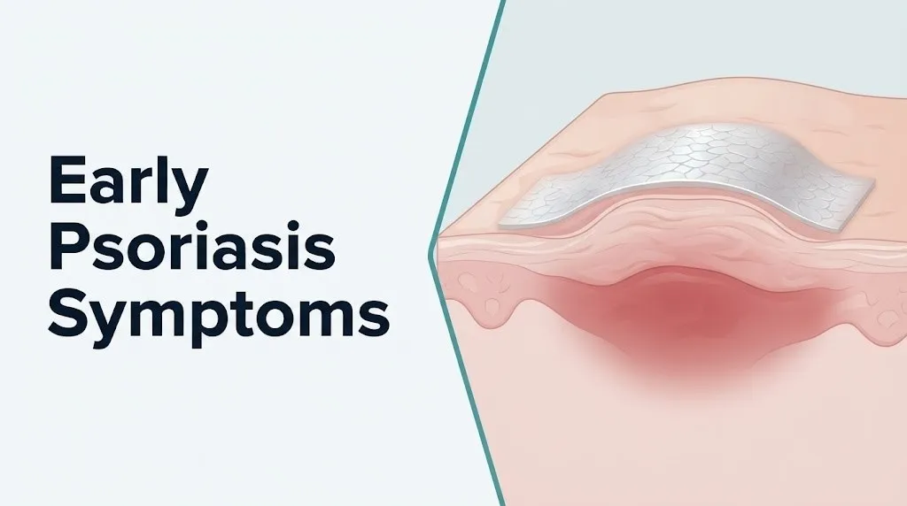
Early psoriasis symptoms typically begin as small, red or discolored, slightly raised patches of skin covered by silvery-white or grayish flaking scale, most often appearing first on the elbows, knees, lower back, or scalp, and they represent the visible result of an immune system malfunction that is accelerating skin cell production by nearly six times the normal rate. Recognizing these signs early, before full plaques develop and before the condition spreads or involves the joints, gives you the best clinical window for effective management.
According to the National Institute of Arthritis and Musculoskeletal and Skin Diseases (NIAMS), psoriasis affects approximately 2 to 3% of the United States population, roughly 7.5 million people, making it one of the most prevalent immune-mediated skin diseases in the country. The National Psoriasis Foundation reports that psoriasis is the most common autoimmune condition in the United States, and most people experience their first symptoms between the ages of 15 and 35, though onset can occur at any age.
This article covers every clinically recognized early psoriasis symptom across all major subtypes, explains the specific immune and cellular mechanism producing each sign, distinguishes early presentations from other common skin conditions, addresses how symptoms differ across skin tones, age groups, and trigger patterns, and tells you exactly which presentations require urgent or emergency medical evaluation.
Early Psoriasis Symptoms: The Complete Clinical Picture
Early psoriasis symptoms are defined by three overlapping features: localized raised skin lesions with a characteristic surface scale, an inflammatory reddish or discolored base beneath that scale, and a tendency for those lesions to appear at specific body sites and to enlarge or multiply if the underlying immune activation is not addressed.
The full spectrum of early symptoms depends on which psoriasis subtype is developing, which body site is affected, and the individual’s skin tone. The table below maps the primary early symptoms across the main clinical presentations:
| Symptom | Subtype Association | Body Site | Clinical Significance |
|---|---|---|---|
| Small red or violet raised patch with silver scale | Plaque psoriasis | Elbows, knees, scalp, lower back | Most common early presentation; scale is pathognomonic |
| Teardrop-shaped small red spots | Guttate psoriasis | Trunk, thighs, upper arms | Often post-streptococcal; appears suddenly |
| Flaking, itchy scalp with adherent scale | Scalp psoriasis | Scalp, hairline, behind ears | Frequently mistaken for dandruff or seborrheic dermatitis |
| Pitting, oil drop discoloration of nail | Nail psoriasis | Fingernails, toenails | Can appear before skin symptoms; precedes psoriatic arthritis |
| Smooth, shiny red rash in skin folds | Inverse psoriasis | Groin, underarm, under breasts | No classic scale due to moisture; frequently misdiagnosed |
| Pinpoint bleeding when scale removed | Auspitz sign (any plaque subtype) | Any plaque site | Indicates dilated dermal capillary loops beneath suprapapillary plate |
| White ring surrounding a healing plaque | Woronoff ring | Around plaque margin | Vasoconstriction around inflamed tissue; resolves with treatment |
People with a family history of psoriasis are at substantially higher risk of early onset. Research published in the Journal of Investigative Dermatology has documented that approximately 35 to 40% of people with psoriasis have a first-degree relative with the condition, indicating a strong genetic component mediated in part by the HLA-Cw6 allele, which is present in roughly 65% of patients with early-onset plaque psoriasis.
What Is Psoriasis and How Does It Start?
Psoriasis is a chronic, immune-mediated inflammatory disease in which dysregulated activation of the adaptive immune system drives keratinocytes in the epidermis to proliferate at a dramatically accelerated rate, producing the thickened, scaling plaques that define the condition’s clinical appearance.
Understanding how psoriasis starts requires knowing what normally happens in the skin. In a healthy epidermis, keratinocytes are generated in the deepest layer, the stratum basale, and migrate upward through the stratum spinosum, stratum granulosum, and stratum corneum over approximately 28 to 30 days before being shed as part of normal skin turnover. In psoriatic skin, this entire cycle is compressed to 3 to 5 days. The result is a skin surface that cannot shed cells fast enough and accumulates them into the raised, thickened plaques with adherent silvery scale that characterize the disease.
The initiating event in most cases involves plasmacytoid dendritic cells in the dermis being activated by complexes of cathelicidin LL-37 (an antimicrobial peptide produced by keratinocytes) with self-DNA or self-RNA, through Toll-like receptor 9 (TLR9) and TLR7/8 signaling. This triggers production of type I interferons. Subsequent activation of myeloid dendritic cells produces interleukin-23 (IL-23) and interleukin-12 (IL-12), which drive differentiation of naive T-lymphocytes into the Th17 and Th1 subsets, respectively. IL-23 is now recognized as the master regulator of psoriatic inflammation, based on the clinical success of IL-23 inhibitors documented in studies published in the New England Journal of Medicine.
Think of the immune cascade like a fire alarm system that has been triggered by a false alarm and cannot be switched off. The alarm (IL-23) keeps calling in firefighters (Th17 cells). Those firefighters release chemicals (IL-17A, IL-22) that cause the building (the skin) to overproduce construction materials (keratinocytes) at a rate that fills the hallways faster than anyone can clear them out. The result is the visible buildup that is psoriasis.
First Signs of Psoriasis on Skin
The first signs of psoriasis on skin are typically one or more small, clearly demarcated, slightly raised patches that are covered with a dry, silvery or grayish-white scale and rest on an erythematous (red, pink, or violet) base that feels warmer than surrounding skin when touched.
Most people notice these initial patches on the outer surface of the elbows, the front of the knees, the lower back, or the scalp. These sites are clinically preferred because they experience more friction and minor trauma than other body areas, and trauma to the skin in a genetically susceptible person can trigger the Koebner isomorphic response (discussed in a later section), where new psoriatic lesions develop at sites of skin injury.
Early plaques typically have these characteristics:
- Clearly defined borders where normal skin ends and the plaque begins, a feature called “well-demarcated” that distinguishes psoriasis from eczema’s more blurred margins
- A surface scale that is dry, layered, and loosely adherent in the center but may be more tightly attached at the plaque margins
- A base color that ranges from bright pink-red on lighter skin tones to violet, dark brown, or slate gray on darker skin tones
- A slightly raised, indurated (firm) texture compared to surrounding normal skin
- A tendency to form satellite lesions, smaller spots appearing around an initial larger patch
The American Academy of Dermatology states that early psoriasis plaques most commonly measure between 1 and 10 centimeters in diameter at initial presentation, though this varies by subtype and individual immune response. Guttate psoriasis produces much smaller spots (under 1 cm) while inverse psoriasis presents as larger, confluent areas in skin folds.
For postmenopausal women, early psoriasis plaques may appear or worsen as estrogen levels decline, because estrogen has a general immunomodulatory effect that tends to suppress the Th1-driven inflammatory pathway. Reduced estrogen removes this partial brake on T-cell activity, and some women experience their first psoriasis diagnosis after menopause.
Key Takeaway: The earliest psoriasis lesions are small, well-demarcated, raised patches with silvery scale on a reddish or discolored base, most often appearing at elbows, knees, scalp, or lower back, and early recognition allows intervention before plaques enlarge or multiply.
What Does Early Psoriasis Look Like?
Early psoriasis looks like a raised, clearly bordered skin patch covered with a dry, flaky, silver-white or grayish scale that sits on a base that appears red or pink on lighter skin and violet, brown, or dark gray on darker skin, with the scale tending to flake off in layers rather than in fine powder-like particles.
Two clinical signs that are specific to psoriasis plaques and help distinguish them from other rashes are worth knowing. The Auspitz sign describes the appearance of pinpoint bleeding when a psoriasis scale is gently removed. This happens because the dilated and tortuous capillary loops in the dermal papillae beneath the thinned suprapapillary epidermis are extremely close to the surface, and removing the scale ruptures them. The Woronoff ring is a pale, blanched ring visible in the skin surrounding a psoriasis plaque, produced by localized vasoconstriction. Neither of these signs should be tested deliberately at home, but their presence or absence can be clinically informative during a dermatologist’s examination.
Early psoriasis plaques may look subtly different depending on location:
| Body Location | Typical Early Appearance | Distinguishing Feature |
|---|---|---|
| Elbows / Knees | Thick, adherent silvery scale on reddish raised patch | Scale is thickest at pressure points |
| Scalp | Flaking white to gray scale extending slightly beyond the hairline | Scale may clump hair shafts together |
| Lower back / Sacral area | Large, flat plaque with prominent scale | Often first noticed as persistent “dry patch” |
| Nails | Pitting (small ice pick-like depressions), oil drop yellowing | Scale does not appear; pitting is the key early sign |
| Skin folds (inverse) | Smooth, shiny, non-scaled reddish or dark patch | Moisture prevents scale formation |
| Trunk (guttate) | Multiple 0.5 to 1 cm teardrop spots with fine scale | Appears suddenly, often after sore throat |
For men, who account for a slightly higher proportion of psoriasis diagnoses in some epidemiological studies, early plaques tend to appear most frequently on the scalp and trunk. For women, the lower extremities and hands are more commonly reported as initial sites in data compiled by the National Psoriasis Foundation.
Does Early Psoriasis Itch and What Does It Feel Like?
Early psoriasis does itch in the majority of cases, with studies estimating that approximately 70 to 90% of people with plaque psoriasis report pruritus (itching) as a symptom, and the itch in psoriasis is physiologically distinct from the itch of eczema or allergic reactions in both its character and its neural mechanism.
Psoriatic itch is driven primarily by the release of substance P, calcitonin gene-related peptide (CGRP), and IL-31 in the inflamed skin. IL-31, produced by Th2 and Th22 cells in the psoriatic dermis, directly activates itch-transmitting C-nerve fibers in the skin. Substance P, a neuropeptide released from sensory nerve endings in the dermis, contributes to both itch and the amplification of mast cell degranulation and the local inflammatory response. This neurogenic component partly explains why psychological stress, which triggers the release of substance P through the hypothalamic-pituitary-adrenal (HPA) axis and the sympathetic nervous system, reliably worsens psoriatic itch and can precipitate flares.
What the itch actually feels like varies by individual and by the stage of the lesion. Early-stage psoriasis most commonly produces:
- A dry, persistent itching sensation localized to the plaque itself
- Occasional burning or stinging at the lesion margins, particularly when the plaque is actively expanding
- Sensitivity to friction from clothing over the affected area
- A feeling of skin tightness across the plaque surface, especially after bathing
- In some cases, no itch at all, particularly with early scalp psoriasis where some individuals report only visible flaking without discomfort
Approximately 10 to 30% of people with psoriasis report minimal or no itch, particularly in early-stage or mildly inflamed lesions. This is why the absence of itch does not rule out psoriasis and why a rash that looks consistent with psoriasis but does not itch still warrants clinical evaluation by a primary care physician or board-certified dermatologist.
People with atopic dermatitis (eczema) as a co-existing condition, which occurs in a subset of psoriasis patients, typically report more intense and more generalized itch than those with psoriasis alone, because atopic dermatitis involves stronger IL-4 and IL-13 signaling that amplifies the pruritic neural response.
Early Guttate Psoriasis Symptoms
Early guttate psoriasis symptoms appear as a sudden eruption of multiple small, teardrop-shaped, salmon-pink or reddish spots covered with a fine, thin scale, distributed across the trunk, thighs, upper arms, and sometimes the face and scalp, typically appearing two to four weeks after a streptococcal pharyngitis (strep throat) infection.
The name “guttate” derives from the Latin word for drop, which describes the characteristic shape and size of these lesions, ranging from 0.5 to 1.5 centimeters. Unlike plaque psoriasis, where plaques are thick and adherent, guttate lesions are thinner, with a lighter, finer scale. The eruption can involve dozens to hundreds of individual spots appearing over the course of days, producing a dramatic change in skin appearance that many people find alarming.
The mechanism connecting streptococcal infection to guttate psoriasis involves molecular mimicry. Certain epitopes of the streptococcal M-protein share structural similarity with keratin peptides in the skin. In genetically susceptible individuals (particularly those carrying HLA-Cw6), streptococcal antigen-reactive T-cells cross-react with keratinocyte antigens, triggering the same IL-17A-driven inflammatory cascade that produces plaque psoriasis but doing so suddenly and throughout the skin rather than at localized friction sites.
Quick Tip:
- Anyone who develops a sudden widespread spotty rash two to three weeks after a sore throat should bring both the rash and the sore throat history to a primary care physician’s attention at the same appointment. A throat swab or anti-streptolysin O (ASO) titer blood test can confirm recent streptococcal infection, which is directly relevant to both the diagnosis and the management approach.
- Guttate psoriasis in a first episode resolves spontaneously in many cases within three to four months, but approximately one-third of people with guttate psoriasis go on to develop chronic plaque psoriasis over subsequent years, particularly those who are HLA-Cw6 positive and have recurrent streptococcal infections.
- Children and young adults under age 30 are most susceptible to guttate psoriasis, making early recognition in this age group particularly important for long-term monitoring.
Key Takeaway: A sudden eruption of multiple small teardrop spots two to four weeks after a strep throat infection is the defining early pattern of guttate psoriasis, and it warrants evaluation by a primary care physician because both the infection and the skin condition need to be addressed.
Early Scalp Psoriasis Symptoms
Early scalp psoriasis symptoms include persistent flaking from the scalp, localized areas of scale that adhere to hair shafts and clump them together, itching or tenderness at the scalp surface, and in many cases a visible reddish or discolored border at the hairline where the scalp psoriasis extends slightly onto the forehead, behind the ears, or onto the back of the neck.
Scalp psoriasis is reported to affect approximately 45 to 56% of people with psoriasis at some point, making it one of the most common sites of involvement, and it is frequently the first site where psoriasis becomes clinically apparent. The scalp’s sebaceous gland density and hair follicle distribution create a distinct microenvironment that influences how psoriatic plaques form there, with the scale appearing differently, often yellower and more oily-appearing in sebum-rich areas, compared to the classic dry silvery scale of elbow or knee plaques.
Early scalp psoriasis symptoms to distinguish from ordinary dandruff or seborrheic dermatitis:
- Scale that is thicker, more adherent, and chunkier than the fine white powder of ordinary dandruff
- Scale that extends beyond the hairline onto the forehead, behind the ears, or at the nape of the neck, a feature not typical of dandruff alone
- Scalp surface that feels rough and slightly raised under fingertip palpation compared to unaffected areas
- Localized redness or discoloration visible at the scalp when the hair is parted, with a clearly defined plaque border
- Temporary hair thinning at heavily scaled areas, not from the psoriasis itself but from the force of scratching or from dense scale creating traction on hair shafts
- Tenderness or pain when the scalp is touched or when a hat or headband is worn over an affected area
According to the American Academy of Dermatology, scalp psoriasis is often significantly undertreated because patients treat it as dandruff with over-the-counter antifungal shampoos for months or years before seeking dermatological evaluation. This delay allows the condition to progress and can result in significant quality of life impact from visible flaking and discomfort.
Adults over 60 should be aware that seborrheic dermatitis (which involves Malassezia yeast overgrowth) becomes more common with age and can co-exist with scalp psoriasis, making clinical differentiation by a board-certified dermatologist more important in this age group.
Early Nail Psoriasis Signs
Early nail psoriasis signs include pitting of the nail surface (small ice pick-like depressions in the nail plate), a yellowish-brown discoloration under the nail called the oil drop sign or salmon patch, and separation of the nail plate from the nail bed beginning at the distal edge, a process called onycholysis.
Nail involvement in psoriasis is clinically important beyond its cosmetic effect because it is one of the strongest predictors of developing psoriatic arthritis. A study published in JAMA Dermatology found that nail psoriasis is present in approximately 40 to 45% of people with plaque psoriasis and in up to 80 to 90% of those who develop psoriatic arthritis. Nail changes frequently appear years before joint symptoms, making them an early warning sign worth recognizing.
The mechanism of nail psoriasis differs by the structure involved. Pitting originates in the proximal nail matrix (the tissue under the base of the nail that produces the nail plate surface). Small foci of parakeratotic cells (incompletely keratinized cells produced by the accelerated proliferation process) are shed as the nail plate grows forward, leaving the characteristic pit depressions. The oil drop sign originates in the nail bed (the tissue beneath the nail plate from mid-nail to the tip), where the same inflammatory infiltrate and capillary dilation seen in skin plaques produces the characteristic yellowish discoloration visible through the overlying nail.
Early nail psoriasis signs in order of typical appearance:
- Pitting: one to several small depressions in the nail surface, most visible in bright light held at an angle to the nail
- Oil drop or salmon patch: yellowish, brownish, or orange discoloration visible through the nail plate, typically near the free edge
- Onycholysis: whitish-yellow discoloration at the free edge of the nail where it separates from the nail bed, beginning as a small area and potentially spreading
- Nail plate thickening (subungual hyperkeratosis): buildup of scale-like material under the nail plate that can raise and deform the nail
- Leukonychia: white patches in the nail plate related to matrix involvement
People with nail psoriasis should specifically inform both their dermatologist and their primary care physician about nail changes, and ask whether a rheumatological assessment is warranted, because early psoriatic arthritis management is significantly more effective when initiated before irreversible joint damage occurs.
Early Inverse Psoriasis Symptoms
Early inverse psoriasis symptoms appear as smooth, shiny, clearly demarcated patches of bright red or darkly discolored skin in the body’s skin folds, specifically the groin and inner thighs, the skin under the breasts, the armpits, and the area around the navel, and these patches are notably absent of the characteristic silvery scale seen in plaque psoriasis because the enclosed, moist environment of skin folds prevents scale formation and drying.
This absence of scale is precisely what makes inverse psoriasis one of the most frequently misdiagnosed forms of early psoriasis. Without scale, the condition looks more like a fungal infection (tinea cruris, jock itch), intertrigo (friction-related skin inflammation), or candidiasis (yeast infection). Many patients receive multiple courses of antifungal treatment before the correct diagnosis is made.
The mechanism of scale absence in skin folds is straightforward. Scale in psoriasis forms because the hyperproliferated, parakeratotic stratum corneum is exposed to ambient air, which allows the abnormal cells to dry and crack into the visible white flakes. In skin folds, the same keratinocyte hyperproliferation occurs, but the enclosed, humid, occluded environment keeps the skin surface perpetually moist, so the parakeratotic cells cannot dry into scale. The plaque therefore appears as a wet, shiny, smooth patch with a vivid border.
Key features distinguishing early inverse psoriasis from other skin fold conditions:
- A very sharply defined border between the affected and unaffected skin, more defined than the blurred margins of intertrigo or candidiasis
- The affected area follows the skin fold line precisely
- The surface is smooth and glistening rather than rough or scaly
- Minimal or no satellite lesions (tiny spots around the main patch), which distinguishes it from candidiasis where satellite lesions are characteristic
- The patch does not improve with antifungal treatment over a two to four week trial
- There may be concurrent psoriasis lesions at classic sites (elbows, knees) that together form a diagnostic pattern
Obese individuals are at substantially higher risk for inverse psoriasis because deeper, more enclosed skin folds create a larger surface area of occluded, friction-prone skin that both triggers the Koebner response and prevents scale formation, making inverse psoriasis more likely and harder to identify. A board-certified dermatologist should evaluate any skin fold rash that does not respond to antifungal therapy within four weeks.
Key Takeaway: Inverse psoriasis in skin folds has no visible silver scale, which makes it easy to mistake for a fungal infection. A smooth, sharply bordered reddish patch in a skin fold that does not improve with antifungal treatment over four weeks needs dermatological evaluation.
Psoriasis Koebner Phenomenon and Early Triggers
The Koebner phenomenon (also called the isomorphic response) in psoriasis describes the development of new psoriasis lesions at sites of skin trauma, injury, or irritation in someone who is genetically susceptible to psoriasis, and it explains why psoriasis frequently appears first at high-friction sites like the elbows and knees and can be triggered by events as seemingly minor as a sunburn, a scratch, a vaccination injection site, or a new waistband rubbing the skin.
The mechanism underlying the Koebner phenomenon involves the release of damage-associated molecular patterns (DAMPs) from physically injured keratinocytes. These DAMPs, along with self-DNA and RNA released from damaged cells, activate the same TLR9 and TLR7/8 pathways in plasmacytoid dendritic cells that initiate psoriasis at any site, effectively converting a zone of mechanical trauma into a new inflammatory focus. In genetically predisposed individuals, this local activation is sufficient to trigger the full IL-23/Th17 cascade and produce a new psoriatic plaque within days to weeks.
Beyond mechanical trauma, established clinical triggers for early psoriasis onset or first-episode flares include:
- Streptococcal pharyngitis: the most well-documented infectious trigger, particularly for guttate psoriasis in those under 30
- Psychological stress: activates the HPA axis, releases substance P from sensory nerve endings, causes mast cell degranulation, and amplifies the IL-17 inflammatory axis; a systematic review published in the British Journal of Dermatology found that more than 68% of people with psoriasis report stress as a consistent flare trigger
- Certain medications: beta-blockers (such as propranolol and metoprolol), lithium, antimalarial drugs (hydroxychloroquine), interferons, and rapid withdrawal of systemic corticosteroids are all documented to trigger or worsen psoriasis through various inflammatory mechanisms
- Tobacco use: nicotine activates nicotinic acetylcholine receptors on keratinocytes and T-cells, amplifying epidermal inflammatory signaling; observational data indicates smoking approximately doubles the risk of psoriasis onset
- Heavy alcohol use: disrupts the skin’s barrier function and modulates T-cell activity, increasing psoriasis susceptibility and worsening existing disease severity
- Skin injury from sunburn, cuts, or insect bites: direct Koebner trigger
For people with a family history of psoriasis who have not yet developed the condition, being aware of these triggers is directly actionable: a strep throat or a new medication from the list above in the context of new skin changes together deserve early evaluation by a primary care physician.
Early Signs of Psoriatic Arthritis
Early signs of psoriatic arthritis (PsA) include morning stiffness lasting more than 30 minutes in one or more joints, asymmetric joint swelling (affecting corresponding joints on different sides of the body inconsistently, unlike rheumatoid arthritis’s typically symmetric pattern), pain and swelling of an entire finger or toe producing a sausage-like appearance called dactylitis, and heel pain at the insertion of the Achilles tendon or the bottom of the foot at the plantar fascia, a pattern called enthesitis.
PsA develops in approximately 30% of people with psoriasis, and the joint disease can precede, coincide with, or follow the skin symptoms. According to the National Psoriasis Foundation, the average time between first psoriasis skin symptoms and first PsA diagnosis is approximately ten years, but PsA can appear simultaneously with or even before visible skin disease in a meaningful proportion of patients.
Early joint signs that should prompt evaluation by both a board-certified dermatologist and a board-certified rheumatologist:
- Morning joint stiffness lasting more than 30 minutes that improves with activity but returns after rest
- Swelling of an entire finger or toe (dactylitis), making it look like a small sausage
- Pain at the back of the heel or at the bottom of the foot that is worst in the morning (enthesitis at the Achilles tendon insertion or plantar fascia)
- Back pain and stiffness, particularly if present in a young adult, which can indicate psoriatic spondylitis (spinal involvement)
- Swollen, warm, or stiff joints in the fingers closest to the fingernails (distal interphalangeal joints), a pattern more specific to PsA than to rheumatoid arthritis
- Nail pitting or onycholysis alongside any of the above joint symptoms
Men with psoriasis develop PsA at approximately equal rates as women, but men appear more likely to develop spinal involvement (psoriatic spondylitis), while women are more likely to develop hand and foot joint involvement, based on data published in the Journal of Investigative Dermatology.
Early Psoriasis Symptoms by Skin Tone
Early psoriasis symptoms look distinctly different depending on a person’s skin tone, a clinical reality that is documented in dermatological literature but largely absent from mainstream patient-facing health content, and this difference is a primary reason why psoriasis is diagnosed later and less accurately in people with darker skin tones.
On lighter skin tones (Fitzpatrick types I through III), the inflammatory base of psoriatic plaques appears bright pink to red, and the silvery-white scale provides high visual contrast against the erythematous background, making the characteristic appearance relatively easy to recognize.
On darker skin tones (Fitzpatrick types IV through VI), the same inflammatory process produces a base color that appears violet, dark purple, reddish-brown, or slate gray rather than red. The silvery scale may be less visible or may appear white against a darker background without the prominent erythematous contrast. Additionally, as psoriatic plaques heal on darker skin, they frequently leave behind areas of post-inflammatory hyperpigmentation (darkened skin) or post-inflammatory hypopigmentation (lightened skin) that can persist for months and may be more distressing visually than the plaque itself.
The clinical implications of these differences:
| Skin Tone (Fitzpatrick Type) | Plaque Base Color | Scale Appearance | Post-Lesion Change | Diagnostic Challenge |
|---|---|---|---|---|
| Types I-III (lighter) | Bright pink to red | Prominent silver-white | Minimal post-inflammatory change | Lower; classic appearance is well-described |
| Types IV-V (medium to darker) | Violet to reddish-brown | Less prominent; may appear grayish | Hyperpigmentation common | Moderate; may be confused with other conditions |
| Type VI (darkest) | Dark violet, slate gray, or brownish | May be subtle or hard to distinguish | Significant hypo or hyperpigmentation | Higher; underdiagnosis well-documented |
A 2021 analysis published in JAMA Dermatology found that Black patients with psoriasis were more likely to receive a delayed diagnosis, more likely to have uncontrolled disease at first presentation to a specialist, and less likely to receive biologic therapy than white patients with comparable disease severity. A board-certified dermatologist experienced with psoriasis across diverse skin tones should evaluate any persistent, scaling, raised skin change regardless of the classic “red and silvery” appearance that most general health resources emphasize.
Key Takeaway: Early psoriasis symptoms on darker skin tones appear violet, brown, or gray rather than red, and the silver scale may be subtle, making clinical evaluation by a board-certified dermatologist the only reliable path to accurate diagnosis across all skin tones.
Psoriasis Symptoms in Children and Young Adults
Psoriasis in children and young adults, defined as onset before age 18 for pediatric cases and between 15 and 35 for young adult cases, presents with the same fundamental immune mechanism as adult psoriasis but with several clinically distinct differences in site of involvement, subtype distribution, and trigger pattern that make early recognition in this population specifically important.
The American Academy of Dermatology estimates that approximately one-third of all psoriasis cases begin in childhood or adolescence. Pediatric psoriasis often presents differently from the classic adult plaque pattern:
- Scalp psoriasis is the most common first site in children, more so than elbows or knees
- The perianal region (around the anus) is a characteristic early site in young children that is rarely mentioned in general psoriasis content
- Facial involvement (forehead, around the eyebrows, nose, and ears) is more common in children than in adults
- Guttate psoriasis is the second most common subtype in children and adolescents, strongly linked to recurrent streptococcal throat infections
- Plaques in children tend to be thinner, softer, and less scaly than adult plaques, sometimes making them look more like eczema
- The diaper region in infants and toddlers can be a first presentation site, appearing as persistent diaper rash that does not respond to standard barrier creams
Young adults aged 15 to 35 with early psoriasis frequently experience their first episode in the context of a major life stress, a significant illness, or first-time use of a triggering medication. The HLA-Cw6-positive early-onset subgroup (Type I psoriasis) tends to have more severe disease, a stronger family history, and a greater lifetime risk of psoriatic arthritis compared to those with later-onset disease.
Any persistent, scaling rash in a child that does not respond to standard eczema treatment within four weeks, or any rash appearing around the scalp, ears, nails, or perianal area in a child with a family history of psoriasis, should be evaluated by a board-certified pediatric dermatologist or general dermatologist experienced with pediatric skin disease.
Erythrodermic and Pustular Psoriasis Warning Signs
Erythrodermic psoriasis and generalized pustular psoriasis (von Zumbusch type) are rare but potentially life-threatening forms of psoriasis whose early warning signs must be recognized immediately, as both conditions can progress to systemic medical emergencies within hours.
Erythrodermic psoriasis occurs when psoriatic inflammation spreads to cover 90% or more of the body’s surface area, and the physiological consequences extend far beyond the skin. The stratum corneum functions as the body’s primary barrier against fluid loss and thermoregulation. When nearly the entire skin surface is inflamed and structurally abnormal, the body loses the ability to regulate heat effectively, retain fluid, and maintain electrolyte balance. The result is a syndrome resembling severe burns physiologically, with high-output cardiac demands from the massively increased cutaneous blood flow driven by widespread vasodilation.
Generalized pustular psoriasis (von Zumbusch) is characterized by sudden-onset widespread formation of sterile pustules (pus-filled blisters that do not contain bacteria) across the trunk and limbs, accompanied by fever, systemic illness, elevated white blood cell count, and severe pain. This form can develop in a person with pre-existing psoriasis whose disease destabilizes, often following rapid withdrawal of systemic corticosteroids, or occasionally as a first psoriasis presentation.
Early warning signs of these emergencies that should not be dismissed:
- Rapidly spreading redness covering large areas of the body (moving from patchy to near-total involvement over hours to days)
- High fever (above 101 degrees Fahrenheit) occurring alongside widespread skin involvement
- Widespread small pustules appearing across previously normal or mildly affected skin
- Chills, malaise, and systemic illness developing in the context of worsening psoriasis
- Skin that feels hot to the touch over large areas of the body
- Swelling of the legs or feet from protein loss through the compromised skin barrier
- Difficulty regulating body temperature: feeling alternately excessively hot and excessively cold
These presentations can indicate erythrodermic psoriasis or generalized pustular psoriasis, both of which require emergency hospital admission, not an urgent care visit or a phone call to a dermatologist.
Emergency Symptoms: When to Call 911 or Go to the ER
Certain symptoms associated with psoriasis require immediate emergency evaluation. Do not wait to see if these resolve on their own.
Call 911 or go to the nearest emergency room immediately if you experience:
- Redness and inflammation spreading rapidly to cover most of your body surface within hours to days, accompanied by fever, chills, or feeling severely unwell: this can indicate erythrodermic psoriasis, which impairs thermoregulation and fluid balance and can cause high-output cardiac failure
- Widespread sudden-onset pustules (small pus-filled blisters) covering large areas of the trunk and limbs, with high fever, severe pain, and systemic illness: this pattern indicates generalized pustular psoriasis (von Zumbusch type), a dermatological emergency requiring IV fluids, electrolyte management, and specialist care
- Difficulty breathing or shortness of breath in the context of known severe psoriasis, which can indicate high-output cardiac strain from the massively increased cutaneous blood flow of erythrodermic disease
- Signs of serious secondary skin infection over psoriatic plaques, including rapidly spreading redness with warmth, streaking red lines from a lesion, high fever, and extreme tenderness, which can indicate cellulitis or sepsis
These presentations require emergency medical assessment and hospital admission, not an urgent care appointment or a phone call to a dermatologist or primary care physician.
Early Psoriasis vs. Eczema and Other Skin Conditions
Early psoriasis is most frequently confused with atopic dermatitis (eczema), seborrheic dermatitis, tinea corporis (ringworm), and pityriasis rosea, and distinguishing between these conditions matters because their treatments differ and misidentification delays effective care.
The table below maps the key differentiating features:
| Feature | Plaque Psoriasis (Early) | Atopic Dermatitis (Eczema) | Tinea Corporis (Ringworm) | Seborrheic Dermatitis |
|---|---|---|---|---|
| Border | Well-defined, sharp | Poorly defined, blurred | Clear raised ring edge | Diffuse, oily-appearing |
| Scale | Thick, dry, silvery | Fine, dry, or absent | Fine scale at ring edge | Oily, yellowish, greasy |
| Location | Elbows, knees, scalp, sacrum | Flexural creases, antecubital, popliteal | Anywhere; expanding ring pattern | Scalp, face, central chest |
| Itch character | Moderate, localized to plaque | Intense, widespread | Mild to moderate | Mild |
| Associated features | Family history psoriasis, nail pitting | Family history atopy, allergies, asthma | Positive KOH preparation for fungal hyphae | Responds to antifungal shampoo |
| Response to antifungal treatment | No response | No response | Resolves | Partial response |
The KOH (potassium hydroxide) preparation is the most useful immediate diagnostic tool for ruling out tinea corporis (ringworm). A scraping from the active edge of a suspicious ring-shaped lesion is placed in KOH solution, which dissolves human cells but not fungal cell walls, and examined under a microscope. The presence of fungal hyphae confirms tinea and rules out psoriasis at that site.
Pityriasis rosea, which produces an initial large oval patch (the “herald patch”) followed by a widespread rash two to four weeks later, is frequently confused with guttate psoriasis because both involve multiple spots appearing across the trunk. Pityriasis rosea lesions typically follow the skin’s cleavage lines in a “Christmas tree” pattern on the back, a distribution pattern not typical of guttate psoriasis, and pityriasis rosea is self-limiting within six to ten weeks without treatment.
Patients with a personal or family history of inflammatory bowel disease (Crohn’s disease or ulcerative colitis) should mention this to their dermatologist when presenting with early psoriasis symptoms, because psoriasis, Crohn’s disease, and uveitis (eye inflammation) share overlapping IL-23/Th17 immune pathway dysregulation, and the co-occurrence of these conditions in the same patient changes both the diagnostic approach and treatment considerations.
When to See a Dermatologist for Early Psoriasis Symptoms
See a board-certified dermatologist for early psoriasis symptoms when a new rash, scaling skin patch, or persistent skin change has not resolved after two to four weeks despite basic skin care, when you have a family history of psoriasis and develop any scaling or raised skin change, when your nails develop unexplained pitting, oil drop changes, or separation from the nail bed, or when any skin change is accompanied by joint pain, morning stiffness, or swollen fingers or toes.
A primary care physician can appropriately evaluate early psoriasis as a first step. The referral to a board-certified dermatologist is warranted when the diagnosis is uncertain, when the rash affects the face, genitals, or nails, when more than approximately 3% of body surface area is involved (roughly the area of one hand equals 1% of BSA), or when initial treatment has not produced improvement.
To prepare for a dermatology appointment for a suspected early psoriasis presentation:
- Photograph the rash at the time it first appears and at any subsequent changes in size, color, or distribution, as psoriasis can fluctuate between appointments
- Write down when the rash first appeared, any preceding illness (particularly a sore throat), any new medications started within the previous three months, and any recent significant stress events
- List all topical products currently being applied to the area, including moisturizers, antifungal creams, or over-the-counter hydrocortisone
- Bring a list of all current medications including supplements, as several drug classes (beta-blockers, lithium, antimalarials) are known psoriasis triggers
- Note any family members with psoriasis, psoriatic arthritis, Crohn’s disease, or uveitis
- Describe any joint symptoms: morning stiffness, swollen digits, heel pain, or back pain
According to the American Academy of Dermatology, the average delay between first psoriasis symptom and confirmed diagnosis is two to three years, largely because patients and sometimes primary care physicians do not recognize early presentations. The two to four week self-monitoring window before seeking care is appropriate for a single mild patch. It is not appropriate for rapidly spreading lesions, widespread eruptions, nail changes, or any joint symptoms, all of which warrant early professional evaluation.
Frequently Asked Questions About Early Psoriasis Symptoms
What does psoriasis look like when it first starts?
Psoriasis typically starts as one or more small, raised, clearly defined patches covered with a dry silvery or grayish-white scale on a reddish, pinkish, or (on darker skin tones) violet or brownish base.
These early patches most commonly appear first on the elbows, knees, scalp, or lower back, and they may start as small as a dime before slowly enlarging or multiplying over weeks.
Guttate psoriasis, which is a distinct subtype, starts differently as multiple teardrop-shaped small spots spread across the trunk, usually appearing suddenly two to four weeks after a streptococcal throat infection.
Can you have psoriasis without it being itchy?
Psoriasis can occur without significant itching in approximately 10 to 30% of people, particularly in early-stage or mildly inflamed lesions.
The itch in psoriasis is driven by IL-31 signaling and neuropeptide substance P release in the inflamed skin, and the intensity of that itch varies substantially between individuals depending on the degree of immune activation and the location of plaques.
The absence of itch does not rule out psoriasis, and a scaling, raised, well-demarcated skin patch that shows no response to antifungal treatment over four weeks should be evaluated by a board-certified dermatologist regardless of whether it itches.
How quickly does psoriasis spread in the early stages?
The rate of spread in early psoriasis varies widely depending on the subtype and the individual’s immune response, ranging from very slow (a single plaque remaining static for months) to rapid (dozens of guttate spots appearing over days following a streptococcal infection).
Plaque psoriasis typically enlarges gradually, with individual plaques growing centimetrically over weeks to months, and new plaques forming at additional sites over the same period.
Any rash spreading noticeably over days to weeks, covering increasingly larger areas of the body, or producing systemic symptoms like fever warrants urgent evaluation rather than continued home monitoring.
What triggers psoriasis to start for the first time?
First-episode psoriasis is most commonly triggered by streptococcal pharyngitis (strep throat), psychological stress, trauma to the skin (Koebner phenomenon), or initiation of a triggering medication such as beta-blockers, lithium, or antimalarial drugs.
Tobacco use approximately doubles psoriasis risk, and heavy alcohol consumption is also associated with both onset and severity, based on observational data from multiple epidemiological studies.
In genetically susceptible individuals (particularly those carrying the HLA-Cw6 allele), any of these triggers can be sufficient to initiate the IL-23/Th17 inflammatory cascade that produces the first visible psoriatic lesion.
How do doctors tell if a rash is psoriasis or eczema?
Doctors distinguish psoriasis from eczema primarily through clinical examination of lesion characteristics: psoriasis produces well-defined plaques with thick silvery scale at friction sites, while eczema produces poorly defined patches with fine scale or no scale in flexural creases.
A skin biopsy (punch biopsy) provides definitive histopathological distinction, with psoriasis showing Munro microabscesses (neutrophil collections in the stratum corneum), acanthosis, and parakeratosis, while eczema shows spongiosis (intercellular edema) without Munro microabscesses.
In ambiguous cases, the patient’s history (family history of psoriasis versus atopy and allergies), nail findings (pitting favors psoriasis), and the distribution pattern together guide the clinical diagnosis before biopsy is considered.
Is early-stage psoriasis ever a medical emergency?
Early-stage mild plaque psoriasis is not a medical emergency, but two rare forms of psoriasis, erythrodermic psoriasis and generalized pustular psoriasis (von Zumbusch type), are genuine medical emergencies requiring immediate hospital evaluation.
Erythrodermic psoriasis involves more than 90% body surface area coverage with active psoriatic inflammation, which impairs thermoregulation, fluid balance, and cardiac output; generalized pustular psoriasis involves widespread sterile pustules, high fever, and systemic illness that can progress rapidly to sepsis-like deterioration.
Call 911 or go to the nearest emergency room immediately if psoriasis-related inflammation is spreading rapidly across most of the body or if pustules, high fever, and systemic illness appear alongside known or suspected psoriasis.
A Practical Final Word
Every psoriasis case begins with a first lesion. That single patch or cluster of spots is the clearest clinical signal you will ever have that something is happening at the level of your immune system’s T-cell regulation, and it is the moment when diagnostic evaluation and early management have the greatest potential impact on long-term outcomes.
If a scaling, raised, persistent skin change matches any of the patterns described in this article, bring it to a primary care physician within two to four weeks of noticing it. If it involves your nails, joints, or a large proportion of your skin, or if it appeared suddenly after a sore throat, bring it to medical attention sooner. A board-certified dermatologist can confirm the diagnosis with a clinical examination, and if necessary a punch biopsy, in a single appointment.
Psoriasis is a condition that clinical medicine now understands in precise molecular detail and can manage effectively for the majority of people who receive an accurate, timely diagnosis.