Cracked Tongue Causes: Why Your Tongue Develops Fissures
The most common cracked tongue causes include genetic predisposition, systemic inflammatory conditions such as psoriasis, salivary gland dysfunction from medications or autoimmune disease, and nutritional deficiencies in riboflavin, niacin, iron, or zinc. The clinical picture ranges from a lifelong benign variation in tongue architecture to a new finding that signals an underlying condition requiring investigation.
Fissured tongue, formally classified under ICD-10 code K14.5, affects an estimated 2 to 5 percent of the general population according to the National Institute of Dental and Craniofacial Research. That prevalence figure climbs substantially in certain groups: it affects up to 80 percent of people with Down syndrome and occurs in the majority of patients with Melkersson-Rosenthal syndrome. The condition itself is not contagious and does not become cancerous, but new-onset fissuring in an adult can reflect systemic disease that deserves clinical attention.
This article covers every named cause of tongue fissuring with the specific anatomical and physiological mechanism behind each one. It explains who is most at risk, what distinguishes benign congenital fissuring from disease-associated fissuring, how to distinguish a cracked tongue from related conditions like geographic tongue, and the specific circumstances that warrant evaluation by a named provider type.
Cracked Tongue Causes: What Is Actually Happening to the Tongue
Cracked tongue causes produce fissures by disrupting the structural integrity of the tongue’s surface layer, the dorsal lingual epithelium, through one of several distinct physiological pathways.
The tongue’s top surface is covered in stratified squamous epithelium, a multi-layered sheet of cells that is constantly regenerated from basal cells in the deepest layer. Beneath this epithelium sits the lamina propria, a layer of connective tissue rich in collagen fibers that gives the tongue surface its mechanical strength and elasticity. When this architecture is intact and well-hydrated by the thin salivary film covering the oral cavity at all times, the tongue surface remains smooth or gently contoured.
Fissures form when any of these processes fail. The epithelium may lose tensile strength because the underlying lamina propria collagen is poorly organized from birth. The surface may dry and crack because salivary film is inadequate. Keratinocytes in the epithelium may hyperproliferate abnormally, producing thickened, rigid plaques that develop surface cracks under the mechanical stress of speaking and swallowing. Or the submucosa below the lamina propria may swell from granulomatous inflammation, distorting the surface geometry above it into folds and fissures.
Understanding which mechanism is active in a specific person’s tongue determines both the likely cause and the appropriate response.
| Fissure-Forming Mechanism | Named Cause Category | Structural Layer Affected |
|---|---|---|
| Congenital lamina propria underdevelopment | Genetic/idiopathic | Lamina propria connective tissue |
| Salivary film deficiency and surface desiccation | Xerostomia, dehydration | Dorsal lingual epithelium surface |
| Keratinocyte hyperproliferation | Psoriasis, pustular psoriasis | Stratified squamous epithelium |
| Granulomatous submucosal inflammation | Melkersson-Rosenthal, Crohn’s, orofacial granulomatosis | Submucosa |
| Epithelial renewal impairment | Nutritional deficiency (B2, B3, iron, zinc) | Basal cell layer, lamina propria |
| Autoimmune exocrine gland destruction | Sjogren’s syndrome | Salivary glands; secondary effect on epithelium |
Fissured Tongue Anatomy: The Dorsal Surface and Why It Cracks
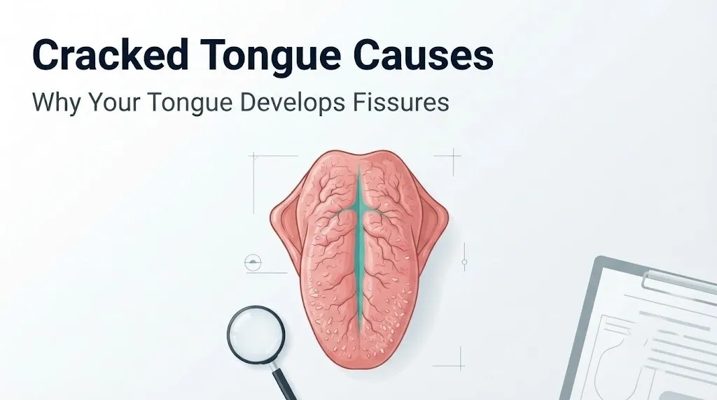
Fissured tongue anatomy centers on the specific architecture of the tongue’s upper surface, where three types of projections called papillae cover the dorsum and give the tongue its textured appearance.
Filiform papillae are the most numerous, covering the anterior two-thirds of the tongue. They are thin, hair-like projections without taste buds, and they contribute to the tongue’s mechanical functions during eating. Fungiform papillae are rounded, mushroom-shaped projections scattered among the filiform papillae that contain taste buds on their upper surface. Circumvallate (vallate) papillae are the large, circular structures arranged in a V-shaped row near the posterior tongue, also containing taste buds.
Fissures in fissured tongue primarily occur in the spaces between papillae on the anterior two-thirds of the dorsum. The central groove running front to back along the midline of the tongue is the most constant feature. Secondary grooves branch laterally from this central fissure, producing a pattern sometimes described as brain-like or lobulated. The depth of fissures ranges from less than 1 millimeter in mild cases to 6 millimeters or more in severe cases.
The depth of a fissure matters clinically because deep fissures trap food particles, bacteria, and fungi in a protected microenvironment that the tongue’s normal self-cleaning movements cannot reach. According to the American Academy of Oral Medicine, food debris accumulation in deep tongue fissures can produce localized halitosis, mild soreness, and in some cases candidal (yeast) colonization in immunocompromised individuals. The fissures themselves are not infected, but they create conditions that facilitate overgrowth of commensal oral organisms if oral hygiene is inadequate.
Quick Tip:
- Deep tongue fissures require deliberate cleaning during tooth brushing: using a tongue scraper or soft toothbrush to clean into the grooves removes trapped debris that causes odor and soreness
- The tongue’s self-cleaning mechanism relies on adequate salivary flow; reduced saliva allows debris accumulation to accelerate
- Individuals taking immunosuppressive medications should report tongue soreness in fissures to their primary care physician promptly, as candidal fissure infection is possible in this population
What Causes Fissured Tongue: Genetic and Congenital Factors
The most common cause of fissured tongue is genetic predisposition, producing a congenital variation in tongue architecture that is present from early childhood and persists throughout life.
Genetic fissured tongue reflects inherited differences in the development of the lamina propria connective tissue during embryogenesis. The collagen and elastin fiber organization in the lamina propria determines the mechanical compliance of the tongue surface. When this connective tissue framework is less dense or differently organized than average, the tongue surface has reduced resistance to the repeated mechanical stress of speaking, chewing, and swallowing, allowing grooves to form and deepen over time.
Fissured tongue shows a clear familial pattern: multiple members of the same family frequently share the condition. A 2014 study published in the Journal of Oral Pathology and Medicine examining oral mucosal findings across family groups found that fissured tongue occurrence was strongly clustered in first-degree relatives, supporting an autosomal dominant pattern of inheritance with variable expressivity. This means some family members may have only a single central groove while others develop an elaborate network of deep lateral fissures, even within the same family.
The condition is present from birth in these individuals but may not be noticed until childhood when the tongue is large enough for fissures to be apparent on inspection. It does not progress to malignancy, does not affect tongue function in the absence of complicating poor hygiene, and does not require treatment beyond adequate oral hygiene. That said, being diagnosed with congenital fissured tongue does not exclude the possibility of a second contributing cause, such as nutritional deficiency, developing later in life.
People with lifelong fissured tongue should still have any worsening of fissure depth or any new oral symptoms evaluated by a primary care physician or dentist, because changes in a previously stable pattern can indicate a superimposed systemic condition.
What Causes Cracks in Tongue: Systemic Inflammatory Conditions
Several named systemic inflammatory conditions cause cracks in the tongue as an oral manifestation of a broader disease process affecting the mucosal immune system or the submucosal tissue architecture.
Inflammatory bowel disease (IBD), particularly Crohn’s disease, produces oral manifestations in approximately 6 to 20 percent of affected individuals according to research published in the Oral Diseases journal. The mechanism in Crohn’s disease involves transmural granulomatous inflammation that is not limited to the gastrointestinal tract. In the mouth and tongue, this same granulomatous process can occur in the submucosal layer, causing tissue induration and distortion of the tongue’s surface geometry into folds and fissures. Oral Crohn’s lesions sometimes precede gastrointestinal symptoms, making their recognition clinically relevant.
Reactive arthritis (formerly called Reiter’s syndrome) can produce transient tongue and oral mucosal lesions including fissuring as part of its triad of joint inflammation, urethritis, and ocular inflammation. These oral changes are driven by the same CD4+ T-cell mediated inflammatory cascade affecting the joints and mucous membranes.
The oral changes in inflammatory conditions are not cosmetic side effects. They are direct tissue manifestations of the same immune dysregulation driving the systemic disease. Tongue fissuring that appears alongside joint pain, gastrointestinal symptoms, chronic diarrhea, or unexplained fatigue should be evaluated by a primary care physician with referral to a gastroenterologist or rheumatologist depending on the broader clinical picture.
Inflammatory systemic causes of tongue fissuring to discuss with your doctor:
- Crohn’s disease (transmural granulomatous inflammation extending to oral mucosa and tongue submucosa)
- Reactive arthritis (T-cell mediated mucosal inflammation as part of systemic inflammatory triad)
- Systemic lupus erythematosus (immune complex deposition causing mucosal changes including tongue lesions)
- Behcet’s disease (vasculitis affecting oral and genital mucosa, producing ulcers that can produce fissure-like lesions)
Key Takeaway: Genetic fissured tongue is the most common cause and is benign, but systemic inflammatory conditions including Crohn’s disease and psoriasis produce tongue fissuring through distinct immune mechanisms that require medical management of the underlying condition, not just oral hygiene.
Cracked Tongue Psoriasis Connection
Psoriasis causes tongue fissuring through the same immune pathway that drives skin plaque formation, producing keratinocyte hyperproliferation in the lingual epithelium that thickens, hardens, and develops surface cracks.
Plaque psoriasis is driven primarily by an aberrant Th17 immune response, in which CD4+ T helper 17 cells release interleukin-17 and are in turn amplified by interleukin-23 from dendritic cells. This cytokine cascade instructs keratinocytes in the affected epithelium to proliferate far faster than normal. In the skin, this produces the thick, scaly plaques characteristic of psoriasis. When the same immune response involves the oral mucosa, including the dorsal lingual epithelium, it produces thickened, rigid areas that crack under the mechanical stress of tongue movement.
According to a review published in the Journal of Oral Pathology and Medicine, oral mucosal manifestations occur in a subset of psoriasis patients, with fissured and geographic tongue patterns being among the most frequently identified. The precise prevalence of oral involvement in psoriasis remains debated in the literature, but studies have consistently found an association between psoriasis and both fissured tongue and geographic tongue (benign migratory glossitis) at rates higher than in the general population.
Pustular psoriasis, a more severe and less common subtype, can produce discrete pustular lesions on the oral mucosa including the tongue, which may rupture and leave erosions or fissures. This presentation is more painful than the dry fissures of plaque psoriasis and may require evaluation by both a dermatologist and an oral medicine specialist.
People with psoriasis who notice new or worsening tongue fissuring should mention this at their next dermatology appointment. A dermatologist managing psoriasis can assess whether the tongue change is consistent with oral mucosal involvement of psoriasis or represents a separate process. Treatment of systemic psoriasis with biologics targeting the IL-17 or IL-23 pathway may improve oral mucosal involvement concurrently with skin disease.
Melkersson-Rosenthal Syndrome and Fissured Tongue
Melkersson-Rosenthal syndrome is a rare neurocutaneous disorder in which fissured tongue is one of the three defining diagnostic features, alongside episodic facial nerve palsy and recurrent orofacial swelling.
The mechanism of tongue fissuring in Melkersson-Rosenthal syndrome differs from genetic or psoriatic fissuring. In this condition, granulomatous inflammation occurs within the submucosa of the lips, facial soft tissues, and tongue. Histopathology of affected tissue shows collections of epithelioid histiocytes forming non-caseating granulomas, sometimes accompanied by Langerhans giant cells, in a pattern similar to the granulomatous inflammation seen in Crohn’s disease and sarcoidosis. This submucosal swelling distorts the normal surface geometry of the tongue, producing the lobulated, fissured appearance characteristic of the syndrome.
The fissured tongue in Melkersson-Rosenthal syndrome is often described as scrotal tongue due to its markedly lobulated surface. The fissuring in this condition tends to be deeper and more architecturally complex than in genetic fissured tongue. It may be accompanied by lip swelling (cheilitis granulomatosa) and facial swelling that is characteristically episodic, appearing over hours and resolving over days, only to recur.
According to the U.S. National Library of Medicine, Melkersson-Rosenthal syndrome is rare and frequently underdiagnosed because its three features do not always occur simultaneously. Many patients present with only one or two features for years before the complete triad is recognized. The full triad is present in only 8 to 25 percent of affected individuals at the time of initial presentation.
Diagnosis typically requires a biopsy of the affected labial or tongue tissue to demonstrate granulomatous inflammation on histopathology. Management involves collaboration between a neurologist (for facial nerve palsy episodes), an oral medicine specialist or oral and maxillofacial surgeon (for orofacial manifestations), and in some cases a rheumatologist or dermatologist familiar with granulomatous conditions.
Cracked Tongue Causes Related to Sjogren’s Syndrome and Dry Mouth
Sjogren’s syndrome causes tongue fissuring through a fundamentally different mechanism than inflammatory or genetic causes: it destroys the salivary glands themselves, eliminating the continuous salivary film that keeps the lingual epithelium hydrated and structurally intact.
Sjogren’s syndrome is an autoimmune exocrinopathy in which CD4+ T lymphocytes infiltrate and destroy the acinar cells of the parotid, sublingual, and submandibular glands, as well as the minor salivary glands distributed throughout the oral mucosa. The result is a progressive reduction in both stimulated and unstimulated salivary flow rates. The target antigens in Sjogren’s syndrome are the Ro/SSA and La/SSB ribonucleoprotein complexes expressed on the surface of salivary acinar cells, and antibodies against these antigens (anti-Ro/SSA and anti-La/SSB antibodies) are present in a majority of primary Sjogren’s patients.
Think of the tongue’s surface like a rubber gasket that must stay moist to remain pliable. When the salivary glands are progressively destroyed, the rubber gasket dries out, loses flexibility, and develops cracks under the repeated mechanical stress of normal use. The fissures in Sjogren’s-related xerostomia develop gradually as salivary output falls, and they deepen as the condition progresses without treatment.
According to the National Institutes of Health, Sjogren’s syndrome affects an estimated 4 million Americans, with women comprising approximately 90 percent of those affected. The condition frequently goes undiagnosed for an average of 4 to 7 years from the onset of symptoms because dry mouth and dry eyes are attributed to aging or environmental factors rather than recognized as autoimmune disease signs.
Sjogren’s syndrome should be considered in any adult with a dry, fissured tongue accompanied by dry eyes, joint pain, fatigue, or parotid gland swelling. A primary care physician can order initial blood tests including anti-Ro/SSA and anti-La/SSB antibodies, and refer to a rheumatologist for definitive evaluation if results are suggestive.
Key Takeaway: Sjogren’s syndrome destroys the salivary glands that keep the tongue surface hydrated, producing progressive fissuring from mucosal desiccation. A dry, fissured tongue in a woman with dry eyes and fatigue warrants a blood test for anti-Ro/SSA antibodies before attributing the finding to aging or dehydration.
Fissured Tongue Causes From Medication Side Effects
Medications cause tongue fissuring by inducing xerostomia (dry mouth) through blockade of muscarinic M3 acetylcholine receptors in the salivary glands, reducing salivary output and allowing the lingual epithelium to desiccate and develop fissures.
Saliva is produced by the parotid, sublingual, and submandibular glands in response to parasympathetic nervous system signals delivered via muscarinic acetylcholine receptors on acinar cells. When medications block these receptors as part of their intended or unintended mechanism, salivary production drops. Over weeks to months of chronic medication use, the resulting persistent oral dryness allows the tongue surface to develop the same desiccation-related fissuring seen in Sjogren’s syndrome, but through a pharmacological rather than autoimmune mechanism.
The drug classes most commonly responsible for medication-induced xerostomia include:
- Tricyclic antidepressants (amitriptyline, nortriptyline, imipramine): strong anticholinergic M3 blockade as a class-wide side effect
- First-generation antihistamines (diphenhydramine, chlorpheniramine): anticholinergic activity causing marked salivary reduction
- Anticholinergic bladder medications (oxybutynin, tolterodine, solifenacin): specifically target M3 receptors, producing profound dry mouth in a substantial proportion of users
- Antipsychotic medications (older typical antipsychotics including chlorpromazine, haloperidol, and some atypical agents): varying anticholinergic burden producing dry mouth
- Benztropine (used for Parkinson’s disease and antipsychotic-induced extrapyramidal effects): potent M3 blockade causing severe xerostomia
- Diuretics (furosemide, hydrochlorothiazide): reduce salivary gland perfusion through systemic fluid reduction rather than receptor blockade
- Oral isotretinoin: reduces mucous gland secretion throughout the body, including minor salivary glands in the oral mucosa
Older adults are the population most vulnerable to medication-induced tongue fissuring. Polypharmacy is common in this group, and the combined anticholinergic burden from multiple medications acting simultaneously can produce severe xerostomia even when no single drug in the regimen is a primary anticholinergic agent. Any adult who has noticed worsening tongue fissuring after starting a new medication, or whose tongue fissuring worsened gradually alongside a medication regimen, should bring a complete medication list (including over-the-counter antihistamines and supplements) to their primary care physician and specifically ask about the anticholinergic burden of their current regimen.
Tongue Fissures and Nutritional Deficiency Causes
Nutritional deficiencies cause tongue fissuring and surface changes by impairing the continuous renewal of the stratified squamous epithelium and the synthesis of collagen in the underlying lamina propria, both of which are metabolically demanding processes requiring specific vitamins and minerals as cofactors.
Riboflavin (vitamin B2) deficiency impairs the flavin-dependent oxidative phosphorylation pathways that power rapidly dividing cells, including the basal keratinocytes that continuously regenerate the tongue’s epithelial surface. B2 deficiency produces a characteristic glossitis (tongue inflammation) with magenta discoloration, swelling, and surface changes including fissuring, alongside angular cheilitis (cracking at the corners of the mouth). The combination of tongue fissuring and corner-of-mouth cracking in the same patient should prompt consideration of riboflavin deficiency.
Niacin (vitamin B3) deficiency causes pellagra, a systemic condition that produces the classic triad of dermatitis, diarrhea, and dementia. Tongue involvement in pellagra includes bright red glossitis and fissuring of the dorsal surface. Niacin deficiency severe enough to produce oral manifestations is now rare in developed countries with food fortification programs, but it occurs in individuals with alcohol use disorder, malabsorption syndromes, and severely restricted diets.
According to the National Institute of Dental and Craniofacial Research, iron deficiency is one of the most common nutritional causes of oral mucosal change, producing atrophic glossitis in which the tongue becomes smooth from papillary atrophy and may develop fissures and a burning sensation. Iron is required for the oxygen delivery needed by rapidly proliferating epithelial cells and for the enzyme ribonucleotide reductase involved in DNA synthesis in basal keratinocytes.
Zinc deficiency impairs collagen synthesis in the lamina propria (zinc is a cofactor in collagenase regulation and in multiple zinc-dependent metalloenzymes required for tissue repair) and reduces the immune barrier function of the lingual epithelium. Signs of zinc deficiency can include tongue surface changes, taste alterations, poor wound healing in the oral mucosa, and increased susceptibility to oral candidiasis in fissures.
| Nutrient | Mechanism of Tongue Fissuring | Associated Signs | Who Is at Risk |
|---|---|---|---|
| Riboflavin (B2) | Impairs keratinocyte energy metabolism via FAD-dependent pathways | Magenta tongue, angular cheilitis | Restricted diets, alcohol use disorder, malabsorption |
| Niacin (B3) | Impairs NAD-dependent cell replication; pellagra glossitis | Red glossitis, skin rash, diarrhea | Alcohol use disorder, malabsorption, severely restricted diets |
| Iron | Impairs oxygen delivery and DNA synthesis in basal keratinocytes | Smooth atrophic tongue, pallor, fatigue | Menstruating women, vegetarians, celiac disease |
| Zinc | Impairs lamina propria collagen synthesis; reduces mucosal barrier | Taste changes, poor mucosal healing | Older adults, inflammatory bowel disease, alcohol use disorder |
| Vitamin B12 | Impairs DNA synthesis in rapidly dividing epithelial cells | Beefy red glossitis, neurological symptoms | Vegans, pernicious anemia, metformin users |
Cracked Tongue Causes Related to Dehydration
Dehydration causes tongue fissuring by reducing total body water to a level where salivary gland output falls measurably, allowing the normally moist dorsal lingual epithelium to lose its surface hydration and develop or worsen existing surface cracks.
The salivary glands are sensitive to systemic hydration status. Unstimulated whole saliva flow rate, measured by clinical sialometry, begins to decrease when total body water deficit reaches approximately 1 to 2 percent. At this level of dehydration, which can occur after even one day of inadequate fluid intake in a hot environment or during illness, the thin salivary film covering the dorsal tongue surface thins. The stratified squamous epithelium of the tongue, which depends on this salivary film for surface moisture in the way a contact lens depends on tear film to remain functional, begins to dry at its surface.
In someone with no pre-existing fissured tongue, mild transient dehydration rarely produces new deep fissures because the healthy lamina propria maintains enough structural support to resist cracking. However, in individuals with any degree of pre-existing genetic fissuring, even mild salivary flow reduction from dehydration can deepen existing fissures and cause discomfort or food trapping that was not previously present.
Mouth breathing, whether from nasal obstruction, sleep apnea, or habitual patterns, produces a local desiccation effect on the tongue independent of systemic hydration status. The continuous airflow across the tongue during mouth breathing evaporates the salivary film far faster than it can be replenished by normal unstimulated salivary output. People who wake with a particularly dry, fissured, and sore tongue each morning should be evaluated for sleep-disordered breathing, including obstructive sleep apnea, by a primary care physician who can order a polysomnography study if indicated.
Quick Tip:
- The goal for salivary gland support is consistent hydration throughout the day, not large boluses of water in the evening
- Sipping water regularly during waking hours maintains salivary flow rates more effectively than catching up on fluids at one time
- Older adults have a blunted thirst perception relative to their actual hydration needs, making deliberate scheduled fluid intake more clinically appropriate than relying on thirst as the only prompt
Key Takeaway: Dehydration worsens pre-existing tongue fissures by reducing salivary output from as little as 1 to 2 percent total body water deficit, and mouth breathing from sleep apnea produces local lingual desiccation that presents as a dry, painful, and more deeply fissured tongue on waking.
Cracked Tongue Causes by Population: Age, Sex, and Health Status
The causes and clinical significance of cracked tongue vary by age group, biological sex, and underlying health status in ways that change both the likely diagnosis and the appropriate clinical response.
Older adults have the highest population prevalence of fissured tongue, with some epidemiological surveys finding rates of 20 to 30 percent in adults over 60 compared to under 5 percent in younger adults. Multiple converging factors explain this: lifetime accumulation of mechanical stress on the tongue surface, polypharmacy-related xerostomia from anticholinergic medication burden, declining salivary gland output with aging (independent of medication use), nutritional deficiencies related to reduced appetite and absorption, and higher prevalence of systemic conditions including Sjogren’s syndrome and inflammatory bowel disease.
Males show slightly higher rates of fissured tongue than females in several epidemiological studies, though the difference is modest and may reflect reporting or examination bias in some studies. The mechanistic reason for any sex difference in idiopathic fissured tongue is not established.
Postmenopausal women face an elevated risk of Sjogren’s syndrome-related and xerostomia-related tongue fissuring. Estrogen affects salivary gland function: estrogen receptors are present on the acinar cells of major salivary glands, and estrogen decline after menopause is associated with reduced salivary output in some women. This hormonal salivary change, superimposed on the immune dysregulation that favors autoimmune conditions in women, makes postmenopausal women the demographic most likely to develop Sjogren’s syndrome and its associated fissured, dry tongue.
Children with Down syndrome (trisomy 21) have fissured tongue rates reported as high as 80 percent by age 10. The mechanism involves congenital differences in tongue musculature (macroglossia relative to oral cavity size) and lamina propria architecture associated with trisomy 21. The protruding tongue position in some children with Down syndrome also exposes the dorsal surface to ambient air desiccation. Dental and oral medicine evaluation is recommended for this population.
| Population Group | Primary Risk Factor | Most Likely Cause | Clinical Action |
|---|---|---|---|
| Adults over 60 | Polypharmacy, aging salivary gland decline | Medication-induced xerostomia | Review anticholinergic medication burden |
| Postmenopausal women | Estrogen decline, autoimmune risk | Sjogren’s syndrome, xerostomia | Test anti-Ro/SSA antibodies if dry eyes present |
| Children with Down syndrome | Trisomy 21 anatomy, macroglossia | Congenital structural fissuring | Oral medicine evaluation, hygiene guidance |
| People with Crohn’s disease | Transmural granulomatous inflammation | Orofacial granulomatosis | Gastroenterology and oral medicine co-management |
| Psoriasis patients | IL-17/IL-23 pathway activation in oral mucosa | Psoriatic lingual involvement | Report to dermatologist at next visit |
| Long-term anticholinergic users | M3 receptor blockade in salivary glands | Medication-induced xerostomia | Discuss medication alternatives or saliva substitutes |
What Causes Cuts on Tongue Versus Chronic Fissured Tongue
The distinction between cuts on the tongue and chronic fissured tongue matters because the two presentations have different causes, different clinical significance, and different appropriate responses.
Acute cuts on the tongue arise from specific mechanical insults: biting the tongue during eating or sleep (which is common during a seizure or with bruxism-related nocturnal jaw clenching), direct trauma from sharp food edges or dental restorations, thermal injury from very hot food or liquids, and ill-fitting dentures or orthodontic appliances causing repetitive friction. These cuts are acute lesions in otherwise healthy lingual mucosa, they have a specific onset moment the person can identify, they produce sharp pain rather than the diffuse aching discomfort of fissured tongue, and they heal within 7 to 14 days in healthy individuals with an intact immune response.
Chronic fissured tongue has no single onset moment the person can identify. It was always there, or it developed gradually over months. The fissures are bilateral and patterned rather than single and random. They do not bleed spontaneously without physical provocation and they do not heal because they are not wounds: they are a persistent structural change in the tongue’s surface architecture.
The clinical distinction becomes practically important when someone presents with what they describe as a “cut” on the tongue that is not healing after two weeks. Any oral mucosal lesion that does not heal within 14 days warrants evaluation by a primary care physician or a dentist, who should refer to an oral medicine specialist or oral and maxillofacial surgeon if the lesion persists or has concerning features. Non-healing oral lesions can represent oral leukoplakia (white patches with premalignant potential), erythroplakia (red patches with high premalignant or malignant risk), or early oral squamous cell carcinoma. This is the one scenario in the cracked tongue clinical picture where the word urgency is appropriate without qualification.
Key things to tell your doctor if you’re concerned about a tongue lesion:
- When you first noticed it and whether it has changed in size, color, or character since then
- Whether it bleeds spontaneously or only when touched
- Whether it is painful, numb, or associated with changes in how food tastes
- Whether you use tobacco in any form or consume alcohol regularly, as these are the primary risk factors for oral squamous cell carcinoma
- Whether you have had any prior biopsies of oral lesions
Fissured Tongue and Geographic Tongue: Overlapping Causes
Geographic tongue (clinical name: benign migratory glossitis, ICD-10 K14.1) and fissured tongue frequently coexist, and they share several contributing causes, which explains why clinicians and patients often confuse or conflate the two conditions.
Geographic tongue is characterized by irregular smooth red patches on the dorsal and lateral tongue surface where filiform papillae have been lost (a process called papillary atrophy), surrounded by slightly raised whitish-yellow borders. The patches change location over days to weeks (hence “migratory”), giving the tongue a map-like appearance. The mechanism involves focal immune dysregulation at the mucosal surface, driven in part by the same Th2 and Th17 immune pathways implicated in atopic dermatitis and psoriasis.
The two conditions co-occur in a subset of patients at rates higher than chance would predict. A 2016 study published in the Oral Surgery Oral Medicine Oral Pathology Oral Radiology journal found that fissured tongue and geographic tongue co-occurred in approximately 30 percent of patients who had either condition, compared to rates that would be expected by independent chance occurrence alone. Both conditions share associations with psoriasis, reactive arthritis, and genetic predisposition, suggesting overlapping immune and genetic mechanisms.
The practical clinical implication of their co-occurrence is that geographic tongue patches can settle into tongue fissures, producing a particularly pronounced visual appearance and sometimes an increased risk of discomfort from food sensitivity (spicy or acidic foods often trigger burning in geographic tongue). Individuals with both conditions often describe a burning sensation with certain foods that varies in intensity with the geographic tongue’s migratory pattern. A dentist or primary care physician can visually distinguish the two conditions on examination, but laboratory testing is rarely needed for the combined benign presentation.
Individuals with psoriasis are a specific population who should know this connection: psoriasis has been associated with geographic tongue at rates substantially higher than the general population, and a dermatologist managing psoriasis should be informed of any tongue surface changes alongside skin disease.
When Fissured Tongue Causes Require Medical Evaluation
Fissured tongue causes require medical evaluation when the fissuring is new or worsening in an adult, when it is accompanied by systemic symptoms, when the fissures are painful, bleeding, or infected, or when associated oral lesions are not healing within two weeks.
The clinical triage framework below distinguishes situations appropriate for monitoring at home from those requiring a healthcare provider appointment and those requiring same-day or urgent evaluation.
| Clinical Situation | Appropriate Response | Provider Type |
|---|---|---|
| Lifelong fissured tongue, no new symptoms, adequate oral hygiene | Monitor at home; mention at next dental visit | Dentist or primary care physician |
| New tongue fissuring in adult over 40 with no prior tongue changes | Schedule primary care physician appointment within 2 to 4 weeks | Primary care physician |
| Fissured tongue with persistent dry mouth and dry eyes | Schedule primary care physician appointment within 2 weeks; request anti-Ro/SSA antibody test | Primary care physician, rheumatologist referral |
| Fissured tongue with facial swelling, lip swelling, or facial nerve weakness | Schedule evaluation within 1 week with primary care physician | Primary care physician, oral medicine specialist, neurology referral |
| Fissured tongue with Crohn’s disease history and worsening oral pain | Schedule gastroenterology and oral medicine co-evaluation | Gastroenterologist, oral medicine specialist |
| Non-healing tongue sore or lesion present for more than 14 days | Schedule dental or primary care evaluation within 1 week | Dentist, oral medicine specialist, or oral and maxillofacial surgeon |
| Tongue swelling, difficulty swallowing or breathing, rapid-onset facial swelling | Emergency evaluation immediately | Emergency medicine physician: Call 911 or go to the ER |
The last scenario in this table warrants clear emphasis. Although fissured tongue itself is not a medical emergency, any tongue or oral swelling that develops rapidly alongside difficulty breathing or swallowing may indicate angioedema (deep tissue swelling from histamine or bradykinin-mediated vascular permeability) or a severe allergic reaction. Call 911 or go to the nearest emergency room immediately if tongue swelling develops rapidly, particularly if accompanied by throat tightness, difficulty swallowing, or any breathing change.
When to Seek Emergency Evaluation for Tongue-Related Symptoms
Tongue fissuring alone is not a medical emergency. Certain symptoms occurring alongside or in the context of tongue changes require immediate emergency evaluation.
Call 911 or go to the nearest emergency room immediately if you experience:
- Rapid swelling of the tongue or throat that develops over minutes to hours, with or without associated tongue fissures: this pattern indicates angioedema or a severe allergic reaction requiring emergency airway management
- Difficulty swallowing or breathing accompanying any tongue swelling: indicates potential airway compromise, a life-threatening emergency
- A new, non-healing sore or growth on the tongue with sudden onset of severe pain, uncontrolled bleeding, or rapid change in size: while oral cancer is not an emergency in its early stages, rapidly bleeding or rapidly enlarging oral lesions require same-day emergency evaluation
- High fever with a severely swollen, painful tongue that makes it impossible to swallow: may indicate Ludwig’s angina (a severe deep space infection of the floor of the mouth) or another deep oral infection requiring emergency surgical evaluation
These presentations require emergency medical assessment, not a dental appointment scheduled for next week or a call to a nurse advice line.
Fissured Tongue Causes and Treatment Approaches
Fissured tongue treatment is determined entirely by the underlying cause, and managing the cause is the only approach that produces meaningful improvement in fissure depth and associated symptoms.
For genetic or idiopathic fissured tongue with no associated systemic condition, no specific treatment is needed. The standard clinical guidance from the American Academy of Oral Medicine is adequate oral hygiene: thorough tongue cleaning during tooth brushing, using a soft-bristled brush to clean into fissures and remove food debris, and ensuring adequate salivary flow through hydration. Fissures in this group are permanent architectural features that do not close with any treatment, but symptoms from food trapping and odor are entirely preventable with consistent oral hygiene.
For medication-induced xerostomia fissuring, the primary approach is a review of the medication regimen with the prescribing physician. Where medically appropriate, switching from a high-anticholinergic agent to a lower-anticholinergic alternative in the same therapeutic class can reduce dry mouth substantially. Where medication changes are not feasible, over-the-counter saliva substitutes (products containing carboxymethylcellulose or hydroxyethylcellulose as the salivary film substitute) can help maintain mucosal hydration. These products are described here for clinical context, not as a treatment recommendation for any individual: a prescribing physician should guide these decisions.
For nutritional deficiency fissuring, correction of the specific identified deficiency under medical guidance is the appropriate approach. A complete blood count (CBC), serum ferritin, serum zinc, and serum vitamin B12 level measured by a primary care physician or internist can identify the most common nutritional contributors.
For systemic disease-associated fissuring (Sjogren’s syndrome, psoriasis, Crohn’s disease, Melkersson-Rosenthal syndrome), treatment of the underlying systemic disease is the primary approach. Oral manifestations often improve when systemic disease activity is controlled, though fissuring may not completely resolve if structural changes have become established over time.
Key steps to prepare for a medical appointment about tongue fissuring:
- Note when you first noticed the fissuring and whether it was always present or is a new change
- List all medications you take, including over-the-counter antihistamines, supplements, and herbal products
- Note any other symptoms that developed around the same time: dry eyes, joint pain, fatigue, diarrhea, skin rash
- Describe your usual daily fluid intake and whether you breathe through your mouth at night
- Note whether the fissuring causes any discomfort, bleeding, bad breath, or taste changes
- Bring any prior dental records or notes from previous examinations of your tongue
Key Takeaway: Fissured tongue from genetic causes requires only consistent oral hygiene, while fissuring from medications, nutritional deficiency, Sjogren’s syndrome, psoriasis, or Crohn’s disease requires identification and management of the specific cause, not just surface-level treatment of the tongue itself.
Frequently Asked Questions About Cracked Tongue Causes
Is a cracked tongue a sign of something serious?
A cracked tongue is most often a benign genetic variation requiring no treatment beyond good oral hygiene.
When fissuring is new or worsening in an adult, or accompanied by dry eyes, joint pain, facial swelling, or gastrointestinal symptoms, it can indicate a systemic condition such as Sjogren’s syndrome, Crohn’s disease, or Melkersson-Rosenthal syndrome that warrants evaluation by a primary care physician.
Any non-healing sore or growth on the tongue that persists beyond 14 days requires evaluation by a dentist or oral medicine specialist, regardless of whether fissured tongue is also present.
What vitamin deficiency causes a cracked tongue?
Riboflavin (vitamin B2), niacin (vitamin B3), iron, zinc, and vitamin B12 deficiencies are each associated with tongue surface changes including fissuring and glossitis.
Riboflavin deficiency characteristically produces magenta tongue discoloration alongside fissuring, while iron deficiency produces smooth atrophic tongue changes alongside fissuring and burning.
A primary care physician can identify the specific deficiency through a complete blood count, serum ferritin, serum zinc, and serum vitamin B12 level rather than treating all possibilities simultaneously.
Can dehydration cause a fissured tongue?
Dehydration reduces salivary flow rates and dries the lingual epithelium, which can worsen pre-existing fissures and cause soreness in people who already have genetic fissured tongue.
Dehydration alone is unlikely to produce deep new fissures in a previously smooth tongue in a healthy adult, but it is a common exacerbating factor when other contributing causes are already present.
Mouth breathing during sleep, which desiccates the tongue surface regardless of systemic hydration, is a specific cause of worsening tongue fissuring and morning soreness that should prompt evaluation for sleep apnea by a primary care physician.
What is the difference between a cracked tongue and geographic tongue?
A cracked tongue (fissured tongue) has persistent deep grooves or furrows on the dorsal surface that do not change location, while geographic tongue features smooth red patches with slightly raised borders that migrate across the tongue surface over days to weeks.
The two conditions frequently co-occur and share genetic and immune predispositions, including associations with psoriasis and reactive arthritis.
A dentist or primary care physician can distinguish the two conditions by visual examination, and laboratory testing is not typically needed unless systemic disease is suspected.
Can medications cause your tongue to crack?
Yes, medications that block muscarinic M3 acetylcholine receptors in the salivary glands, a category that includes tricyclic antidepressants, first-generation antihistamines, and overactive bladder medications such as oxybutynin, reduce salivary output and allow the tongue surface to desiccate and develop fissures over weeks to months.
Diuretics produce a similar effect by reducing total body fluid volume, which reduces salivary gland perfusion.
Any adult whose tongue fissuring began or worsened after starting a new medication should bring their complete medication list to their primary care physician and ask specifically about the anticholinergic burden of their regimen.
Should I see a doctor if my tongue has deep cracks and hurts?
A painful fissured tongue warrants evaluation by a primary care physician or dentist, particularly if the pain is new, worsening, or accompanied by bleeding, white patches, or a sore that is not healing.
Pain in tongue fissures can result from food trapping and bacterial or fungal overgrowth, which improves with thorough hygiene, or from an underlying systemic condition such as Sjogren’s syndrome or nutritional deficiency, which requires clinical investigation.
If tongue pain is accompanied by facial swelling, difficulty swallowing, or breathing difficulty, go to the nearest emergency room immediately because these symptoms can indicate a severe allergic reaction or deep oral infection.
Closing
The most actionable insight from the clinical picture of cracked tongue causes is this: the appearance of fissures on the tongue’s surface is a common and usually benign finding in people with a lifelong history of it, but new-onset fissuring in an adult is a different clinical situation that deserves a clear explanation. Medications, salivary gland disease, nutritional deficiencies, and systemic inflammatory conditions each produce fissuring through distinct mechanisms, and each has a specific investigation and management pathway.
Your next step is straightforward. If your tongue has always looked this way, focus on oral hygiene: a soft toothbrush used to clean into the grooves daily removes the food debris and bacterial load that causes odor and soreness. If your tongue fissuring is new, worsening, or accompanied by any systemic symptom, book an appointment with your primary care physician and bring a complete medication list and a description of any other symptoms present.
A primary care physician can order the blood tests that will identify the most common systemic and nutritional contributors within one visit. That single appointment distinguishes the benign from the clinically relevant, and it gives you a specific answer rather than a general category.