Middle Back Pain Causes in Females: 16 Reasons (2026)
Middle back pain causes in females span a wider range of physiological origins than in males, including not only thoracic spine and muscle problems but also reproductive organ conditions, hormonal fluctuations, and kidney pathology that produce pain through mechanisms entirely absent in male anatomy. Understanding which category your pain falls into is the first step toward the right diagnosis and the right type of care.
The National Institute of Arthritis and Musculoskeletal and Skin Diseases (NIAMS) reports that women are disproportionately affected by several conditions that produce mid-back pain, including osteoporosis, endometriosis, and fibromyalgia, each of which involves a distinct hormonal or anatomical pathway that general back pain articles rarely address in adequate depth. Research published in the Spine journal has consistently found that sex-based differences in pain processing, hormonal modulation of nociception, and reproductive anatomy create genuinely different back pain profiles between men and women.
This article covers all 16 evidence-based causes of middle back pain in females, the specific physiological mechanism behind each one, which causes require urgent or emergency evaluation, and exactly what type of provider to see based on your symptom pattern.
Middle Back Pain Causes Female: Why This Pain Needs a Female-Specific Explanation
Middle back pain causes in females cannot be fully explained by general back pain guidelines because female anatomy includes reproductive organs, hormonal cycles, and bone health patterns that produce thoracic and mid-back pain through mechanisms that do not exist in male physiology.
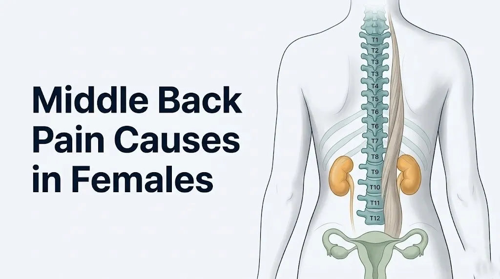
The middle back refers to the thoracic spine, which spans the T1 through T12 vertebral levels, running from the base of the neck to the bottom of the rib cage. In the general population, thoracic spine pain is less common than lumbar pain but still affects a substantial proportion of adults. A 2022 systematic review published in the Spine journal found that thoracic pain prevalence ranges from 13 to 35 percent across adult populations, with higher rates reported in women, particularly those of reproductive age and those in the perimenopausal and postmenopausal years.
The anatomical reason female middle back pain requires its own clinical framework is straightforward. The uterus, ovaries, and fallopian tubes share sympathetic afferent nerve pathways with the T10 through L1 thoracolumbar spinal segments, meaning pain originating in these organs is frequently perceived in the mid-to-lower back rather than exclusively in the pelvis. No male organ shares this referral pathway. The kidneys sit in a retroperitoneal position at approximately T12 to L2, and because women have a shorter urethra than men (roughly 4 cm versus 20 cm), ascending urinary tract infections reaching the kidney are far more common in women, making pyelonephritis a female-predominant cause of costovertebral angle pain.
| Cause Category | Female-Specific? | Primary Spinal Levels Affected |
|---|---|---|
| Musculoskeletal (muscle, disc, facet) | No (but posture and bra weight add sex-specific loading) | T1-T12 broadly |
| Reproductive organ referred pain | Yes | T10-L1 |
| Hormonal bone loss (osteoporosis) | Predominantly female | T6-T12 most affected |
| Kidney infection/stones | Female predominant | T12-L2 |
| Endometriosis | Female only | T10-L1 |
| Menstrual cycle pain referral | Female only | T10-L1 |
| Pregnancy-related | Female only | T8-L1 |
Why Females Experience Middle Back Pain Differently Than Males
Females experience middle back pain differently than males because of three converging physiological realities: sex-based differences in spinal biomechanics and muscle architecture, hormonal modulation of pain perception and connective tissue laxity, and the presence of pelvic reproductive organs that refer pain to the thoracolumbar junction.
From a biomechanical standpoint, women on average have a wider pelvis relative to their shoulder width, a narrower thoracic spinal canal diameter, and a higher proportion of thoracic lordotic curvature variation. These structural differences alter how load is distributed across the costovertebral joints (the joints connecting each rib to its corresponding thoracic vertebra) and the zygapophyseal (facet) joints at each thoracic level. Women who carry significant breast weight without adequate support also place asymmetric anterior loading on the thoracic spine, which increases compensatory paraspinal muscle tension in the upper and mid thoracic region.
Hormonally, estrogen influences pain sensitivity at the level of the dorsal horn of the spinal cord. Research published in the journal Pain has found that estrogen modulates the activity of mu-opioid receptors and the density of substance P receptors in the dorsal horn, meaning that pain processing in the central nervous system fluctuates with the menstrual cycle. Women consistently show lower pain thresholds in experimental pain studies compared to men, and this difference is measurably altered by hormonal contraceptive use and menopausal status.
The third distinction is anatomical and has no equivalent in male physiology. The visceral pain referral network connecting the uterus, ovaries, and fallopian tubes to the mid and lower back via the T10-L1 sympathetic chain creates an entire category of middle back pain causes that a woman’s body produces and a man’s does not.
Quick Tip:
- Tracking when your mid-back pain occurs relative to your menstrual cycle gives a primary care physician or obstetrician-gynecologist critical diagnostic information. Note the cycle day on which pain begins, peaks, and resolves.
- Women taking hormonal contraceptives may have suppressed prostaglandin-driven menstrual pain but still experience mid-back pain from structural causes. Do not assume hormonal contraceptive use rules out all gynecological sources.
- Postmenopausal women should inform any provider evaluating their back pain of their last menstrual period date and whether they use hormone therapy, as both affect bone density interpretation.
Muscle Strain and Thoracic Spine Overload in Women
Muscle strain of the thoracic paraspinal muscles, particularly the erector spinae group (comprising the iliocostalis thoracis, longissimus thoracis, and spinalis thoracis) and the multifidus muscle, is the most common cause of middle back pain in women of all ages, produced when these muscles sustain a load greater than their current capacity and develop localized micro-injury, ischemia, and nociceptor sensitization.
The mechanism works like this: each paraspinal muscle fiber maintains the upright thoracic spine against gravity and resists rotational and flexion forces during movement. When overloaded by a single event (lifting with a flexed thoracic spine, sudden rotational movement) or by cumulative daily demand (prolonged desk sitting with forward thoracic flexion), the myofibers develop small tears. The local release of bradykinin, prostaglandin E2, and potassium ions from the injured tissue activates the group III and IV muscle afferent nociceptors (also called free nerve endings) in the muscle belly and fascia. These nociceptors send pain signals up the thoracic dorsal root ganglia to the spinal cord, where the pain is perceived as a deep, aching, or burning sensation localized to the mid-back.
The thoracolumbar fascia, a broad connective tissue sheet that envelops the paraspinal muscles from the thoracic to the lumbar spine, is a common source of mid-back pain in women who sit for extended periods with poor thoracic posture. When this fascia is chronically under-stretched and under-loaded, its collagen fibers lose the organized cross-linking pattern that provides tensile strength, and it becomes a source of diffuse aching pain across the mid-back that does not localize sharply to one vertebral level.
According to the NIAMS, muscle-related back pain in women between the ages of 25 and 55 is the most frequently reported musculoskeletal complaint in primary care settings in this demographic. Women in physically demanding jobs (nursing, childcare, manual labor) and those with sedentary office-based work are both high-risk groups, albeit for different biomechanical loading patterns.
- Pain that begins within 24 to 48 hours of a specific physical activity or awkward movement: consistent with acute muscle strain
- Pain that is worse with specific movements (twisting, bending, lifting) but not present at rest: suggests musculoskeletal origin
- Pain that resolves within 2 to 4 weeks with relative rest and activity modification: consistent with self-limiting muscle strain
- Pain that persists beyond 4 weeks, worsens with rest, or is accompanied by systemic symptoms: warrants evaluation by a primary care physician
Key Takeaway: Middle back pain in females is not just a spinal or muscle problem. The uterus, ovaries, kidneys, and hormonal cycle all produce pain perceived at the mid-back through distinct nerve pathways, and identifying which category your pain belongs to determines which provider and which diagnostic path is appropriate.
Thoracic Facet Joint and Disc Problems Causing Mid Back Pain
Thoracic disc herniation and facet joint (zygapophyseal joint) degeneration are structural causes of mid-back pain that occur when the nucleus pulposus of a thoracic intervertebral disc herniates through the annulus fibrosus and compresses a nearby thoracic nerve root, or when the cartilage surfaces of the thoracic facet joints wear and produce local joint inflammation.
Thoracic disc herniation is less common than lumbar disc herniation, accounting for roughly 0.25 to 0.75 percent of all disc herniation cases per data from the Journal of Bone and Joint Surgery. However, when it does occur, it is clinically serious. The thoracic spinal canal is narrower relative to the spinal cord diameter than the lumbar canal. A thoracic disc herniation at T6-T7 or T11-T12, for example, can compress the thoracic spinal cord directly, producing not just local back pain but also a distinct pattern of neurological changes: band-like tightness or numbness wrapping around the rib cage at the level of the herniation, progressive lower extremity weakness, or changes in bladder and bowel function.
Thoracic facet joint degeneration produces a more localized, deep aching pain that is typically worsened by extension and rotation of the thoracic spine and relieved by forward flexion. The facet joint capsule contains a rich network of nociceptors (innervated by the medial branch of the thoracic dorsal rami), and when the articular cartilage degenerates and the joint capsule becomes inflamed, these nociceptors fire persistently, generating pain perceived as deep aching in the mid-back that is often worse in the morning and after prolonged sitting.
Women with a prior history of cervical or lumbar disc herniation, women who perform repetitive thoracic flexion-extension tasks occupationally, and women with pre-existing thoracic kyphosis (forward rounding of the thoracic spine) face a higher mechanical load on thoracic disc structures. A board-certified orthopedic spine surgeon or board-certified physiatrist should evaluate any mid-back pain accompanied by neurological symptoms (numbness, tingling, or weakness in the legs) to rule out spinal cord involvement.
Endometriosis and Middle Back Pain
Endometriosis causes middle back pain in females when ectopic endometrial tissue implants in retroperitoneal locations posterior to the uterus or on structures adjacent to the thoracolumbar spinal nerve roots, releasing prostaglandin E2 and triggering a localized inflammatory cascade that directly irritates nearby somatic nerves at the T10 through L1 spinal segments.
The American College of Obstetricians and Gynecologists (ACOG) states that endometriosis affects approximately 10 percent of reproductive-age women, and back pain is reported as a symptom in a substantial proportion of those with moderate to severe disease. The reason mid-back pain occurs in endometriosis is not simply referred pelvic pain. When endometriosis implants grow in the retroperitoneal space, specifically behind the uterus near the uterosacral ligaments and along the posterior parietal peritoneum, they are anatomically close to the sympathetic nerve roots that share afferent pathways with the T10-L1 dermatomal region. Prostaglandin E2 secreted by these implants sensitizes nearby nociceptors, generating pain perceived as deep mid-to-lower back aching or cramping.
What makes endometriosis-related mid-back pain particularly difficult to identify from symptom pattern alone is that it fluctuates with the menstrual cycle. Pain is typically worst in the late luteal phase (the days before menstruation begins) and during menstruation itself, as prostaglandin E2 production peaks in response to progesterone withdrawal. After menstruation ends, the implants temporarily reduce inflammatory output and the pain often diminishes, leading many women to attribute the back pain to menstruation rather than a chronic structural disease.
A 2021 study published in Obstetrics and Gynecology found that women with endometriosis and back pain had a mean delay of 7 to 10 years from symptom onset to diagnosis. Women who notice mid-back pain that follows a consistent menstrual cycle pattern, is accompanied by painful periods (dysmenorrhea), painful intercourse (dyspareunia), or difficulty conceiving, should discuss this pattern with a board-certified obstetrician-gynecologist and ask specifically about endometriosis as a possible diagnosis. A pelvic ultrasound and MRI pelvis can identify some forms of endometriosis, though laparoscopic surgery remains the gold-standard diagnostic procedure.
Ovarian Cysts and Uterine Fibroids Causing Mid Back Pain
Ovarian cysts and uterine fibroids (leiomyomata uteri) cause middle back pain in females by exerting mass effect on retroperitoneal structures adjacent to the T10-L1 nerve roots or by producing a sustained increase in intrauterine or pelvic pressure that is referred to the mid-back through shared sympathetic afferent pathways.
An ovarian cyst is a fluid-filled sac developing within or on the surface of an ovary. Functional cysts are common in premenopausal women (formed during normal ovulation cycles), while endometriomas (chocolate cysts) are specific to women with endometriosis. When an ovarian cyst grows large enough (typically above 5 cm in diameter) or when it is positioned posteriorly near the broad ligament or uterosacral structures, it can compress adjacent retroperitoneal nerves or pelvic vasculature, producing a persistent deep ache in the lower-to-middle back region that is not clearly related to movement or posture.
Uterine fibroids are benign smooth muscle tumors of the myometrium (the muscular wall of the uterus). They are more common in women of African descent, affecting an estimated 70 to 80 percent of women by age 50 per Mayo Clinic data, though most cause no symptoms. Large subserosal fibroids (those growing on the outer uterine surface) can press posteriorly against the sacrum, lumbosacral plexus, and retroperitoneal structures, generating a chronic, dull, sometimes pressure-like mid-to-lower back pain that is often worse during menstruation when uterine contractile activity increases.
The distinction between fibroid-related back pain and musculoskeletal back pain is clinically important. Fibroid-related pain does not change with spinal position changes (does not worsen with forward flexion or improve with lying flat) and is frequently accompanied by heavy menstrual bleeding (menorrhagia), pelvic pressure, or urinary frequency from bladder compression. Any woman with heavy periods and mid-back pain that does not clearly relate to physical activity should have a pelvic ultrasound ordered by a primary care physician or obstetrician-gynecologist.
Quick Tip:
- Ovarian cyst pain that becomes suddenly severe, is accompanied by nausea and vomiting, and causes a woman to feel faint may indicate ovarian torsion, a surgical emergency. Go to the emergency room immediately in this scenario.
- Women with fibroids who are perimenopausal should note that fibroids often shrink after menopause as estrogen levels decline; however, back pain from fibroid-related structural changes may persist if nerve irritation was chronic.
- Post-menopausal women who develop a new ovarian cyst require urgent evaluation by a gynecologist, as functional cysts do not form after menopause.
Key Takeaway: Endometriosis, ovarian cysts, and uterine fibroids all produce mid-back pain through retroperitoneal nerve pressure or visceral referral via the T10-L1 sympathetic chain, meaning the back pain itself may be the most prominent symptom even when the root cause is entirely gynecological.
Menstrual Cycle and Hormonal Middle Back Pain
Menstrual cycle-related middle back pain occurs when prostaglandin E2 released by the shedding endometrium during menstruation acts on uterine smooth muscle to produce rhythmic contractions, and these uterine contractions generate pain signals that travel through the T10-L1 sympathetic afferent pathways and are perceived as deep cramping or aching in the mid-to-lower back.
The physiological sequence is specific and well-documented. During the late luteal phase of the menstrual cycle, progesterone levels drop as the corpus luteum regresses. This progesterone withdrawal signals the endometrium to begin shedding. As the endometrial lining breaks down, arachidonic acid is released from membrane phospholipids and converted by cyclo-oxygenase-2 (COX-2) enzymes to prostaglandin E2 and prostaglandin F2-alpha. These prostaglandins bind to receptors on uterine smooth muscle cells, triggering strong myometrial contractions. Those contractions, combined with the prostaglandins’ direct sensitizing effect on uterine nociceptors, generate pain signals that ascend through the T10-L1 sympathetic chain and are interpreted by the brain as cramping or aching in both the lower abdomen and the mid-to-lower back.
Women with primary dysmenorrhea (painful periods with no underlying structural cause) typically experience mid-back pain that begins 12 to 24 hours before menstruation and resolves within 2 to 3 days of flow starting, corresponding to the prostaglandin production curve. Women with secondary dysmenorrhea (painful periods caused by an underlying condition such as endometriosis, fibroids, or adenomyosis) often experience back pain that starts earlier in the cycle, lasts longer, and is more severe, because the prostaglandin load from ectopic tissue adds to that from the endometrium.
According to the ACOG, approximately 50 to 90 percent of women of reproductive age experience dysmenorrhea, making prostaglandin-mediated menstrual back pain one of the most prevalent causes of recurrent mid-back pain in reproductive-age females. Women who find that over-the-counter nonsteroidal anti-inflammatory drugs (NSAIDs, which inhibit COX-2 and reduce prostaglandin synthesis) do not adequately control menstrual back pain, or whose pain has worsened over time, should be evaluated by a board-certified obstetrician-gynecologist to screen for secondary causes such as endometriosis or adenomyosis.
Middle Back Pain During Pregnancy
Middle back pain during pregnancy is caused by the combined effect of relaxin-mediated ligamentous laxity in the spinal and costovertebral joints, a progressively shifting center of gravity that increases thoracolumbar paraspinal muscle demand, and expanding uterine size that compresses retroperitoneal structures and alters the mechanical relationship between the pelvis and thoracic spine.
Relaxin, a hormone produced first by the corpus luteum and later by the placenta throughout pregnancy, acts on the relaxin receptor family (RXFP1 and RXFP2) expressed in fibrocytes and smooth muscle cells of ligaments throughout the body. Its primary function is to remodel the pubic symphysis and sacroiliac ligaments to allow pelvic expansion during delivery, but it acts on all spinal ligaments simultaneously, including the posterior longitudinal ligament, interspinous ligaments, and costovertebral joint capsules of the thoracic spine. The result is a thoracic spine that is less mechanically stiff than usual, shifting more of the spinal stabilization demand to the paraspinal muscles, which fatigue faster and develop nociceptor sensitization earlier with daily activity.
As the uterus expands through the second and third trimesters, the abdominal wall musculature stretches and the center of gravity shifts anteriorly and inferiorly. The thoracic paraspinal muscles compensate by increasing their continuous background contraction to maintain upright posture, generating cumulative muscle overload and the characteristic mid-to-upper back aching that peaks in the third trimester.
Research published in the European Spine Journal found that approximately 50 to 80 percent of pregnant women experience clinically notable back pain, with thoracic and mid-back pain most commonly reported in the second and third trimesters. Pregnant women with mid-back pain that is accompanied by urinary burning, fever, or blood in the urine require same-day evaluation by their obstetrician or midwife to rule out pyelonephritis, which carries a risk of preterm labor if untreated. Thoracic back pain in a pregnant woman that is tearing in quality and radiates to the chest requires immediate emergency evaluation.
Osteoporosis and Vertebral Compression Fractures in Women
Osteoporosis causes middle back pain in women primarily through vertebral compression fractures at the T6 through T12 thoracic vertebral levels, produced when declining estrogen removes its inhibitory effect on osteoclast activity via the RANKL-RANK-OPG signaling pathway, accelerating trabecular bone resorption in the vertebral bodies until the cortical shell can no longer support normal compressive load and the vertebral body collapses.
The RANKL-RANK-OPG pathway is the central regulator of osteoclast (bone-resorbing cell) activity. Estrogen normally suppresses osteoclast differentiation and activity by upregulating osteoprotegerin (OPG), a decoy receptor that prevents RANKL from binding to its activating receptor RANK on osteoclast precursor cells. When estrogen levels decline at perimenopause and menopause, this OPG-mediated suppression is removed, osteoclast activity accelerates, and trabecular bone in the vertebral bodies is resorbed faster than osteoblasts can replace it. The thoracic vertebral bodies, which have a higher proportion of trabecular bone than the lumbar vertebrae, are preferentially affected.
According to JAMA Internal Medicine, approximately 25 percent of postmenopausal women will sustain a vertebral compression fracture during their lifetime, and at least two-thirds of these fractures are clinically silent initially, meaning the woman may not realize a fracture has occurred until mid-back pain develops. The pain from a vertebral compression fracture is characteristically acute in onset (often described as a sudden sharp pain in the mid-back after a minimal trauma such as bending forward, lifting a light object, or even coughing), worsens with sitting or standing, and is relieved somewhat by lying flat.
Postmenopausal women, women who have used oral corticosteroids long-term (which suppress osteoblast activity independently of estrogen), women with a low body mass index, and women with a family history of hip or vertebral fracture are at highest risk. The DEXA scan (dual-energy X-ray absorptiometry) is the gold-standard screening tool for bone mineral density, and the USPSTF (United States Preventive Services Task Force) recommends DEXA screening for all women aged 65 and older, and for younger postmenopausal women with risk factors. A primary care physician or board-certified endocrinologist can order and interpret a DEXA scan and discuss treatment options if osteoporosis or osteopenia is identified.
Key Takeaway: Vertebral compression fractures from osteoporosis occur silently in about two-thirds of cases, meaning persistent mid-back pain that began after a minor movement or lifting incident in a postmenopausal woman should be evaluated with thoracic spine imaging before attributing it to simple muscle strain.
Kidney Infection and Kidney Stones Causing Middle Back Pain in Females
Acute pyelonephritis (kidney infection) and nephrolithiasis (kidney stones) both cause middle-to-lower back pain in females through distinct mechanisms, and women face a disproportionately higher risk of urinary tract infections ascending to the kidney than men because the female urethra is approximately 4 cm in length compared to approximately 20 cm in males, providing a significantly shorter anatomical barrier to ascending bacterial colonization.
The kidneys are retroperitoneal organs positioned bilaterally at approximately the T12 to L2 vertebral levels, nestled within the renal fascia (Gerota’s fascia) against the posterior abdominal wall. Because of this posterior anatomical position, inflammation or obstruction of the kidney produces pain perceived at the costovertebral angle: the triangular area between the lateral border of the erector spinae muscles and the inferior edge of the 12th rib. Clinically, this is the angle at the junction of the mid and lower back. Most women describe kidney pain as a deep, persistent, often constant ache on one side of their mid-to-lower back that does not change significantly with body position.
The Centers for Disease Control and Prevention (CDC) reports that urinary tract infections affect approximately 8 million women annually in the United States, and pyelonephritis develops when an ascending bladder infection reaches the renal pelvis and renal parenchyma. The bacterial pathogen most commonly involved is Escherichia coli, responsible for approximately 80 to 85 percent of cases. When the renal parenchyma becomes inflamed, the swelling within the fibrous renal capsule activates stretch-sensitive nociceptors, generating the characteristic dull-to-severe unilateral costovertebral angle tenderness that a clinician elicits by gently tapping the costovertebral angle with a closed fist.
Kidney stones produce a different quality of pain. The muscular contractions of the renal pelvis and ureter attempting to move a calculus downstream generate intermittent, severe, colicky pain (called renal colic) that typically radiates from the costovertebral angle toward the groin. The pain of renal colic is characteristically one-sided, comes in waves, and is often accompanied by nausea and vomiting. Women with diabetes, recurrent urinary tract infections, or a personal or family history of kidney stones are at elevated risk. Both pyelonephritis and severe renal colic require evaluation by a primary care physician or emergency medicine physician: pyelonephritis typically requires antibiotic therapy, and symptomatic kidney stones may require urological intervention.
Gallbladder and Pancreatic Causes of Mid Back Pain in Women
The gallbladder and pancreas both produce referred pain in the middle back through visceral afferent pathways that carry nociceptive signals to the thoracic spinal cord at levels overlapping with mid-back dermatomal distributions, and women are at higher risk for gallstone disease than men due to estrogen’s effect on bile composition and cholesterol saturation.
The gallbladder sits beneath the right lobe of the liver and refers pain to the right shoulder and the right mid-back at approximately the T6 through T9 dermatomal levels via the right phrenic nerve (C3-C5) and right thoracic splanchnic nerves. When a gallstone obstructs the cystic duct, pressure builds within the gallbladder wall and activates stretch-sensitive visceral nociceptors. The resulting pain, called biliary colic, is typically felt as a severe, cramping pain under the right rib cage that radiates to the right shoulder blade area and sometimes to the right mid-back. The pain is classically worst after fatty meals and lasts 30 minutes to several hours before resolving.
Estrogen increases cholesterol secretion into bile while reducing bile acid secretion, increasing the lithogenicity (stone-forming tendency) of bile. This is why gallstone disease is roughly twice as common in premenopausal women as in men of the same age, and why pregnancy and oral contraceptive use are both recognized risk factors for gallstone formation, as both states involve elevated estrogen levels.
Pancreatitis (inflammation of the pancreas) produces a more diffuse mid-to-upper back pain that is characteristically described as a band-like, boring pain across the mid-back, often rated as the most severe pain the patient has experienced. According to Mayo Clinic, pancreatitis pain is classically relieved slightly by leaning forward or curling into a fetal position and is worsened by lying flat. Acute pancreatitis is a medical emergency requiring hospitalization and should not be managed with watchful waiting. Any woman with severe mid-back or upper abdominal pain accompanied by nausea, vomiting, fever, and pain that is not relieved by any position should be evaluated in an emergency room.
Anxiety, Stress, and Muscle Tension as Mid Back Pain Causes
Anxiety and chronic psychological stress cause middle back pain in women through cortisol-mediated and sympathetic nervous system-driven increases in resting paraspinal muscle tone, which sustain low-level isometric contraction in the erector spinae and multifidus muscles of the thoracic spine, producing cumulative muscle ischemia and nociceptor sensitization without any mechanical injury.
The mechanism begins in the hypothalamic-pituitary-adrenal (HPA) axis. Psychological stressors activate the hypothalamus to release corticotropin-releasing hormone (CRH), which stimulates the pituitary to release ACTH, which drives the adrenal cortex to secrete cortisol. Concurrently, the sympathetic nervous system releases epinephrine and norepinephrine, which increase muscle fiber excitability. In states of chronic anxiety, this sympathetic activation remains persistently elevated, maintaining a background elevation in paraspinal muscle tension throughout the day. The muscle never fully relaxes between demands. Over days to weeks, this sustained low-level contraction produces the same ischemia and prostaglandin release cascade as acute mechanical overload, generating diffuse mid-back aching without a clear precipitating injury.
Women are diagnosed with anxiety disorders at approximately twice the rate of men, according to the Anxiety and Depression Association of America, a pattern that reflects both biological sex differences in HPA axis reactivity and social stressor patterns. Women with generalized anxiety disorder frequently report mid-back and upper back tension as a prominent physical symptom, sometimes preceding the recognition of the psychological component of their condition.
An important clinical distinction: anxiety-related mid-back pain typically fluctuates with stress levels (worse during high-stress periods, improved during vacation or low-demand periods), responds partially to heat and stretching applied to the thoracic paraspinal region, and is not position-dependent in the same way that structural spinal pain is. However, anxiety and structural back pain commonly coexist, and a primary care physician evaluating mid-back pain should screen for both. Women using corticosteroid medications long-term for inflammatory conditions should inform their provider, as chronic corticosteroid use independently increases osteoporosis risk, creating a potential overlap of anxiety-related muscle tension and osteoporosis-related bone vulnerability.
Key Takeaway: Anxiety and chronic stress cause real, measurable middle back pain through cortisol and sympathetic nervous system-driven paraspinal muscle tension, and this mechanism is more prevalent in women than men due to higher rates of anxiety disorder diagnosis and sex-based HPA axis reactivity differences.
Scoliosis and Postural Causes of Middle Back Pain in Women
Scoliosis, a lateral curvature of the spine with rotation of the vertebral bodies, causes middle back pain in women through asymmetric loading of the thoracic facet joints and costovertebral joints, unequal tension in the paraspinal muscles on the concave versus convex sides of the curve, and progressive disc degeneration at the levels of greatest curvature.
Adolescent idiopathic scoliosis is the most common form of scoliosis and occurs at a 4:1 female-to-male ratio for curves requiring treatment, per data cited by the American Academy of Orthopaedic Surgeons (AAOS). The reason for this sex-predominant pattern is not fully established, but research published in the Spine journal points to differences in thoracic growth plate sensitivity to mechanical loading during the adolescent growth spurt, potentially modulated by estrogen receptor signaling in vertebral growth plate chondrocytes. A Cobb angle (the standard radiographic measurement of scoliosis curve magnitude) above 10 degrees defines scoliosis, while curves above 25 to 30 degrees typically warrant orthopedic evaluation and above 40 to 50 degrees may be considered for surgical intervention.
Adults with previously untreated mild adolescent scoliosis often develop progressive mid-back pain in their 30s to 50s as the asymmetric joint loading from even a small curve accumulates over decades and accelerates facet joint degeneration on one side of the curve. Women who were told as adolescents that their scoliosis was “mild enough not to need treatment” should mention this history to any provider evaluating their adult mid-back pain, as it changes the differential diagnosis and the value of upright thoracic spine X-rays in the workup.
Postural middle back pain from habitual thoracic kyphosis (forward rounding) is distinct from scoliosis but similarly produces asymmetric loading of the posterior thoracic structures. Women who work at computers for extended hours frequently develop this pattern, characterized by anterior head translation, rounded shoulders, and increased thoracic kyphotic angle, which loads the posterior thoracic facet joints in sustained extension and compresses the posterior annulus of the thoracic discs. A board-certified physiatrist or physical therapist can evaluate thoracic postural alignment and design a specific corrective exercise and movement program.
Red Flags for Middle Back Pain in Women
Red flags for middle back pain in women are specific symptom features that indicate the pain may be caused by a serious or life-threatening condition rather than a benign musculoskeletal problem, and their presence requires prompt medical evaluation rather than watchful waiting or self-management.
The following red flags require evaluation by a primary care physician within 24 to 48 hours at minimum, or emergency evaluation as specified:
- Fever above 101°F (38.3°C) with mid-back pain: raises strong concern for pyelonephritis (kidney infection), epidural abscess, or vertebral osteomyelitis. Fever with chills, rigors, nausea, and costovertebral angle tenderness in a woman constitutes a probable acute pyelonephritis presentation and warrants emergency evaluation the same day.
- Unintentional weight loss of more than 10 pounds over weeks to months alongside new mid-back pain: raises concern for malignancy with vertebral metastasis, most commonly from breast, lung, or thyroid primary tumors in women.
- Neurological changes in the legs, including progressive weakness, numbness, or tingling below the waist: suggests thoracic spinal cord compression from disc herniation, tumor, or abscess. This is a time-sensitive emergency.
- Loss of bladder or bowel control in combination with back pain: indicates possible thoracic cord compression with myelopathy. Call 911 or go to the emergency room immediately.
- Mid-back pain that is constant, present at rest, and worse at night, not relieved by any position: inconsistent with muscle strain (which improves with rest). Suggests an inflammatory process, infection, or malignancy.
- Mid-back pain after a minor trauma in a postmenopausal woman: raises concern for vertebral compression fracture. Requires thoracic spine imaging (X-ray at minimum, MRI if neurological symptoms are present).
- History of cancer with new onset mid-back pain: warrants urgent evaluation for vertebral metastasis. Do not attribute new back pain to muscle strain in a woman with a cancer history without imaging.
| Red Flag Feature | Concern | Action Required |
|---|---|---|
| Fever and costovertebral angle tenderness | Pyelonephritis, spinal infection | Emergency evaluation same day |
| Neurological deficit below mid-back | Spinal cord compression | Call 911 immediately |
| Night pain, rest pain, weight loss | Malignancy | Primary care within 24 hours; imaging urgently |
| Bladder or bowel loss of control | Thoracic myelopathy | Call 911 immediately |
| New pain in postmenopausal woman after minor movement | Vertebral compression fracture | Primary care within 48 hours; imaging required |
Emergency Symptoms: When to Call 911 or Go to the ER
Certain middle back pain presentations in women require immediate emergency evaluation. Do not wait to see if these resolve on their own.
Call 911 or go to the nearest emergency room immediately if you experience:
- Sudden severe tearing or ripping pain in the mid-to-upper back, possibly radiating to the chest or between the shoulder blades: this presentation is consistent with Type B aortic dissection (tearing of the descending thoracic aorta), which is a life-threatening vascular emergency. Women with connective tissue disorders such as Marfan syndrome or Ehlers-Danlos syndrome and women with uncontrolled hypertension are at elevated risk.
- Middle or lower back pain that becomes suddenly and severely worse, is accompanied by nausea and vomiting, and causes dizziness or near-fainting in a woman of reproductive age: this is consistent with ovarian torsion, a surgical emergency requiring immediate intervention to prevent permanent ovarian damage.
- Middle back pain with progressive weakness in both legs, numbness spreading down one or both legs, or inability to walk properly: this indicates possible thoracic spinal cord compression, a neurological emergency where delay in treatment risks permanent paralysis.
- Middle back pain accompanied by loss of bladder control (inability to urinate or uncontrolled leakage) or loss of bowel control: this constitutes a spinal cord emergency. Do not attempt to schedule an outpatient appointment.
- Middle back pain with high fever (above 101.5°F), severe chills, rigors, and vomiting in a pregnant woman: this is consistent with acute pyelonephritis during pregnancy, which can trigger preterm labor and requires immediate hospital evaluation and intravenous antibiotic therapy.
- Severe mid-back pain of sudden onset in a woman with a known history of cancer: this raises concern for pathological vertebral fracture from metastatic disease and requires emergency imaging.
These presentations require emergency medical assessment in a hospital emergency department, not an urgent care clinic and not a phone call to a primary care office.
How Middle Back Pain in Women Is Diagnosed
Middle back pain in women is diagnosed through a structured clinical evaluation combining a targeted medical history (including menstrual cycle pattern, gynecological history, reproductive hormone status, and urinary symptoms), a physical examination of the thoracic spine, paraspinal musculature, and costovertebral angles, followed by targeted laboratory and imaging testing chosen based on the most probable cause category identified in the history and exam.
To describe your symptoms most effectively to a primary care physician or obstetrician-gynecologist:
- Note the exact location of the pain (is it bilateral or one-sided? does it radiate to the front, the ribs, or the groin?), as location significantly narrows the cause category.
- Note the onset pattern (sudden onset with a specific triggering movement suggests musculoskeletal; gradual onset without a clear trigger suggests visceral or systemic).
- Note the relationship to your menstrual cycle (does pain begin before your period, during it, or is it unrelated to cycle timing?).
- Note associated symptoms: urinary burning or frequency suggests kidney; nausea and right upper quadrant pain after meals suggests gallbladder; fever and chills suggests infection.
- Note what makes the pain better or worse (position-dependent pain suggests musculoskeletal; pain unchanged by position suggests visceral or inflammatory).
- Bring a list of all current medications (including hormonal contraceptives and corticosteroids), any prior imaging results for spine or pelvis, and your date of last menstrual period to every appointment.
| Suspected Cause | First-Line Test | Provider Who Orders It |
|---|---|---|
| Muscle strain or facet joint | Clinical examination; MRI thoracic spine if neurological signs | Primary care physician; physiatrist |
| Kidney infection | Urinalysis; urine culture and sensitivity | Primary care physician; ER |
| Kidney stones | CT scan of abdomen and pelvis without contrast | ER; urologist |
| Endometriosis or ovarian cyst | Pelvic ultrasound (transvaginal); MRI pelvis | OB-GYN |
| Uterine fibroids | Pelvic ultrasound (transabdominal and transvaginal) | OB-GYN; primary care |
| Osteoporosis and vertebral fracture | DEXA scan; thoracic spine X-ray; MRI if neurological signs | Primary care; endocrinologist |
| Gallbladder disease | Right upper quadrant abdominal ultrasound; liver function tests | Primary care; gastroenterologist |
| Pancreatitis | Serum lipase; CT abdomen with contrast | ER |
| Aortic dissection | CT angiography of chest and abdomen | ER (immediate) |
| Fibromyalgia or anxiety-related | Clinical diagnosis via ACR 2016 criteria; PHQ-9; GAD-7 | Primary care; rheumatologist |
Key Takeaway: The single most diagnostic piece of information you can give a provider about female mid-back pain is its relationship to your menstrual cycle. Cycle-linked pain shifts the differential immediately toward gynecological causes; cycle-independent pain that worsens with movement points toward musculoskeletal causes; cycle-independent pain with urinary symptoms points toward kidney.
Frequently Asked Questions About Middle Back Pain Causes in Females
What causes middle back pain in females specifically?
Middle back pain causes in females include all the musculoskeletal causes that affect men (thoracic muscle strain, disc herniation, facet joint degeneration) plus female-specific causes including endometriosis, ovarian cysts, uterine fibroids, menstrual prostaglandin referral, pregnancy-related ligamentous laxity, and postmenopausal osteoporosis-related vertebral compression fractures.
The T10-L1 sympathetic afferent nerve pathways shared between the uterus, ovaries, and the mid-back dermatomal region mean that gynecological conditions frequently produce back pain as a primary or even sole symptom.
A primary care physician or board-certified obstetrician-gynecologist can help identify which cause category applies based on your symptom pattern, cycle timing, and associated findings.
Can endometriosis cause middle back pain?
Endometriosis causes middle back pain when ectopic endometrial tissue implants in retroperitoneal locations near the uterosacral ligaments or posterior parietal peritoneum, releasing prostaglandin E2 that sensitizes adjacent nociceptors connected to the T10-L1 spinal nerve roots.
This pain typically fluctuates with the menstrual cycle, worsening in the days before menstruation and during the period itself, and is often accompanied by severe menstrual cramps, painful intercourse, or difficulty conceiving.
According to the American College of Obstetricians and Gynecologists, women with endometriosis experience an average diagnostic delay of 7 to 10 years from symptom onset, making it worth specifically asking your OB-GYN about this diagnosis if your back pain follows a consistent menstrual cycle pattern.
What does kidney pain feel like in the middle back in women?
Kidney pain in women typically presents as a deep, persistent, constant aching or pressure on one side of the mid-to-lower back at the costovertebral angle, which is the junction between the last rib and the outer edge of the paraspinal muscles.
Unlike muscle strain pain, kidney pain from infection does not change significantly with body position, and it is frequently accompanied by fever, chills, nausea, urinary burning, or blood-tinged urine.
Kidney stone pain differs by being intermittent and severely colicky, often radiating from the mid-back toward the lower abdomen and groin, which reflects the ureter’s muscular contractions trying to move the stone downward.
Can ovarian cysts cause mid back pain?
Ovarian cysts can cause mid-back pain when they grow large enough to exert pressure on retroperitoneal structures or compress nerves near the broad ligament that share afferent pathways with the T10-L1 spinal dermatomes.
The pain tends to be a persistent deep ache on one side (corresponding to the affected ovary) that is not clearly related to body position or physical activity.
Sudden severe worsening of ovarian cyst pain accompanied by nausea, vomiting, and dizziness may indicate ovarian torsion, which requires immediate emergency room evaluation.
Why does my middle back hurt more during my period?
Middle back pain during your period is caused by prostaglandin E2 released by the shedding endometrium, which triggers uterine smooth muscle contractions that send pain signals through the T10-L1 sympathetic afferent pathways to the mid-back.
This is the same physiological mechanism responsible for menstrual cramping in the lower abdomen, but the shared nerve pathways mean the pain is simultaneously perceived in the low-to-mid back.
If your menstrual back pain has become progressively more severe over time, starts earlier in your cycle than before, or is accompanied by pain during intercourse or between periods, evaluation by a board-certified obstetrician-gynecologist for endometriosis or adenomyosis is warranted.
When should a woman go to the ER for middle back pain?
A woman should go to the emergency room immediately for middle back pain that is sudden and severe with a tearing quality (possible aortic dissection), accompanied by fever above 101°F with chills and nausea (possible kidney infection requiring IV antibiotics), or accompanied by leg weakness, numbness, or loss of bladder or bowel control (possible spinal cord compression).
Severe acute mid-back pain with nausea and vomiting in a woman of reproductive age could indicate ovarian torsion, which is a surgical emergency with a narrow treatment window for ovarian preservation.
These presentations require emergency department evaluation: urgent care facilities and phone consultations cannot provide the imaging and interventional capabilities that these diagnoses require.
Closing
The range of conditions that cause middle back pain in females is wider and more anatomically specific than most general back pain resources acknowledge. Muscle and spine causes are real and common. But so are endometriosis, ovarian cysts, prostaglandin-driven menstrual pain, kidney infections, osteoporosis fractures, and gallbladder disease, each of which produces pain through a completely different physiological mechanism and requires a different diagnostic path.
The most actionable step you can take right now is to track your back pain in relation to your menstrual cycle for one full month before your next appointment. Note the cycle day when pain begins, how it changes through the month, and what associated symptoms appear alongside it. This single piece of information shifts the entire diagnostic conversation.
If your mid-back pain has lasted more than 3 to 4 weeks, is accompanied by any red flag feature listed in this article, or is affecting your ability to work or sleep, schedule an evaluation with your primary care physician and ask specifically about both musculoskeletal and gynecological causes. You deserve a diagnosis that matches the actual anatomy, not a generic recommendation to rest and stretch