BVD Symptoms: Signs of Binocular Vision Dysfunction 2026
BVD symptoms include persistent headaches, dizziness, eye strain, double vision, reading difficulty, and anxiety that many people carry for years without a correct diagnosis. Binocular vision dysfunction occurs when the two eyes fail to align precisely enough for the brain to produce a single, stable image without sustained muscular and neurological effort.
According to the American Optometric Association, binocular vision problems affect an estimated 56% of the general population to some degree, with a subset experiencing symptoms severe enough to impair daily function, reading, and driving. Many of these individuals have been told their eyes are “normal” by general eye exams that do not measure binocular alignment under the right conditions.
This article covers every clinically recognized symptom of BVD, the specific anatomical and neurological mechanisms behind each one, how symptoms differ across age groups and health contexts, what the driving experience reveals about the condition, and exactly what kind of eye care evaluation identifies it.
Binocular Vision Disorder Symptoms: The Full Clinical Picture
Binocular vision disorder symptoms span the visual, neurological, vestibular, and musculoskeletal systems, making BVD one of the most commonly misidentified conditions in outpatient medicine.
The sheer range of symptoms surprises most people. Headaches, dizziness, nausea, anxiety, difficulty reading, light sensitivity, and neck pain all trace back to a single underlying mechanism: the brain forcing misaligned eyes to work together through sustained neurological override. That override has a cost, and the cost shows up across multiple body systems.
The core symptom categories in binocular vision dysfunction include:
- Headaches, typically frontal or temporal, worsening with sustained visual tasks
- Dizziness and spatial disorientation, particularly in visually busy environments
- Eye strain and visual fatigue after short periods of reading or screen use
- Double vision (diplopia), either intermittent or sustained
- Difficulty reading, including losing place on the page and rereading lines
- Nausea and motion sickness in vehicles or moving visual environments
- Anxiety, panic-like sensations, and difficulty in crowded spaces
- Light sensitivity (photophobia), particularly to fluorescent or overhead lighting
- Neck and shoulder pain without orthopedic cause
- Difficulty judging distance, particularly while driving
The College of Optometrists in Vision Development (COVD) recognizes that these symptoms are produced by the prolonged, high-demand effort of the oculomotor system attempting to maintain binocular fusion against a persistent misalignment. When that effort fails, even temporarily, the visual, vestibular, and autonomic systems all respond.
Older adults may report a gradual worsening of several of these symptoms over years as the fusional vergence reserves naturally diminish with age. Children may present with none of the headache or dizziness symptoms and instead show only behavioral and academic changes, discussed in detail in a later section.
What Is BVD Eye Condition and How Does It Affect Vision?
BVD, or binocular vision dysfunction, is a condition in which the two eyes fail to maintain precise alignment during sustained visual tasks, forcing the oculomotor system into continuous, energy-costly compensatory work.
Think of binocular vision like a perfectly matched stereo system. Both speakers have to produce the same frequency at the same time for the sound to appear centered and clear. If one speaker is even slightly out of phase, the brain hears something uncomfortable and tries to compensate. BVD is that misalignment in the visual system: the two cameras are not perfectly synchronized, and the brain’s visual cortex is working overtime to correct the mismatch every second you are looking at anything.
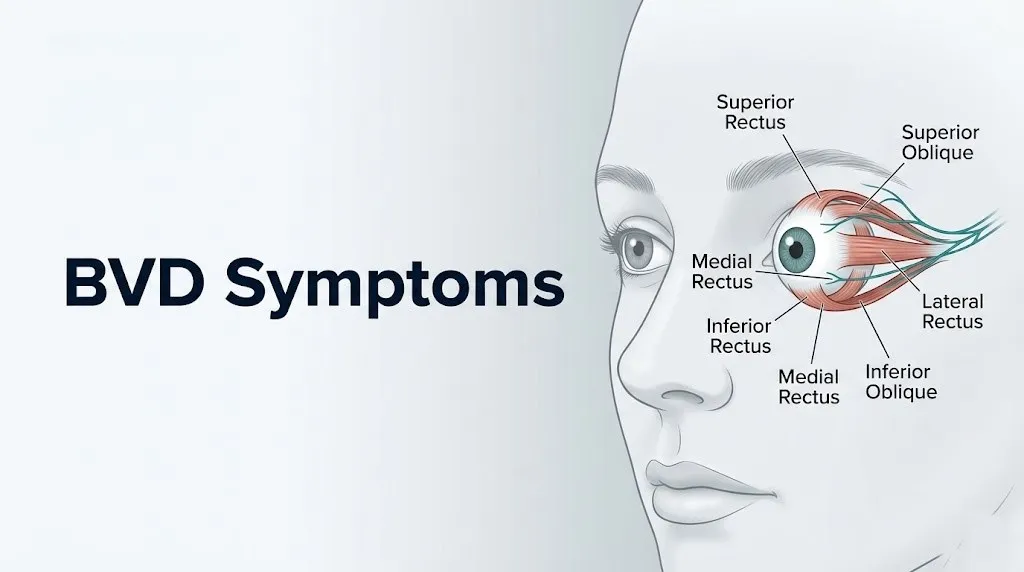
The anatomy is precise. Six extraocular muscles control each eye’s movement: the superior rectus, inferior rectus, medial rectus, lateral rectus, superior oblique, and inferior oblique. These muscles receive instruction from three cranial nerves: the oculomotor nerve (cranial nerve III), the trochlear nerve (cranial nerve IV), and the abducens nerve (cranial nerve VI). For binocular vision to work perfectly, all twelve extraocular muscles across both eyes must coordinate with millimeter precision.
In BVD, there is a consistent, slight offset in this coordination, called a heterophoria: a latent tendency for one or both eyes to drift from the correct alignment position. Most people have some degree of heterophoria. The problem arises when the fusional vergence reserve, the neurological capacity to correct the offset, is insufficient for the demands of the visual task.
According to the National Eye Institute, the Convergence Insufficiency Treatment Trial (CITT), a randomized controlled study involving 221 children, confirmed that measurable vergence dysfunction produces real, quantifiable symptoms that respond to targeted treatment. This established the physiological reality of vergence dysfunction symptoms in controlled human research.
Individuals with pre-existing neurological conditions, including multiple sclerosis and Parkinson’s disease, are at higher risk of developing secondary BVD because the demyelination and dopaminergic pathway dysfunction in these conditions directly impair the oculomotor nuclei responsible for vergence control.
BVD Symptoms: The Complete List Explained
BVD symptoms are the direct result of the oculomotor system operating under sustained neurological stress to maintain a binocular image the visual system is not built to produce without effort.
The full symptom inventory is wider than most people expect. Patients often present to primary care physicians, neurologists, and even psychiatrists before BVD is considered, because the symptom spread mimics anxiety disorder, vestibular disorder, chronic migraine, and cervicogenic headache simultaneously.
| Symptom | Physiological Mechanism | Clinical Significance |
|---|---|---|
| Headache (frontal/temporal) | Sustained oculomotor muscle tension; trigeminal nerve (CN V) pain signaling | Present in approximately 80% of BVD patients; typically worsens with screen or book time |
| Dizziness/spatial disorientation | Vestibulo-ocular reflex receives conflicting visual input from misaligned eyes | Often described as feeling “off balance” without true vertigo |
| Eye strain (asthenopia) | Continuous vergence effort exceeds fusional vergence reserve | Worsens after 20 to 30 minutes of near work |
| Intermittent double vision (diplopia) | Fusional vergence overcomes alignment offset inadequately; images separate | Typically horizontal or vertical splitting; intermittent in early stages |
| Reading difficulty | Binocular misalignment disrupts saccadic tracking along text lines | Lines appear to move; reader loses place repeatedly |
| Nausea | Vestibular-visual mismatch triggers autonomic nausea response via the vomiting center (area postrema) | Particularly in moving vehicles, supermarkets, and busy public spaces |
| Anxiety / panic sensations | Persistent, unresolved sensory conflict activates the autonomic nervous system’s sympathetic branch | Often misdiagnosed as generalized anxiety disorder |
| Light sensitivity (photophobia) | Pupillary light reflex is dysregulated under sustained oculomotor stress; ciliary muscle spasm | Fluorescent and overhead lighting particularly provocative |
| Neck and shoulder pain | Compensatory head tilt; trigeminal-cervical complex sensitization | Bilateral or unilateral; often attributed incorrectly to muscle tension |
| Poor depth perception | Disruption of stereopsis, the binocular disparity processing in visual cortex V2 and V3 areas | Affects driving, ball sports, stair navigation |
Women of reproductive age may report that BVD symptoms fluctuate with hormonal cycles. Estrogen influences vestibular sensitivity through estrogen receptor expression in the labyrinth of the inner ear, which can amplify vestibular-visual mismatch symptoms during the luteal phase of the menstrual cycle.
BVD Eye Symptoms: What Your Eyes Are Actually Experiencing
BVD eye symptoms are the direct sensory experience of the two eyes failing to maintain precise alignment: strain, fatigue, intermittent splitting of images, and an inability to sustain comfortable vision during close or prolonged visual work.
Most people with BVD describe an eye experience they struggle to put into words. The eyes don’t hurt exactly. They feel tired after far too short a period of reading or screen time. Focusing feels effortful in a way it never used to. Some people describe a sense of visual “pulling” or the feeling that one eye is working harder than the other.
The primary BVD eye symptoms include:
- Blurred vision that clears temporarily with blinking or rubbing
- Eyes that feel sore or aching after 20 to 30 minutes of near work
- The sense that words or images are slightly “unstable” or vibrating
- Intermittent double vision, especially late in the day or after sustained reading
- Difficulty shifting gaze between near and far distances smoothly
- Excessive blinking or eye rubbing as an unconscious compensation mechanism
- Eyes that water or itch without allergic cause, from sustained accommodative and vergence effort
- One eye drifting or turning inward or outward in photographs, often noticed by others before the individual is aware
The ciliary muscle, which controls the crystalline lens’s shape during accommodation (focusing), operates in close neurological coordination with the vergence system through the vergence-accommodation reflex. In BVD, the vergence demand placed on the extraocular muscles conflicts with the accommodative signal sent to the ciliary muscle. This produces accommodative spasm, a sustained and uncomfortable contraction of the ciliary muscle that blurs vision at near distance and contributes to the sensation of visual fatigue.
Children under age 10 rarely describe these symptoms in clinical language. They more commonly rub their eyes frequently, close one eye when reading, hold books very close, or complain that reading “hurts their brain.” These behavioral expressions represent the same underlying oculomotor stress.
Symptoms of BVD Eyes: How Misalignment Affects Visual Comfort
The symptoms of BVD eyes are directly proportional to the size of the misalignment and the individual’s remaining fusional vergence reserve: the smaller the reserve relative to the demand, the more severe and frequent the symptoms.
Visual comfort depends entirely on the brain receiving two nearly identical images from both eyes and fusing them seamlessly in the visual cortex (primary visual area V1 in the occipital lobe) and the binocular disparity processing areas (V2 and V3). When the two images arrive at slightly different angles due to heterophoria, the visual cortex must process the discrepancy and the vergence system must actively correct it, every second, for as long as the person is using their eyes for any visual task.
According to a 2022 study published in Optometry and Vision Science, the discomfort rating associated with symptomatic convergence insufficiency was statistically comparable to the discomfort ratings reported in chronic dry eye disease, challenging the clinical assumption that vergence dysfunction is a minor complaint.
How visual comfort breaks down in BVD:
- The fusional vergence reserve handles small alignment offsets easily during short visual tasks.
- Sustained near work (reading, screens) depletes the vergence reserve progressively.
- As the reserve drops, the extraocular muscles work harder, producing increasing strain.
- When the reserve is exhausted temporarily, the images separate: intermittent diplopia occurs.
- The brain may suppress the image from one eye to avoid diplopia, a process called suppression, which reduces 3D perception and depth accuracy.
- After the visual task ends, recovery is slow: symptoms often persist 30 to 60 minutes after the provocative activity stops.
Older adults over age 60 experience this progression more quickly because presbyopia reduces the accommodative range of the crystalline lens, placing greater demand on vergence control to maintain a stable near image. This can cause BVD symptoms to emerge or worsen significantly in the sixth decade of life even without any change in the underlying alignment offset.
BVD Headaches Symptoms: Why Your Head Hurts
BVD headaches occur because sustained extraocular muscle tension activates the trigeminal nerve pathway, converting oculomotor fatigue into referred pain felt in the forehead, temples, and behind the eyes.
The trigeminal nerve (CN V) is the largest cranial nerve and carries sensory information from the face, orbit, scalp, and meninges. The ophthalmic branch of the trigeminal nerve (V1) innervates the orbital structures including the periorbital tissues and the superior orbital fissure where the oculomotor nerves travel. When extraocular muscles sustain prolonged contraction during the vergence override process, nociceptive signals travel through the trigeminal pathway to the trigeminal nucleus caudalis in the brainstem, producing referred pain in the forehead and temporal regions.
Characteristics of BVD-associated headache:
- Location: frontal (forehead), temporal, or retro-orbital (behind the eyes)
- Timing: typically begins 20 to 40 minutes into a sustained near visual task and worsens progressively
- Trigger: reading, screen use, fluorescent lighting, visually complex environments
- Relief pattern: often improves within 30 to 60 minutes of closing the eyes or resting in a dark room
- Character: pressure, aching, or a “band” around the forehead, rather than the pulsing quality of migraine
- Frequency: daily or near-daily in moderate to severe BVD; less frequent in mild presentations
This headache pattern is clinically distinct from migraine in that it is consistently provoked by visual effort and reliably relieved by visual rest. Migraine headache, by contrast, is typically accompanied by photophobia, phonophobia, and nausea as independent features rather than as direct consequences of the visual task duration.
The American Optometric Association acknowledges that BVD-associated headache is one of the most frequent reasons patients initially present to an optometrist with no prior diagnosis.
Individuals on medications that impair accommodation, including tricyclic antidepressants and first-generation antihistamines, may experience amplified BVD headache symptoms because anticholinergic effects on the ciliary muscle increase the vergence-accommodation conflict.
Key Takeaway: BVD headaches occur through the trigeminal nerve pathway and are reliably triggered by sustained visual tasks, which distinguishes them clinically from migraine and tension-type headache.
BVD and Dizziness: The Vestibular Connection
Dizziness in BVD occurs because the vestibulo-ocular reflex receives conflicting spatial information from misaligned eyes, causing the brain’s balance centers to generate a persistent, unresolved mismatch signal experienced as dizziness or spatial disorientation.
The vestibulo-ocular reflex (VOR) is a high-speed stabilization reflex that keeps your visual field stable during head movement. It works by detecting head rotation in the inner ear’s semicircular canals and generating a compensatory eye movement in the opposite direction, at equal speed, so your visual world appears stable even as your head moves. This reflex operates through a pathway connecting the vestibular apparatus to the oculomotor nuclei in the brainstem.
In BVD, the eyes are not in the position the VOR expects them to be. The VOR sends a corrective signal based on the assumption of normal binocular alignment. The actual image received by the visual cortex does not match the predicted stable image. The cerebellum, which cross-references vestibular input with visual input to maintain spatial accuracy, detects the discrepancy and generates a persistent error signal.
That error signal is the subjective experience of dizziness in BVD: not the spinning sensation of benign paroxysmal positional vertigo (BPPV), but a more chronic sense of being “off,” slightly unsteady, or as if the world is slightly delayed in responding to head movement.
According to the National Institute of Neurological Disorders and Stroke (NINDS), disruptions in visual-vestibular integration are a recognized mechanism in chronic dizziness disorders, and oculomotor dysfunction is identified as a contributing factor in a subset of patients with persistent postural perceptual dizziness (PPPD).
Individuals with a prior history of inner ear conditions, including Meniere’s disease or labyrinthitis, face a compounded risk: pre-existing vestibular vulnerability combined with the visual-vestibular mismatch of BVD can produce dizziness that is disproportionately severe relative to the degree of binocular misalignment.
BVD and Anxiety Symptoms: Why BVD Can Mimic Panic Disorder
BVD can produce anxiety, panic-like sensations, and avoidance behaviors because the persistent unresolved sensory conflict between the visual and vestibular systems activates the sympathetic branch of the autonomic nervous system in the same way a genuine threat would.
The autonomic nervous system does not distinguish between an external physical threat and an internal unresolved sensory conflict. Persistent vestibular-visual mismatch signals, sustained without resolution, are processed by the amygdala and hypothalamus as an environment that cannot be spatially oriented. The result is a low-level but continuous activation of the sympathetic branch: elevated heart rate, shallow breathing, increased sweating, a sense of unreality or derealization, and the cognitive experience of anxiety.
BVD-associated anxiety typically presents with these distinguishing features:
- Anxiety that is worse in visually overwhelming environments (supermarkets, shopping malls, busy streets) and better in quiet, low-stimulus settings
- A sense of being “not quite present” or detached in busy public spaces (depersonalization or derealization)
- Avoidance of crowds, large open spaces, or moving environments without a clear social explanation
- Anxiety that does not respond well to standard cognitive behavioral therapy because the sensory trigger is not a thought pattern but a physiological mismatch
- Panic-like sensations, including nausea, heart racing, and the urge to leave a space, triggered reliably by fluorescent lighting or complex visual environments
A 2019 study published in the Journal of Behavioral Optometry found that a subset of patients diagnosed with generalized anxiety disorder had undiagnosed vergence dysfunction, and that targeted treatment of the vergence dysfunction reduced self-reported anxiety symptoms without any psychiatric intervention.
Women are disproportionately diagnosed with anxiety disorder before BVD is identified. This likely reflects the compounding effect of estrogen-mediated vestibular sensitivity on the vestibulo-visual mismatch, producing anxiety presentations that are clinically indistinguishable from generalized anxiety disorder without a binocular vision evaluation.
BVD Symptoms Driving: Why Roads Become Overwhelming
BVD symptoms while driving include difficulty maintaining lane position, visual fatigue at speed, trouble judging distance from other vehicles, and an overwhelming anxiety response to highway driving, all produced by the failure of the binocular visual system under the high spatial-processing demands of driving.
Driving is one of the most demanding visual tasks the human brain performs. It requires simultaneous operation of the dorsal visual stream, the “where” pathway running from the visual cortex (occipital lobe) through the parietal lobe, which handles spatial navigation, depth perception, speed estimation, and object location in three-dimensional space. It also requires rapid, precise vergence shifts between near targets (dashboard, mirrors) and far targets (road, oncoming cars), repeated hundreds of times per hour.
In BVD, the dorsal visual stream receives slightly inconsistent binocular input at every moment. At low speeds, the brain can compensate. At highway speeds, the visual information arrives and demands processing faster than the compensatory vergence system can manage. The result is a progressive, measurable increase in visual fatigue, anxiety, and spatial disorientation with driving duration.
BVD-specific driving symptoms include:
- Difficulty judging the distance of oncoming vehicles, particularly at night
- Struggling to maintain lane position on wide highways without lane markers as reference
- Visual fatigue that builds progressively: the first 20 minutes feel manageable, but longer drives become overwhelming
- Sensitivity to oncoming headlights at night, producing a halo or spreading glare effect
- Difficulty reading road signs, particularly when driving at speed
- An anxious, urgent desire to stop driving or get off a highway without a clear external reason
- Difficulty in complex visual environments: roundabouts, highway merges, tunnel exits
The Mayo Clinic notes that any condition affecting depth perception or binocular alignment poses a safety concern for driving and warrants disclosure to the treating clinician and, in severe cases, assessment by a vision specialist before operating a vehicle.
Older adults with BVD face heightened driving risk because age-related reductions in fusional vergence reserve coincide with decreased contrast sensitivity and slower visual processing speed, compounding the spatial disorientation during complex driving scenarios.
Key Takeaway: BVD driving symptoms are produced by the dorsal visual stream receiving inconsistent binocular input under high spatial-processing demand, and they worsen reliably with driving duration and speed.
BVD and Reading Difficulty: When Words Won’t Stay Still
Reading difficulty in BVD occurs because binocular misalignment disrupts the precise saccadic eye movements required to track text lines and the stable fixation required to decode individual words.
Reading requires two specific oculomotor skills working simultaneously. The first is accurate saccades: small, rapid eye movements that jump from one word cluster to the next along a line of text. The second is stable fixation: holding both eyes on a single point long enough for the brain to decode the word or image. In BVD, neither works reliably.
The saccadic system, controlled by the frontal eye fields in the frontal lobe and coordinated through the superior colliculus in the midbrain, depends on a stable binocular reference point to calculate each saccade’s endpoint accurately. When the two eyes are not aligned consistently, the saccade endpoint estimate is slightly wrong on each jump, causing the reader to land in the wrong place on the page repeatedly. This produces the experience of losing your place on the line, rereading the same line without realizing it, or words appearing to jump or shift.
Reading symptoms specific to BVD:
- Losing place on the line after 2 to 3 lines of text
- Words appearing to blur, double, or move after reading for more than 10 to 15 minutes
- Skipping lines or words without awareness
- Slow reading speed despite adequate literacy and intelligence
- Words appearing to “run together” at the ends of lines
- Needing to use a finger or ruler to track lines, a compensatory strategy common in children with BVD
- Comprehension difficulty because the cognitive effort of visual correction depletes attentional resources available for understanding the content
The College of Optometrists in Vision Development identifies reading difficulty as one of the top presenting complaints in both pediatric and adult BVD patients, noting that it is frequently attributed to dyslexia or attention deficit disorder before the visual cause is identified.
Adults with a new reading difficulty that was not present in earlier years, without any change in corrective lens prescription, should consider binocular vision evaluation as a diagnostic step before concluding the problem is cognitive or learning-based.
BVD Light Sensitivity Symptoms: Why Bright Light Hurts
BVD light sensitivity occurs because sustained oculomotor stress produces pupillary and ciliary muscle dysregulation, making the visual system hypersensitive to light intensity and contrast changes.
Photophobia in BVD is not the same as the acute light sensitivity of migraine, although both can coexist. In BVD, the light sensitivity is more chronic and specifically triggered by fluorescent lighting, overhead lighting in large public spaces, and the high contrast created by sunlight alternating with shadows.
The mechanism involves the pupillary light reflex, controlled by the Edinger-Westphal nucleus (a component of the oculomotor nucleus associated with CN III). Under normal conditions, the pupil constricts proportionally to light intensity and dilates in dim conditions, regulating the amount of light reaching the retina. In BVD, the sustained neurological effort required for vergence correction appears to alter the baseline state of the Edinger-Westphal nucleus, producing a reflex that is either hyperresponsive or inconsistently responsive. The result is a pupil that reacts to light intensity changes in a way that feels uncomfortable before the actual pain threshold would normally be reached.
According to the American Optometric Association’s clinical guidance, photophobia associated with binocular vision disorders is a recognized symptom cluster distinct from photophobia in migraine or uveitis, and it typically improves with prismatic correction of the underlying alignment offset.
Light environments that reliably worsen BVD symptoms:
- Fluorescent overhead lighting in offices, supermarkets, and schools
- Sunlight through moving tree cover (the flickering contrast shift is particularly provocative)
- Oncoming headlights at night
- Bright screens against dark backgrounds
- Reflective surfaces in snow or water
Individuals who already have a migraine diagnosis may find that BVD photophobia is being attributed entirely to migraine, delaying the correct binocular vision evaluation. Both conditions can be present simultaneously, and treating the vergence dysfunction often reduces the frequency and severity of migrainous photophobia as a secondary benefit.
Key Takeaway: BVD light sensitivity is produced by pupillary reflex dysregulation under sustained oculomotor stress and is specifically provoked by fluorescent lighting and high-contrast flickering environments.
BVD and Neck Pain: The Trigeminal-Cervical Connection
BVD can produce neck and upper shoulder pain through the trigeminal-cervical complex, a convergent pain pathway in the brainstem where trigeminal and upper cervical nerve signals overlap, causing oculomotor tension to present as neck and shoulder pain.
This connection surprises people. They understand why their eyes might hurt, but not why their neck does. The anatomy explains it. The trigeminal nucleus caudalis in the brainstem extends into the upper cervical spinal cord, where it converges with sensory input from the C1, C2, and C3 cervical nerve roots. This convergence zone is the anatomical basis for referred pain between the head, eyes, and upper neck: called the trigeminocervical complex.
When BVD produces sustained activation of the trigeminal pain pathway through oculomotor muscle tension, the signal passes through the trigeminocervical complex and activates adjacent cervical nerve pathways. The brain interprets some of this signal as originating in the neck and shoulder muscles, even when no cervical injury or muscle pathology is present. The patient experiences this as a genuine, physical ache in the suboccipital region, upper trapezius, and posterior cervical muscles.
BVD-related neck pain characteristics:
- Located in the suboccipital region (base of skull) and upper trapezius
- Typically bilateral, though may be worse on the side of the more active compensating eye
- Does not respond to stretching, massage, or postural correction alone, because the trigger is not muscular in origin
- Accompanied by other BVD symptoms in the same periods of high visual demand
- May be accompanied by a slight, unconscious head tilt: patients often tilt or turn the head to find a gaze angle that reduces the vergence effort needed
A patient who carries chronic neck pain, receives repeated chiropractic or physical therapy treatment without lasting relief, and experiences the same headache and visual fatigue pattern has a clinical profile that warrants a binocular vision evaluation.
Individuals with prior cervical spine injury, including whiplash, face a compounded presentation: cervicogenic pain from the neck injury and BVD-generated trigeminocervical pain both feed the same brainstem convergence zone, making accurate diagnosis without a binocular vision evaluation particularly difficult.
BVD Symptoms in Children: Why It Gets Missed
BVD symptoms in children are frequently mistaken for ADHD, dyslexia, behavioral problems, or low academic motivation, because children lack the vocabulary to describe binocular visual discomfort and instead express it through avoidance, restlessness, and academic difficulty.
A child’s visual system is still developing through approximately age 7 to 8. The critical period of binocular vision development, during which the visual cortex establishes the neural pathways for fusion and stereopsis, means that undetected BVD during these years can produce suppression of one eye’s input as a compensatory mechanism. The brain essentially learns to ignore one eye’s image to avoid the confusion of misalignment. Standard school vision screenings do not detect binocular alignment problems; they test visual acuity (sharpness of a single eye) but not binocular coordination.
Signs of BVD in school-age children that are commonly misattributed:
- Short attention span specifically for reading and desk work, with normal attention during oral activities, physical play, and screen viewing at a distance
- Avoidance of reading, described as disliking books rather than visual discomfort
- Holding books very close to the face or at an unusual angle
- Closing or covering one eye while reading or watching television
- Frequent headaches reported after school, attributed to stress or hunger
- Difficulty copying from the board: the vergence shift from near to far and back is particularly demanding in BVD
- Complaints that the page “moves” or words “jump,” often dismissed as imagination
- Clumsiness in ball sports requiring depth perception (catching, hitting)
The American Academy of Optometry and the College of Optometrists in Vision Development recommend a comprehensive binocular vision evaluation for any child who exhibits reading difficulty, avoidance behavior, or suspected learning disability, before attributing these presentations to purely cognitive or behavioral causes.
Girls with BVD may be identified later than boys because girls are more likely to persist through visual discomfort with avoidance strategies rather than the acting-out behaviors that prompt earlier referral.
BVD Symptoms After Head Injury: Acquired Binocular Dysfunction
BVD symptoms after a head injury, including concussion, represent acquired oculomotor dysfunction: the physical trauma disrupts the brainstem pathways and cranial nerve function that maintain binocular alignment, producing BVD as a direct neurological consequence of the injury.
Post-traumatic vision syndrome (PTVS), a recognized consequence of traumatic brain injury (TBI), is among the most underdiagnosed sequelae of concussion. The oculomotor nuclei in the brainstem (controlling CN III, CN IV, and CN VI), the cerebellum (coordinating eye movement accuracy), and the frontal eye fields (directing voluntary gaze) are all vulnerable to the diffuse axonal injury and microvascular disruption that occurs in TBI, even mild TBI.
According to the National Institute of Neurological Disorders and Stroke (NINDS), oculomotor dysfunction occurs in approximately 30 to 60% of individuals with mild to moderate TBI, and is among the most persistent post-concussive symptoms, often lasting months to years without targeted intervention.
BVD symptoms after head injury that are frequently overlooked:
- Headache that persists beyond the first 2 to 4 weeks post-injury and is specifically worse with reading or screen use
- New-onset dizziness that is worse in visually complex environments, distinct from any vestibular concussive symptoms
- Reading difficulty that is new post-injury, not present before the head trauma
- Double vision, even intermittent, that was not present before the injury
- Light sensitivity, particularly to fluorescent environments, that persists beyond acute injury
- Difficulty returning to work or school despite resolution of other concussion symptoms
A comprehensive neuro-optometric evaluation, including assessment of vergence ranges, saccadic accuracy, smooth pursuit, and accommodative function, is the appropriate next step for any post-TBI patient whose recovery is stalled. Standard neuroimaging (MRI or CT brain) does not identify oculomotor dysfunction and is not sufficient as a standalone assessment.
Military veterans and contact sport athletes represent the highest-risk populations for acquired BVD, given the elevated rates of TBI exposure in both groups.
Key Takeaway: After any concussion or head injury, new-onset headache during reading, dizziness in busy environments, or double vision should prompt a neuro-optometric evaluation, not just standard ophthalmological clearance.
BVD Symptoms Adults: How the Condition Presents in Grown-Ups
BVD symptoms in adults most commonly present as a cluster of chronic, unexplained complaints that have accumulated over years: daily headaches, persistent dizziness, anxiety in public spaces, and reading intolerance that has been gradually worsening without a clear diagnosis.
Adults with BVD often carry the condition through adolescence and early adulthood with manageable symptoms, then experience a threshold crossing: a point at which the fusional vergence reserve can no longer compensate adequately for the underlying heterophoria during the demands of adult life, which typically include sustained screen work, long-distance driving, and higher cognitive load.
Common adult presentations of BVD by life context:
| Adult Context | Dominant BVD Symptoms | Common Initial Misdiagnosis |
|---|---|---|
| Office worker with screen-heavy job | Daily frontal headache, eye strain, blurred screen vision by afternoon | Digital eye strain, dry eye, tension headache |
| Parent managing children and household | Fatigue during reading, dizziness in supermarkets, neck pain | Stress, burnout, cervical tension |
| Long-distance driver (professional or commuting) | Driving anxiety, difficulty judging distance, visual fatigue at speed | Anxiety disorder, fatigue |
| Adult returning to education | Reading difficulty, loss of place on page, comprehension problems | Learning disability, attention disorder |
| Post-menopause women | New-onset dizziness, anxiety in public, worsened reading tolerance | Menopause-related vestibular change, anxiety |
Adults over age 45 face the additional challenge of presbyopia, the natural loss of accommodative flexibility as the crystalline lens stiffens with age. Presbyopia increases the demand on the vergence system precisely as the fusional vergence reserve begins to naturally decline, making symptomatic BVD significantly more likely in midlife even when the underlying alignment offset has not changed.
The appropriate first step for any adult with this symptom cluster is an evaluation by a developmental optometrist or neuro-optometrist who specifically tests vergence ranges, accommodative function, and binocular alignment under the full range of visual demands.
How Is BVD Diagnosed: The Evaluation Process Explained
BVD is diagnosed through a comprehensive binocular vision evaluation that measures vergence ranges, heterophoria type and magnitude, accommodative function, and saccadic accuracy: tests that are not part of a standard refractive eye exam and must be specifically requested or sought from a provider trained in binocular vision assessment.
This is one of the most practically important points in this article. A standard eye exam tests visual acuity (how sharp each eye sees on its own) and refractive error (whether you need glasses or contact lens correction). It does not routinely test vergence ranges, fusional amplitudes, heterophoria magnitude, or the dynamic interaction between accommodation and vergence. A patient can have 20/20 visual acuity in each eye independently and still have clinically significant BVD.
The diagnostic evaluation for BVD typically includes:
- Cover-uncover test and alternate cover test: One eye is covered while the examiner observes the uncovered eye for a realignment movement, indicating a heterophoria or tropia.
- Maddox rod test: A specialized lens that converts a point of light into a line, allowing the clinician to quantify the magnitude and direction of the phoria (alignment offset).
- Prism bar vergence testing: Measures the strength of fusional vergence: how much prism power can be introduced before the binocular image breaks into double vision.
- Near point of convergence testing: Measures the closest point at which the eyes can maintain convergence before one eye diverges, a key finding in convergence insufficiency.
- Accommodative function testing: Assesses the ciliary muscle’s flexibility and the vergence-accommodation relationship.
- Saccadic accuracy and smooth pursuit assessment: Evaluates the precision of reading-related eye movements.
- Convergence Insufficiency Symptom Survey (CISS): A validated, standardized questionnaire used to quantify symptom severity and monitor treatment response, validated by the National Eye Institute’s CITT study.
If the evaluation identifies a clinically significant heterophoria with insufficient fusional vergence reserve, the typical initial treatment approach is prismatic correction: adding prism power to the patient’s lenses to optically realign the images before the vergence system must compensate. Vision therapy, a structured program of eye exercises supervised by a trained optometrist, is the evidence-based treatment for convergence insufficiency specifically, with the strongest evidence base from the NEI’s CITT randomized controlled trial.
Neurological causes of diplopia (double vision) must be ruled out before attributing symptoms to BVD. A sudden onset of double vision in an adult with no prior BVD history, particularly accompanied by other neurological symptoms, requires neuroimaging and neurological evaluation before a functional BVD diagnosis is made.
Emergency Symptoms: When to Call 911 or Go to the ER
Certain presentations involving double vision and visual disturbance require immediate emergency evaluation. Do not wait to see if these resolve on their own.
Call 911 or go to the nearest emergency room immediately if you experience:
- Sudden onset double vision (diplopia) that appears rapidly without prior BVD diagnosis, particularly if it is your first episode of any kind of double vision
- Double vision accompanied by facial drooping, slurred speech, arm weakness, or sudden severe headache: these are warning signs of ischemic stroke affecting the brainstem or a posterior circulation stroke involving the oculomotor pathways
- Sudden loss of vision in one eye, even briefly, combined with dizziness or balance loss
- Double vision accompanied by a severe, sudden (“thunderclap”) headache: this pattern can indicate subarachnoid hemorrhage
- Double vision with eyelid drooping (ptosis) on the same side and a dilated, fixed pupil: this is a classic presentation of a posterior communicating artery aneurysm compressing the oculomotor nerve (CN III) and is a neurovascular emergency
- Diplopia in a child that appears suddenly: sudden onset strabismus in a child requires same-day neurological evaluation to rule out increased intracranial pressure or posterior fossa tumor
- Visual disturbance accompanied by loss of balance, inability to walk straight, or limb incoordination: brainstem or cerebellar stroke must be excluded
These presentations can indicate life-threatening neurological emergencies including intracranial aneurysm, ischemic or hemorrhagic stroke, or increased intracranial pressure. They require emergency medical assessment at a hospital with neuroimaging capability, not an urgent care center, not an optometry appointment, and not a phone call to a clinic.
Long-standing, gradually worsening BVD symptoms without sudden changes do not require emergency evaluation and can be assessed through a scheduled developmental optometrist or neuro-optometrist appointment.
Frequently Asked Questions About BVD Symptoms
What are the most common symptoms of BVD?
The most common BVD symptoms are headaches (particularly frontal and temporal), dizziness, eye strain after sustained near work, and difficulty reading.
These occur because the extraocular muscles sustain continuous compensatory effort through cranial nerves III, IV, and VI to maintain binocular fusion against a persistent alignment offset.
According to the American Optometric Association, headache and eye strain are reported by approximately 80% of patients with symptomatic binocular vision dysfunction.
Can BVD cause dizziness and anxiety?
Yes, BVD can cause both dizziness and anxiety through the vestibulo-visual mismatch pathway and autonomic nervous system activation.
The vestibulo-ocular reflex receives conflicting spatial signals from the misaligned visual system, generating a chronic error signal that the brain experiences as dizziness and spatial disorientation.
This unresolved sensory conflict also activates the sympathetic branch of the autonomic nervous system, producing anxiety, heart racing, and avoidance behaviors that closely resemble generalized anxiety disorder.
How do I know if I have binocular vision dysfunction?
You cannot self-diagnose BVD from symptoms alone: a comprehensive binocular vision evaluation by a developmental optometrist or neuro-optometrist is the only way to confirm the diagnosis.
A standard eye exam does not test vergence ranges or binocular alignment and will not identify BVD.
If you have persistent headaches, dizziness, reading difficulty, or anxiety in visually complex environments that have not been explained by other evaluations, ask specifically for a binocular vision evaluation with vergence range testing and a cover test.
Can BVD cause headaches every day?
BVD can produce daily headaches in individuals whose fusional vergence reserve is insufficient for their daily visual demands, particularly screen-heavy work.
These headaches are produced by sustained trigeminal nerve activation from extraocular muscle tension and are reliably triggered and worsened by near visual tasks.
If daily headache is your primary complaint and it worsens with screen use, reading, or fluorescent lighting and improves with visual rest, a binocular vision evaluation is a clinically appropriate next step before or alongside neurological evaluation.
Do BVD symptoms get worse while driving?
Yes, BVD symptoms reliably worsen with driving because the dorsal visual stream, responsible for spatial navigation and depth perception, is under continuous, high-demand binocular processing stress during driving.
Highway driving, night driving, and driving in visually complex environments (roundabouts, busy intersections, tunnels) are typically the most symptomatic conditions.
If driving produces visual fatigue, lane-keeping difficulty, distance judgment problems, or anxiety that is not present in other situations, this is a clinically informative pattern that warrants discussion with a developmental optometrist or neuro-optometrist.
What kind of doctor diagnoses BVD?
BVD is diagnosed by a developmental optometrist or a neuro-optometrist: eye care providers specifically trained in the evaluation and treatment of binocular vision disorders, vergence dysfunction, and oculomotor rehabilitation.
A general ophthalmologist or standard optometrist may not perform the specific tests (vergence range testing, Maddox rod, prism bar, CISS survey) needed to identify BVD, so you may need to specifically seek out a provider with training in functional vision or vision therapy.
The College of Optometrists in Vision Development maintains a directory of fellowship-trained providers who specialize in this evaluation.
A Clear Path Forward
The symptom profile of BVD, when you understand the anatomy driving it, makes clinical sense. One small, persistent misalignment between the two eyes sets off a chain reaction through the oculomotor nerves, the vestibular system, the trigeminal pain pathway, and the autonomic nervous system. The result is not just blurry vision. It is headaches, dizziness, anxiety, reading problems, neck pain, and driving difficulty, all tracing back to the same two-millimeter failure of binocular coordination.
The most practical next step for anyone who recognizes this symptom cluster in themselves is to schedule a comprehensive binocular vision evaluation with a developmental optometrist or neuro-optometrist. Bring a written log of your symptoms: which activities trigger them, how long into the activity they begin, and how long they persist after you stop. That information shortens the diagnostic process significantly.
Your eyes may have been tested many times and declared normal. That finding reflects acuity, not alignment. The two are not the same thing, and understanding that difference is the first step toward finding the right evaluation