Lung Nodule Symptoms: What They Mean in 2026
Lung nodule symptoms are absent in the vast majority of cases, because most pulmonary nodules sit within the pulmonary parenchyma, away from the bronchial mucosa, nerve endings, and blood vessels that would generate detectable symptoms. When symptoms do occur, they include a persistent cough, chest discomfort, shortness of breath, or in rare and serious cases, coughing up blood, and their presence depends almost entirely on where in the lung the nodule sits and how large it has grown.
The National Cancer Institute estimates that lung nodules are found incidentally in up to 150,000 Americans each year through chest imaging ordered for unrelated reasons. The vast majority of these are benign, caused by old infections or inflammatory reactions. But understanding which symptoms indicate higher concern, and why, requires knowing something about the specific anatomy and physiology that most health content never explains.
This article covers every established lung nodule symptom with the anatomical mechanism behind each one, why some nodules remain completely silent while others produce early and noticeable symptoms, how symptom patterns differ by nodule type and patient population, what the red flag presentations are, and exactly what the clinical pathway looks like from first discovery to confirmed diagnosis.
Lung Nodule Symptoms: The Clinical Reality Most Articles Miss
The established clinical reality of lung nodule symptoms is that most patients with a pulmonary nodule have no symptoms at all, and the presence or absence of symptoms is determined not primarily by whether the nodule is benign or malignant, but by where in the lung the nodule is located.
This is the single most important point that virtually every general health article on this topic glosses over. A malignant nodule sitting in the peripheral pulmonary parenchyma of the right lower lobe, away from any bronchi or pleural surface, can grow to a centimeter or more with zero symptoms. A benign carcinoid tumor within the lumen of a segmental bronchus can produce a persistent, disabling cough when it’s only a few millimeters across.
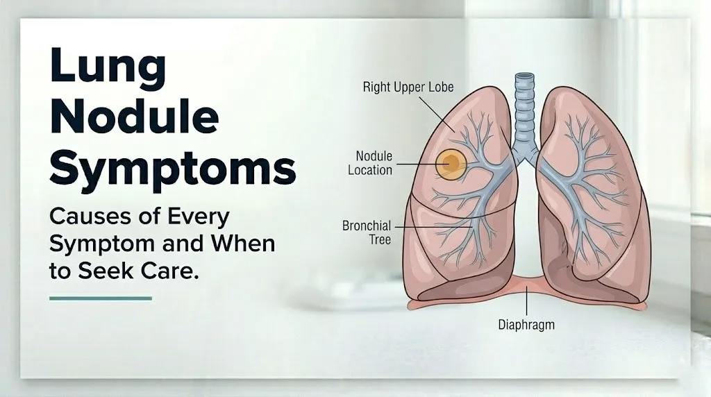
The complete symptom inventory when symptoms do occur includes:
- Persistent cough that does not resolve with typical cold or allergy treatment
- Chest pain or tightness, particularly with deep breathing or position changes
- Shortness of breath with exertion or at rest
- Wheezing or a new monophonic wheeze from one lung region
- Hemoptysis (coughing up blood-tinged or bloody sputum)
- Recurrent chest infections or pneumonia that keeps returning in the same lung area
- Fatigue, unintended weight loss, or loss of appetite in nodules associated with malignancy
- Hoarseness from recurrent laryngeal nerve compression by enlarged mediastinal lymph nodes
The complete absence of symptoms does not mean a nodule is safe to ignore. Conversely, having respiratory symptoms does not confirm a nodule is malignant. Only imaging characteristics, growth rate data, and in many cases a tissue sample can establish what a nodule is. What symptoms do is tell a clinician where to look and how urgently.
What Is a Lung Nodule
A lung nodule (also called a pulmonary nodule) is a discrete, rounded area of increased density within the lung tissue, measuring 3 centimeters or less in diameter on chest imaging, that appears as a spot or shadow on a chest X-ray or computed tomography (CT) scan.
The size threshold of 3 centimeters is clinically defined. Rounded abnormalities larger than 3 centimeters are classified as lung masses rather than nodules, and they carry a substantially higher statistical likelihood of malignancy from the point of initial discovery.
Nodules are further classified by their internal density on CT imaging into three types. Solid nodules appear uniformly dense and opaque on CT. Ground-glass nodules (also called ground-glass opacities, or GGOs) appear as a hazy, semi-transparent area through which underlying lung structures remain partially visible. Part-solid nodules (also called subsolid nodules) contain both a ground-glass component and a denser solid component within the same lesion.
This density classification matters enormously for symptom prediction and risk stratification. Ground-glass nodules are typically earlier-stage lesions with less displacement of lung tissue and less likelihood of endobronchial or pleural involvement, which means they produce symptoms less often. Solid nodules, particularly large ones with irregular margins, are more likely to be in contact with bronchial structures or the pleural surface.
According to the American College of Radiology, incidentally discovered pulmonary nodules are found in approximately 1.6 million Americans annually through CT imaging performed for various reasons. The overwhelming majority, estimated at over 95 percent, are benign.
Patients over 65 years old have a higher baseline rate of incidentally discovered nodules due to the cumulative effect of prior respiratory infections, occupational dust exposures, and age-related parenchymal changes. This population also has a higher proportion of calcified granulomas, which are essentially scars from healed prior infections and have no malignant potential.
Symptoms of Lung Nodules: Asymptomatic vs Symptomatic
Most lung nodules produce no symptoms whatsoever, and this is the direct result of their anatomical location within the pulmonary parenchyma, where they are surrounded by alveolar air sacs and interstitial connective tissue that contain few pain receptors, cough receptors, or mechanoreceptors capable of generating detectable symptoms.
Think of the lung parenchyma like the interior of a sponge. A small marble pushed into the center of a sponge creates minimal immediate disturbance to the surface. Only when the marble grows large enough to deform the outer edge, or is placed near a hollow internal channel, does the sponge’s structure change perceptibly. A lung nodule behaves the same way: its impact on function depends almost entirely on what structures it contacts or compresses.
The transition from asymptomatic to symptomatic occurs through four anatomical mechanisms:
| Mechanism | Nodule Location | Symptom Produced | Required Condition |
|---|---|---|---|
| Bronchial mucosal irritation | Endobronchial (within airway lumen) | Persistent cough, wheezing | Nodule contacts bronchial epithelium |
| Visceral pleural activation | Peripheral, pleural-adjacent | Chest pain (pleuritic) | Nodule reaches or involves visceral pleura |
| Bronchial mucosal vascular erosion | Any location with mucosal contact | Hemoptysis | Nodule erodes bronchial capillary bed |
| Airway lumen compression | Peribronchial (external to bronchus) | Dyspnea, wheezing, recurrent infection | Nodule large enough to compress lumen |
The majority of incidentally discovered nodules never progress through any of these mechanisms. They remain entirely within the parenchyma, stable or slowly enlarging, and are discovered only because imaging was performed for an unrelated reason.
Patients who are immunocompromised, including those receiving biological immunosuppressant medications like anti-TNF agents used for rheumatoid arthritis or inflammatory bowel disease, may develop infectious nodules from fungi like Aspergillus or Cryptococcus that grow more rapidly than typical benign nodules and may reach symptom-producing size or involve bronchial structures more quickly than in immunocompetent individuals.
Lung Nodule Cough Symptoms
A lung nodule produces a cough when it is located within or immediately adjacent to a bronchial lumen, where it can physically contact or irritate the bronchial epithelium and activate rapidly adapting receptors (RARs) and mechanoreceptors in the bronchial mucosa that trigger the cough reflex via vagal afferent fibers.
The cough reflex arc begins in the bronchial wall. Rapidly adapting receptors and C-fiber nociceptors in the bronchial epithelium send signals along vagal afferent fibers (branches of cranial nerve X traveling in the bronchial wall) to the nucleus tractus solitarius in the medulla oblongata. From there, motor output through the phrenic nerve, intercostal nerves, and laryngeal nerves coordinates the forced expiratory effort of a cough. An endobronchial nodule activates this pathway directly.
The cough from an endobronchial nodule has a specific clinical character that distinguishes it from a typical upper respiratory infection cough. It is persistent, typically lasting weeks rather than days, without the usual associated symptoms of viral illness (nasal congestion, sore throat, low-grade fever that resolve progressively). It may be dry and non-productive, or it may produce small amounts of clear or blood-tinged sputum. Importantly, it does not respond to typical over-the-counter cough suppressants or antihistamines the way an allergy or post-nasal drip cough would.
The American Thoracic Society notes that a new, persistent cough lasting more than 3 weeks in an adult over 40 years old, particularly in the context of a smoking history or other pulmonary risk factors, warrants chest imaging as part of the evaluation.
Patients with pre-existing asthma may interpret an endobronchial nodule-related cough as an asthma exacerbation and treat it accordingly, potentially delaying the recognition of a new pulmonary lesion. A new cough that does not respond to usual asthma management in a person with known asthma should prompt re-evaluation including chest imaging.
Key Takeaway: A persistent cough that lasts more than 3 weeks without resolving, especially without other cold or allergy symptoms, is the primary indicator that a lung nodule may be located within a bronchial airway rather than in the deeper lung tissue, and it warrants chest imaging and evaluation by a primary care physician.
Lung Nodule Chest Pain Symptoms
A lung nodule produces chest pain when it reaches or involves the visceral pleura, the thin, smooth membrane covering the outer surface of each lung, where nociceptive nerve fibers supplied by the intercostal nerves and phrenic nerve are activated by stretching, inflammation, or direct invasion of the pleural tissue.
The visceral pleura itself has limited pain-sensitive innervation. Pain from pleural irritation is primarily generated when inflammation or tumor extension reaches the parietal pleura, the corresponding membrane lining the inner chest wall, which is richly innervated by somatic intercostal nerves. This is why pleuritic chest pain from a peripheral nodule is typically sharp, localized to one side of the chest, and characteristically worsens with deep inspiration, coughing, or twisting movements that stretch the pleural surfaces against each other.
A benign nodule in the peripheral lung parenchyma that happens to sit close to the pleural surface does not typically produce chest pain unless it has grown into or inflamed the pleural tissue. Malignant nodules with pleural extension, or infectious nodules causing reactive pleural effusion (fluid accumulation in the pleural space), are more commonly associated with chest pain.
The chest pain pattern associated with a pleural-adjacent nodule is clinically distinct from the diffuse, pressure-type chest pain of cardiac disease or the burning midsternal pain of esophageal origin. It is:
- Sharp rather than dull or pressure-like
- Reproducible with specific movements (deep breath, twisting the torso)
- Localized to a specific chest wall region rather than radiating broadly
- Accompanied in some cases by a shallow breathing pattern as the patient avoids painful deep inspiration
Older adults and patients with concurrent pulmonary fibrosis may have reduced chest wall mobility and altered pain sensitivity that makes the pleuritic character of nodule-related chest pain harder to identify clinically. In these populations, new chest discomfort should prompt early imaging evaluation even when the pain character is atypical.
Lung Nodule Shortness of Breath Symptoms
Shortness of breath from a lung nodule occurs through two specific anatomical mechanisms: bronchial lumen compression reducing airflow to a lung segment or lobe, and large pleural effusion from nodule-associated pleural involvement reducing the functional volume of the affected lung.
The first mechanism involves external compression of a bronchus by a large peribronchial nodule or by enlarged hilar lymph nodes related to a nodule. Airflow through a bronchus follows Poiseuille’s principles: as the lumen narrows from external compression, airflow resistance increases proportionally, requiring greater respiratory effort to move air through the narrowed segment. This produces exertional dyspnea initially, progressing to dyspnea at rest as compression worsens.
The second mechanism involves reactive or malignant pleural effusion. A nodule at or near the pleural surface can stimulate fluid accumulation in the pleural space. As this fluid increases in volume, it compresses the adjacent lung parenchyma, reducing the lung’s functional residual capacity and producing shortness of breath proportional to the effusion volume.
It is worth understanding the scale here. Most pulmonary nodules, even those up to 2 or 3 centimeters in size, occupy a tiny fraction of the total lung volume. A nodule sitting within the parenchyma, away from major bronchi and the pleura, simply does not displace enough lung tissue to produce meaningful reduction in respiratory function. Shortness of breath as an isolated finding from a small parenchymal nodule is exceedingly rare.
According to the National Heart Lung and Blood Institute, new-onset exertional dyspnea in an adult over 50 years old warrants clinical evaluation including pulmonary function testing and chest imaging when no obvious cause such as deconditioning or acute respiratory infection is identified.
Patients with chronic obstructive pulmonary disease (COPD) present a particular diagnostic challenge. Their baseline dyspnea may mask or absorb the incremental reduction in respiratory function from a new nodule, delaying recognition that a new structural lesion is present and contributing to their respiratory symptoms.
Hemoptysis: When a Lung Nodule Causes Coughing Up Blood
Hemoptysis, the coughing up of blood-tinged or frankly bloody sputum, is the most alarming symptom associated with a lung nodule, and it occurs when a nodule erodes into the bronchial mucosal vasculature, the rich capillary network supplying the bronchial epithelium, or in more advanced cases, into a pulmonary or bronchial artery.
The bronchial mucosal vasculature is supplied by the bronchial arteries, which branch from the thoracic aorta and carry systemic arterial pressure. Even small erosions into this capillary network can produce blood-streaked sputum. In more advanced malignant nodules, invasion of larger bronchial arteries or pulmonary vessels can produce massive hemoptysis, defined clinically as the expectoration of 300 to 600 ml of blood within 24 hours, which is a life-threatening emergency because it can cause airway flooding and asphyxia.
The volume and character of hemoptysis provides clinical information about its likely source. Blood-streaked sputum mixed with phlegm is typically from bronchial mucosal capillary erosion and is more commonly associated with endobronchial inflammatory conditions or small endobronchial lesions. Frank blood without mucous, particularly in larger volumes, raises concern for more substantial vascular involvement and warrants immediate evaluation.
Emergency Symptoms: When to Call 911 or Go to the ER
Certain symptoms associated with lung nodules require immediate emergency evaluation. Do not wait to see if these resolve on their own.
Call 911 or go to the nearest emergency room immediately if you experience:
- Coughing up large amounts of blood (more than a teaspoon of frank red blood in a single episode): this indicates potential massive hemoptysis from bronchial or pulmonary vascular involvement and can cause airway obstruction within minutes
- Severe or sudden-onset shortness of breath at rest, with an inability to complete a full sentence: this may indicate airway obstruction, large pleural effusion, or pulmonary embolism from malignancy-associated hypercoagulability
- Chest pain accompanied by rapid breathing, blue-tinged lips or fingernails (cyanosis), and lightheadedness: these findings together suggest acute cardiopulmonary compromise
- Coughing up blood combined with fever and one-sided chest pain: suggests infectious complication with possible septic pulmonary involvement
These presentations require emergency medical assessment, not an urgent care appointment or a scheduled physician visit.
Patients with a known lung nodule who are on anticoagulant medications (warfarin, direct oral anticoagulants) face elevated hemoptysis risk if the nodule contacts any vascular structure, and they should inform their prescribing physician of any new cough or blood-tinged sputum immediately.
Key Takeaway: Any episode of coughing up blood, even blood-streaked sputum, in a person with a known or suspected lung nodule requires same-day evaluation by a primary care physician or emergency medicine physician; a large volume of frank blood requires calling 911 immediately.
Benign Lung Nodule Symptoms
Benign lung nodules typically produce no symptoms at all, because the most common benign nodule types, including pulmonary granulomas from prior infections, pulmonary hamartomas, and inflammatory pseudotumors, are almost exclusively located within the pulmonary parenchyma and do not contact airway mucosa or pleural surfaces.
The most common benign lung nodule is a calcified granuloma, which is the residual scar from a prior infection with organisms like Histoplasma capsulatum (histoplasmosis, endemic in the Ohio and Mississippi River valleys), Coccidioides immitis (coccidioidomycosis, endemic in the southwestern United States), or Mycobacterium tuberculosis (tuberculosis). These organisms triggered a granulomatous immune response, a coordinated macrophage and T-lymphocyte reaction that walled off the infection, which then calcified as the infection resolved. The calcified nodule is physiologically inert and produces no ongoing symptoms.
Pulmonary hamartomas are the most common benign lung tumors, composed of abnormally arranged normal lung tissue elements including cartilage, fat, muscle, and epithelial cells. They grow very slowly, rarely exceed 4 centimeters, and are overwhelmingly parenchymal in location. They produce no symptoms in the vast majority of cases. The classic CT finding of a hamartoma is a “popcorn” calcification pattern within the nodule, which is considered a reliably benign imaging feature.
When a benign nodule does produce symptoms, it is typically because it is located endobronchially (benign carcinoid tumors are a notable example) or because it has grown large enough to produce local mass effect.
Patients with a history of rheumatoid arthritis are at risk for pulmonary rheumatoid nodules, which can be multiple, tend to be peripheral and subpleural in location, and may occasionally cavitate (develop a hollow center). Subpleural rheumatoid nodules can occasionally cause pleuritic chest pain if they involve or inflame the adjacent visceral pleura.
Malignant Lung Nodule Symptoms
Malignant lung nodules are more likely to produce symptoms than benign ones, but this is not because malignancy itself generates immediate symptoms; it is because malignant nodules tend to grow faster, are more likely to develop irregular margins that contact adjacent structures, and have a greater tendency to involve lymphatic and vascular channels.
The specific symptom pattern of a malignant nodule reflects the histological type and growth pattern. Pulmonary adenocarcinoma, the most common type of lung cancer and increasingly common in never-smokers, often grows as a peripheral parenchymal lesion, frequently presenting as a ground-glass or part-solid nodule. In its early stages, it may be completely asymptomatic. Squamous cell carcinoma of the lung has a predilection for central, endobronchial locations, making it more likely to produce cough, hemoptysis, and obstructive symptoms at smaller sizes.
Constitutional symptoms, those that reflect the systemic impact of malignancy rather than local anatomical compression, become more prominent as malignant nodules advance. These include:
- Unintended weight loss, from tumor-driven metabolic reprogramming and elevated tumor necrosis factor-alpha (TNF-alpha) and interleukin-6 (IL-6) affecting appetite regulation and energy metabolism
- Fatigue, from tumor-related systemic inflammation and anemia of chronic disease
- Loss of appetite, driven by the same cytokine cascade
- Hoarseness, from recurrent laryngeal nerve compression by enlarged mediastinal lymph nodes
- Superior vena cava syndrome (facial and arm swelling, venous distension), from mediastinal tumor compression of the superior vena cava
These constitutional and compressive symptoms typically appear in more advanced disease rather than at the stage of a small, isolated nodule. A person with a recently discovered small nodule who is experiencing significant unintended weight loss or persistent hoarseness should be evaluated promptly by a board-certified pulmonologist, as these findings together suggest a more advanced or aggressive process.
Ground Glass Lung Nodule Symptoms
Ground-glass lung nodules (ground-glass opacities, or GGOs) produce symptoms even less frequently than solid nodules, because their characteristic hazy appearance on CT imaging reflects a partial filling of the alveolar air spaces or thickening of the interstitial tissue rather than a dense mass, meaning the surrounding lung architecture is less disrupted and bronchial and pleural structures are less likely to be contacted.
A ground-glass nodule represents an area where the alveolar air spaces are partially filled with fluid, blood, inflammatory cells, or abnormal cells, but the air-to-tissue ratio is still high enough that the underlying lung structures remain partially visible through the GGO on CT. This partial nature means the nodule is less physically dense and less likely to displace, compress, or invade adjacent bronchi or pleural surfaces at equivalent sizes compared to a solid nodule.
According to the Fleischner Society guidelines published in the journal Radiology (2017 update), pure ground-glass nodules smaller than 6 millimeters in low-risk patients do not require routine CT follow-up due to their very low likelihood of malignancy and their minimal risk of symptomatic development. This is a clinically important threshold that most general health content never mentions.
The part-solid variant, containing a denser central or eccentric solid component within a ground-glass background, is the nodule type with the highest statistical risk of malignancy among the three nodule density categories, according to clinical data reviewed in the Journal of Thoracic Oncology. The solid component represents a denser cellular area that behaves more like a conventional solid nodule in terms of symptom production potential.
Patients with pulmonary fibrosis may have multiple areas of ground-glass opacity from the fibrotic process itself, making the detection and characterization of a true ground-glass nodule within a fibrotic background more challenging. These patients should have their CT imaging reviewed by a radiologist with specific experience in interstitial lung disease.
Key Takeaway: Ground-glass nodules are less likely to cause symptoms than solid nodules at equivalent sizes because they displace less lung tissue and are less likely to contact bronchial or pleural structures, but their malignancy risk varies substantially by whether they contain a solid component.
Lung Nodule Symptoms in Females
Lung nodule symptoms in females follow the same anatomical and physiological mechanisms as in males, but the differential diagnosis of a pulmonary nodule in a female patient carries a distinct epidemiological consideration: lung adenocarcinoma is rising in incidence among women who have never smoked, at a rate that has drawn specific attention from the National Cancer Institute.
Research published in the Journal of Thoracic Oncology has found that women who have never smoked now constitute a growing proportion of lung adenocarcinoma cases in the United States, with rates that make lung cancer one of the leading cancer causes of death in female never-smokers. This epidemiological shift means that a pulmonary nodule in a woman with no smoking history cannot be confidently attributed to low malignancy risk on the basis of smoking history alone, as it could have been in prior clinical decades.
The hypotheses for this trend include hormonal influences on lung epithelial cell proliferation (estrogen receptors have been identified on lung adenocarcinoma cells in research published in JAMA Internal Medicine), environmental radon exposure, second-hand smoke exposure, and genetic susceptibility. The practical implication is that risk stratification models for pulmonary nodules in women, particularly those in the 40 to 60 age group who are never-smokers, should account for this changing epidemiology.
Female patients who are pregnant and incidentally discovered to have a pulmonary nodule present a specific clinical challenge. Chest CT, the standard imaging follow-up for pulmonary nodules, exposes the patient to ionizing radiation. While the fetal radiation dose from a properly shielded chest CT is low, decisions about imaging timing and frequency should be made in consultation with the patient’s obstetrician-gynecologist and managing pulmonologist, weighing nodule risk characteristics against radiation exposure timing in pregnancy.
Postmenopausal women who have undergone hormone replacement therapy with combined estrogen-progestogen formulations should inform their pulmonologist, as some research has examined the relationship between hormone use and lung cancer risk, though the evidence remains an area of active investigation rather than confirmed causation.
Lung Nodule Symptoms in Smokers and High-Risk Groups
In current and former smokers, a newly discovered lung nodule carries a higher prior probability of malignancy than in never-smokers, and this changes both the interpretation of symptoms and the aggressiveness of the follow-up strategy recommended by clinical guidelines.
The United States Preventive Services Task Force (USPSTF), in its 2021 updated lung cancer screening recommendation, advises annual low-dose CT (LDCT) lung cancer screening for adults aged 50 to 80 years who have a 20 pack-year or greater smoking history and currently smoke or have quit within the past 15 years. This recommendation is based on evidence from the National Lung Screening Trial (NLST), published in the New England Journal of Medicine in 2011, which found that annual LDCT screening in high-risk smokers reduced lung cancer mortality by approximately 20 percent compared to chest X-ray screening.
Smokers who develop symptoms from a pulmonary nodule are navigating a more complex clinical picture than non-smokers, because the same risk factors (smoking-related airway inflammation, chronic bronchitis, COPD) that increase their nodule malignancy risk also produce baseline respiratory symptoms that can mask nodule-specific symptom changes.
High-risk groups beyond smokers include:
- Individuals with occupational asbestos exposure (construction workers, shipyard workers, insulation workers): asbestos exposure increases risk of both lung adenocarcinoma and mesothelioma, and pleural plaques from asbestos exposure can be mistaken for nodules on imaging
- Individuals in radon-endemic geographic areas: radon gas is the second leading cause of lung cancer in the United States, per the Centers for Disease Control and Prevention
- Patients with a prior history of any malignancy: lung metastases from extrapulmonary cancers (breast, colon, renal cell, melanoma) can present as single or multiple pulmonary nodules
- Immunocompromised individuals: including those with HIV, solid organ transplant recipients, and patients on long-term immunosuppressive therapy, who have elevated risk of fungal and mycobacterial pulmonary infections presenting as nodules
Any smoker or member of a high-risk group who develops a new persistent cough, hemoptysis, or unexplained weight loss alongside a known pulmonary nodule should be evaluated by a board-certified pulmonologist without delay, regardless of the nodule’s most recent follow-up status.
Lung Nodule Symptoms When to Worry
The specific lung nodule symptom patterns that warrant urgent medical evaluation are hemoptysis, new or progressively worsening shortness of breath, chest pain that worsens with breathing over days rather than improving, unexplained weight loss of more than 5 percent of body weight over 6 to 12 months, and persistent hoarseness, particularly when any of these occur in a person with a known pulmonary nodule or significant pulmonary risk factors.
Knowing which symptom features raise concern requires understanding what the concerning scenarios actually are. A nodule that has been growing (as evidenced by interval CT comparison), that has developed new spiculated or irregular margins, or that has developed a new solid component within a previously pure ground-glass opacity, is displaying imaging characteristics associated with higher malignancy probability. Symptoms in this context carry greater clinical weight.
The symptom-to-concern threshold:
| Symptom | Concerning Feature | Action |
|---|---|---|
| Cough | Persistent more than 3 weeks, not resolving | Primary care physician within days |
| Hemoptysis | Any blood-tinged sputum in someone with a known nodule | Same-day evaluation; call 911 for large volume bleeding |
| Shortness of breath | Progressive over weeks, worsening with exertion | Primary care physician evaluation within days |
| Chest pain | Pleuritic (worse with breathing), new and progressive | Primary care physician within days |
| Weight loss | More than 5% body weight unintentionally over 6 months | Pulmonologist evaluation |
| Hoarseness | New, persistent, unexplained | Pulmonologist evaluation within 1 to 2 weeks |
| Recurrent pneumonia | Same lung region repeatedly infected | Pulmonologist evaluation for underlying structural cause |
A critical point that most health content misses: anxiety is itself a documented and common response to lung nodule discovery that can produce physical symptoms including chest tightness, breathlessness, and perceived fatigue. Research published in JAMA Internal Medicine has documented the psychological burden of pulmonary nodule surveillance programs. These symptoms warrant evaluation and support from a primary care physician, not dismissal, but the physician’s evaluation should include appropriate psychological and social support alongside physical assessment.
Lung Nodule Size and Symptom Relationship
The relationship between lung nodule size and symptom production is not linear; it is governed by location, and a small endobronchial nodule may produce more symptoms than a much larger parenchymal nodule, though size does eventually become a determining factor when a nodule grows large enough to compress adjacent bronchi or contact the pleural surface regardless of its exact location.
As a general framework, the Fleischner Society categorizes solid nodule management partly by size: nodules under 6 mm in low-risk individuals require no routine follow-up because their malignancy risk is below 1 percent. Nodules from 6 to 8 mm require a 6 to 12-month follow-up CT. Nodules greater than 8 mm warrant either 3-month CT follow-up or PET-CT or tissue sampling depending on additional risk features. This size stratification reflects both malignancy risk and the size at which a nodule begins to have meaningful interaction with surrounding structures.
At approximately 2 centimeters in diameter, even a purely parenchymal solid nodule begins to have measurable mass effect on adjacent parenchyma, and if located near the pleura, begins to exert tension on the visceral pleural surface. At 3 centimeters, the threshold for reclassification as a lung mass, the likelihood of endobronchial or pleural involvement increases substantially, as does the probability of symptom production.
Volume doubling time is a clinically useful concept that the size-symptom relationship illuminates. A nodule that doubles in volume (which corresponds to a roughly 26 percent increase in diameter) in less than 30 days is growing very rapidly, consistent with an aggressive infectious or malignant process. A nodule that has been stable for 2 or more years on serial imaging is almost certainly benign. Rapid volume growth brings a nodule to symptom-producing size faster and is itself a red flag independent of any current symptoms.
Patients who have had prior thoracic surgery, radiation therapy, or ablation treatment for a lung nodule may have scar tissue that alters the local anatomy and can make a new or residual nodule more likely to be adjacent to bronchi or the pleural surface, potentially producing symptoms at smaller sizes than would be expected in an untreated lung.
Key Takeaway: Nodule size matters for symptom risk, but location matters more: a 5 mm endobronchial nodule can produce a disabling cough while a 2 cm parenchymal nodule produces nothing, which is exactly why symptoms alone cannot determine a nodule’s significance or benignity.
How Lung Nodules Are Diagnosed
Lung nodules are diagnosed primarily through chest computed tomography (CT) scanning, which provides the size, location, density, margin characteristics, and three-dimensional shape information needed for clinical risk stratification, with additional diagnostic tools including PET-CT scanning, bronchoscopy with endobronchial ultrasound (EBUS), and CT-guided needle biopsy used to characterize nodules that require tissue-level diagnosis.
The diagnostic pathway from first discovery to confirmed tissue diagnosis follows a structured sequence:
- Initial discovery: A nodule is identified on a chest X-ray or CT scan performed for any reason. A chest X-ray can detect nodules larger than approximately 8 to 10 mm, but misses smaller lesions and provides no density information. A chest CT is far more sensitive and provides the detailed characteristics needed for risk classification.
- CT characterization: A dedicated chest CT (with thin-slice, 1 to 1.5 mm cuts) provides the nodule’s size in three dimensions, density classification (solid, part-solid, or ground-glass), margin characteristics (smooth, lobulated, or spiculated), presence or absence of calcification, and location within the lung. The radiologist’s report uses the ACR Lung-RADS classification system in screening contexts, rating nodules from Lung-RADS 1 (nothing to report, annual screening recommended) to Lung-RADS 4 (suspicious, tissue sampling recommended).
- Risk stratification and follow-up interval: Based on CT findings and patient risk factors, the managing physician uses the Fleischner Society guidelines (for incidentally discovered nodules) or Lung-RADS (for screening-detected nodules) to determine the appropriate follow-up interval: no follow-up needed, 3-month CT, 6-month CT, 12-month CT, or immediate further investigation.
- PET-CT scanning: For nodules greater than 8 mm with intermediate or high-risk features, a PET-CT (positron emission tomography combined with CT) assesses metabolic activity. Malignant nodules typically show elevated glucose uptake (measured as standardized uptake value, or SUV) compared to benign nodules, because tumor cells have higher metabolic rates. However, inflammatory and infectious nodules can also show elevated uptake, producing false-positive results.
- Tissue sampling: A tissue diagnosis can be obtained through CT-guided needle biopsy (for peripheral, accessible nodules), bronchoscopy with EBUS (for central or hilar nodules accessible from the bronchial tree), or video-assisted thoracoscopic surgery (VATS) resection (for nodules not accessible by other means, or when definitive treatment and diagnosis are combined).
Patients with implanted devices that are not MRI-compatible, or with claustrophobia limiting CT tolerance, should discuss alternative imaging protocols with their managing pulmonologist.
Lung Nodule Follow-Up Guidelines and What to Expect
Lung nodule follow-up is governed by the Fleischner Society guidelines for incidentally discovered nodules and the ACR Lung-RADS system for screening-detected nodules, both of which determine the type of imaging, the interval between scans, and the threshold at which tissue sampling is recommended.
Understanding these guidelines gives patients the context to interpret what their physician is recommending and why. The guidelines are built around the statistical probability that a nodule of a given size, density, and margin type in a patient with specific risk factors will prove to be malignant, balanced against the risk of complications from invasive procedures performed on nodules that would likely prove benign.
The general framework for incidentally discovered solid nodules per Fleischner Society (2017 guidelines, published in Radiology):
| Nodule Size | Low-Risk Patient | High-Risk Patient |
|---|---|---|
| Less than 6 mm | No routine follow-up | Optional 12-month CT |
| 6 to 8 mm | 6 to 12-month CT, then 18 to 24-month CT | 6 to 12-month CT, then 18 to 24-month CT |
| Greater than 8 mm | 3-month CT, PET-CT, or tissue sampling | 3-month CT, PET-CT, or tissue sampling |
High-risk features include: significant smoking history, family history of lung cancer, prior malignancy, upper lobe nodule location, spiculated or irregular margins, and PET-CT showing elevated metabolic activity.
The emotional and psychological experience of lung nodule surveillance is a clinically recognized burden. Research published in JAMA Internal Medicine has documented that patients undergoing pulmonary nodule surveillance frequently experience anxiety, difficulty sleeping, and preoccupation with the nodule between imaging appointments. This is a legitimate medical concern, not a psychological weakness, and patients should be direct with their primary care physician or pulmonologist about the psychological impact of the surveillance period. Many pulmonary nodule programs now incorporate patient support and anxiety management resources into the follow-up pathway.
Patients who have not smoked, are under 40 years old, and have a nodule smaller than 6 mm with no high-risk imaging features on CT are in the lowest risk category and may reasonably expect to be told that no follow-up imaging is needed. They should ask their physician explicitly: “What imaging features would change this recommendation, and what symptoms should prompt me to contact you before my next scheduled appointment?”
Frequently Asked Questions About Lung Nodule Symptoms
Do lung nodules usually cause symptoms?
No, the vast majority of lung nodules produce no symptoms at all, because most are located within the pulmonary parenchyma, away from the nerve receptors, bronchial mucosa, and pleural surfaces that generate detectable symptoms.
Symptoms occur primarily when a nodule is endobronchial (within an airway), pleural-adjacent, or large enough to compress surrounding structures.
The absence of symptoms does not indicate a nodule is benign; it simply reflects its anatomical location within the lung.
What does a lung nodule feel like?
Most people with a lung nodule feel nothing and are unaware of its existence until it is discovered on chest imaging ordered for an unrelated reason.
When symptoms do occur, they may include a persistent dry cough, a sharp chest pain that worsens with deep breathing, or mild shortness of breath, depending on where in the lung the nodule is located.
The absence of any sensation is actually the most clinically typical presentation of a pulmonary nodule, regardless of its size or cause.
Can a benign lung nodule cause symptoms?
Yes, benign lung nodules can cause symptoms, but only when they are located within a bronchial airway or adjacent to the pleural surface, or when they have grown large enough to compress surrounding structures.
The most common benign nodules, calcified granulomas from prior infections, are entirely parenchymal and almost never produce symptoms.
Benign carcinoid tumors located within bronchial lumens are a notable exception, producing persistent cough and occasionally hemoptysis despite being non-cancerous.
What are the warning signs that a lung nodule might be cancerous?
No symptom alone confirms that a lung nodule is cancerous; imaging characteristics, growth rate, and ultimately tissue biopsy are required for a definitive determination.
Symptoms that raise concern in the context of a known nodule include persistent cough, hemoptysis (coughing up blood), unintended weight loss, progressive shortness of breath, and hoarseness from recurrent laryngeal nerve compression.
A nodule with spiculated margins, rapid volume growth on serial CT imaging, or elevated metabolic activity on PET-CT requires urgent evaluation by a board-certified pulmonologist regardless of current symptoms.
When should I be worried about a lung nodule?
You should contact your primary care physician promptly if you develop any new respiratory symptom alongside a known lung nodule, including persistent cough, blood-tinged sputum, progressive shortness of breath, or unexplained weight loss.
Nodule characteristics on imaging, specifically size greater than 8 mm, spiculated margins, solid density, upper lobe location, and growth on serial CT, are the primary drivers of clinical concern, not symptoms alone.
A board-certified pulmonologist should evaluate any nodule with high-risk imaging features, any nodule discovered in a person with a significant smoking history or prior malignancy, and any nodule accompanied by new respiratory symptoms.
How long does it take for a lung nodule to cause symptoms if it is cancerous?
There is no single timeline, because symptom development depends on the nodule’s location within the lung, its growth rate, and its histological type.
Peripheral adenocarcinomas, the most common lung cancer type, often grow slowly and remain symptom-free for years even while progressing from a small ground-glass nodule to a larger lesion.
Central squamous cell carcinomas that grow within bronchial airways may produce cough and hemoptysis at smaller sizes and earlier in their course because of their endobronchial location.
Closing
The single most practically useful thing to carry away from this article is this: a lung nodule’s symptom profile tells you about its location in the lung, not definitively about its benignity or malignancy. A completely silent nodule can be a very early adenocarcinoma. A nodule causing a persistent cough can be a benign carcinoid. Symptoms and imaging characteristics together create a clinical picture that your physician interprets using established guidelines, not either one alone.
If you have been told you have a lung nodule, ask your primary care physician specifically whether your nodule’s size and CT characteristics fall into the Fleischner Society low, intermediate, or high-risk category, what the recommended follow-up interval is and why, and which new symptoms should prompt you to contact them before the next scheduled imaging appointment.
You are the person who lives in your body and notices changes first. Knowing what to watch for, what to report promptly, and what can wait gives you the specific, actionable clarity to participate in your own follow-up effectively.