Warning Signs of Root Canal Symptoms to Know in 2026
Root canal symptoms are your body’s signal that the soft inner tissue of a tooth, the dental pulp, has become inflamed, infected, or is dying, and that signal demands clinical attention before the infection spreads beyond the tooth itself. Recognizing the specific pattern of root canal symptoms early is the difference between straightforward endodontic treatment and a dental emergency.
The American Association of Endodontists (AAE) estimates that more than 15 million root canals are performed annually in the United States, with the vast majority arising from untreated dental caries or trauma that allow oral bacteria to reach the pulp. Many of those cases involve people who experienced warning signs weeks or months earlier and did not act on them.
This article covers every clinically recognized root canal symptom, explains the precise anatomical and physiological reason each one occurs, separates early-stage from advanced-stage presentations, and tells you exactly which symptoms require same-day dental evaluation versus a monitored wait-and-schedule approach. It also covers when a dental infection stops being a dentist’s problem and becomes a hospital emergency room problem.
Root Canal Symptoms: What They Really Mean
Root canal symptoms are the clinical signs that indicate the dental pulp inside a tooth has been damaged severely enough to require endodontic treatment to remove the infected or necrotic tissue. They range from a sharp twinge of pain to a life-altering throbbing ache, and understanding what each symptom means anatomically helps you report them accurately to your dentist.
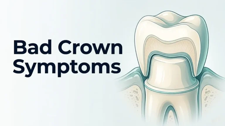
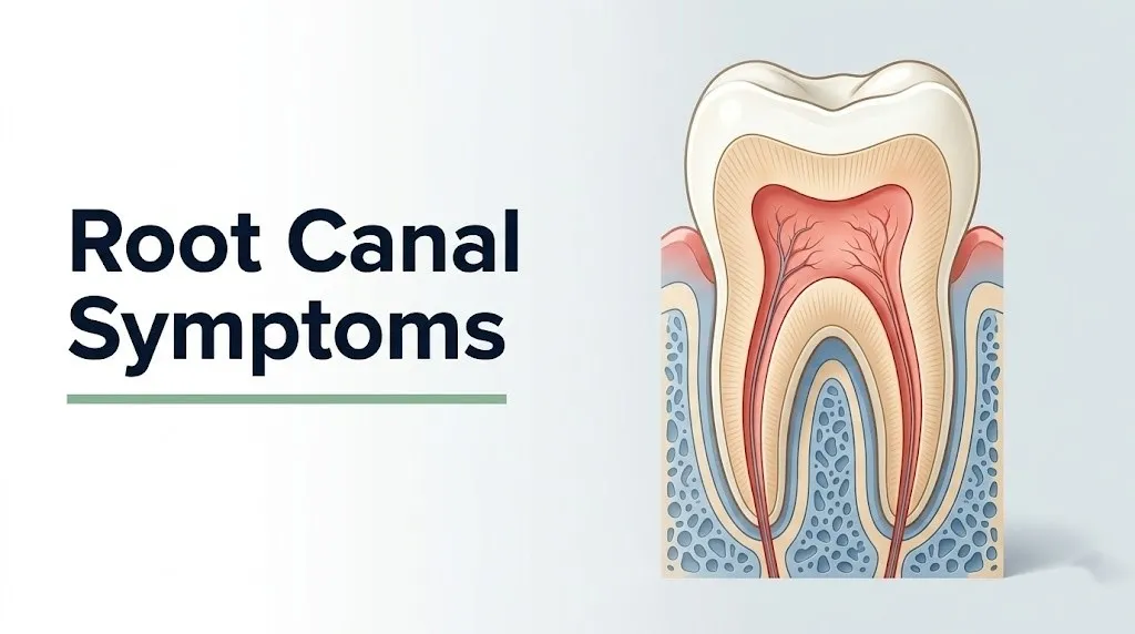
The dental pulp is the soft connective tissue that occupies the interior of every tooth. It runs from the pulp chamber at the center of the crown down through the root canal system to tiny openings at the root tips called apical foramina. The pulp contains blood vessels, lymphatics, and nerve fibers, primarily the unmyelinated C-fiber and thinly myelinated A-delta nociceptors that detect pain.
When bacteria penetrate the pulp through a cavity, crack, or trauma, the immune response that follows is physiologically identical to inflammation elsewhere in the body: vasodilation, increased vascular permeability, and inflammatory cell infiltration. The difference is the location. The pulp sits inside a rigid dentin-walled chamber with zero room to swell. Inflammatory edema has nowhere to expand, so intrapulpal pressure builds rapidly. That pressure is exactly what produces the severe, often spontaneous, throbbing pain that most people recognize as “root canal pain.”
Symptom severity overview:
| Symptom | Stage of Pulp Disease | Urgency Level |
|---|---|---|
| Brief sensitivity to cold | Reversible pulpitis (early) | Schedule appointment |
| Lingering sensitivity to cold or heat | Irreversible pulpitis | Same-day or next-day care |
| Spontaneous throbbing pain | Irreversible pulpitis (advanced) | Same-day care |
| Swollen gum or facial swelling | Periapical abscess | Same-day emergency dentist |
| Fever, neck swelling, difficulty swallowing | Spreading fascial infection | Call 911 / Go to ER |
People taking long-term corticosteroid medications, such as prednisone for autoimmune conditions, may have blunted inflammatory responses that mask the classic pain symptoms, making a tooth with advanced pulp disease appear less symptomatic than it actually is.
What Is a Root Canal and Why Is It Needed
A root canal, formally called endodontic therapy or root canal treatment, is a dental procedure that removes the diseased or dead pulp tissue from inside the tooth’s root canal system, disinfects and shapes the canals, and seals them with an inert filling material called gutta-percha to prevent reinfection.
The procedure is needed when the pulp tissue reaches a state of irreversible damage from which it cannot heal. Pulp tissue has some capacity to defend itself from mild bacterial invasion. Dentinal tubules are partially sealed by odontoblastic processes and the production of tertiary dentin at the pulp surface. But when the bacterial load exceeds the pulp’s defense capacity, or when the blood supply to the pulp is disrupted by trauma, irreversible damage occurs and the tissue either becomes chronically inflamed or dies (necrosis).
A dead pulp is not necessarily a pain-free pulp, and this point confuses many patients. Pulp necrosis eliminates the sensory nerve fibers within the pulp itself, which can reduce or eliminate the spontaneous throbbing pain of active pulpitis. However, the bacterial toxins and breakdown products from the necrotic tissue continue to seep through the apical foramen into the periodontal ligament and periapical bone, causing symptomatic apical periodontitis and periapical abscess, which produce their own distinct pain patterns.
According to the National Institute of Dental and Craniofacial Research (NIDCR), dental pulp disease is one of the most common reasons adults in the United States visit an emergency dentist or hospital emergency department for acute pain management, underscoring how frequently symptoms progress to urgent presentations before treatment is sought.
Children with infected primary (baby) teeth also require endodontic treatment in many cases, typically a procedure called pulpotomy or pulpectomy rather than full root canal therapy, because premature loss of primary teeth disrupts spacing for permanent tooth eruption. Parents should not assume that infected primary teeth can simply be left until they fall out naturally.
Severe Toothache as a Root Canal Symptom
Severe, spontaneous toothache that occurs without any stimulus, particularly pain that wakes you from sleep or throbs in sync with your heartbeat, is one of the most diagnostically important root canal symptoms and indicates irreversible pulp inflammation.
The throbbing character of this pain is mechanistically specific. With every heartbeat, arterial blood pressure in the dental pulp’s vascular supply briefly rises. Inside the rigid, enclosed pulp chamber, that momentary pressure increase has nowhere to go. It compresses the nociceptor nerve endings, producing a pulse of pain that follows your heart rate. This is why patients describe it as “throbbing” or “pulsating” and why it is genuinely difficult to sleep through.
The spontaneous nature distinguishes this from pain caused by a cavity or cracked enamel, which typically requires a stimulus (cold, biting pressure) to provoke pain. When pain occurs without any stimulus, it means the C-fiber nociceptors in the pulp have been sensitized to the point of generating action potentials without external provocation, a state called peripheral sensitization. This is a reliable clinical marker of irreversible pulpitis.
Pain intensity reported by patients does not always correlate with disease severity. Some patients with severe irreversible pulpitis rate their pain at 9 or 10 on the Visual Analog Scale for pain, while others with equally advanced disease report only moderate discomfort due to differences in central pain processing, individual pain threshold variation, and prior analgesic use.
Key characteristics of root canal toothache to report to your dentist:
- Whether the pain is spontaneous or requires a stimulus to start
- Whether the pain lingers after the stimulus is removed (lingering = more serious)
- Whether the pain wakes you from sleep
- Whether over-the-counter ibuprofen or acetaminophen provides meaningful relief or barely touches it
- The precise location, or whether the location is hard to identify
Patients on opioid analgesics for other chronic pain conditions may have altered pain perception that makes the typical presentation of severe toothache less predictable. A general dentist or endodontist should be informed of all analgesic use before diagnosis.
Tooth Sensitivity to Hot and Cold as a Root Canal Symptom
Tooth sensitivity to hot and cold becomes a root canal symptom when it lingers for more than a few seconds after the temperature stimulus is removed, rather than resolving immediately upon removal of the hot or cold item.
Brief, sharp sensitivity to cold that disappears within two to three seconds of removing the cold stimulus is typically a sign of reversible pulpitis: early pulp inflammation that can resolve if the irritating cause (often a new cavity or recent dental work) is addressed. The cold sensation activates the hydrodynamic mechanism in the dentinal tubules, where fluid movement from temperature changes stimulates pulp nerve fibers.
Sensitivity that lingers for 30 seconds, a minute, or longer after removing the cold stimulus is a different clinical picture entirely. Prolonged cold sensitivity indicates that the C-fiber nociceptors in the pulp have become sensitized to the point of sustained firing after the initial stimulus ends. This pattern is a recognized diagnostic indicator of irreversible pulpitis in endodontic clinical guidelines published by the American Association of Endodontists.
Heat sensitivity is clinically even more informative than cold sensitivity. Most patients with simple dentinal sensitivity feel cold more acutely than heat. When heat becomes the primary provocation, or when a patient reports that holding warm water in the mouth reproduces the pain while cold water actually relieves it, this is a specific indicator of advanced irreversible pulpitis. The physiological reason is that heating the inflamed pulp tissue increases the metabolic rate of the inflammatory cascade, raises intrapulpal pressure further, and exacerbates the compression of the neurovascular bundle at the apical foramen.
Elderly patients with sclerotic (calcified) pulp canals may show reduced sensitivity to both cold and heat tests because the calcification narrows the canal lumen and reduces the number of functional nerve fibers, meaning standard thermal testing may underestimate the severity of their pulp disease.
Symptoms of Needing a Root Canal: Pain When Biting or Touching
Pain when biting down on food or when the affected tooth is tapped (percussion sensitivity) is a root canal symptom that indicates inflammation has spread from the pulp to the periodontal ligament and periapical tissues surrounding the root tip.
The periodontal ligament is a dense network of collagen fibers anchoring the tooth root to the surrounding alveolar bone. It is richly innervated with mechanoreceptors and nociceptors. When bacterial toxins or inflammatory mediators from a diseased pulp migrate through the apical foramen into the periodontal ligament space, inflammation develops in that tissue. Any biting force then compresses the inflamed periodontal ligament against the surrounding bone, producing sharp, localized pain at the moment of contact.
This is distinct from the spontaneous throbbing pain of active pulpitis. Biting pain means the disease process has moved beyond the pulp and into the supporting structures of the tooth, a finding called symptomatic apical periodontitis. A tooth that hurts when you bite but does not hurt spontaneously may actually be in a transitional state: the pulp may have partially or fully necrotized (reducing spontaneous pain), while the periapical tissue is now acutely inflamed.
A useful self-assessment: tap the suspected tooth gently with your fingernail and compare the sensation to adjacent teeth. Pain on tapping that is notably greater than the adjacent teeth is a reliable indicator of periapical inflammation worth reporting to your dentist.
Quick Tip:
- Percussion pain that is severe from even light tapping suggests acute periapical pathology requiring prompt evaluation
- Pain that occurs only with hard biting forces and resolves quickly may indicate a cracked cusp rather than pulp involvement, though these conditions frequently coexist
- Patients with dental implants adjacent to the affected tooth should inform their dentist, as periapical infection from natural teeth can occasionally affect adjacent implant osseointegration in close anatomical proximity
Key Takeaway: Lingering heat sensitivity is a stronger indicator of irreversible pulpitis than cold sensitivity, and pain on biting means the infection has spread beyond the pulp into the periodontal ligament, a sign that disease is progressing toward the jawbone.
Root Canal Signs and Symptoms: Tooth Darkening or Discoloration
Tooth discoloration or darkening, particularly a tooth that has become noticeably gray, brown, or yellow compared to adjacent teeth, is a root canal sign that typically indicates the dental pulp has undergone necrosis (death) or that internal resorption is occurring.
When the pulp dies, the blood within the pulpal blood vessels breaks down through a process called hemolysis. The hemoglobin from ruptured red blood cells decomposes into iron compounds and other pigmented byproducts, which penetrate the dentinal tubules and stain the dentin from the inside. This produces the characteristic gray or brown discoloration seen in teeth with necrotic pulps. The staining originates from within the tooth, which is why it cannot be removed by external whitening treatments.
Discoloration following dental trauma deserves particular attention. A tooth that turns gray in the weeks to months after an injury to the face or jaw may be showing delayed pulp necrosis from disruption of the blood supply at the apical foramen. The trauma may have been minor enough that the patient did not initially seek dental care. According to research published in the International Endodontic Journal, traumatic injuries are a leading cause of pulp necrosis in anterior (front) teeth in children and young adults, with color change being one of the first observable signs.
Discoloration can also result from prior dental work. Silver amalgam restorations can leach metallic ions into surrounding dentin over decades, producing a gray appearance that is not related to pulp disease. A dentist must differentiate between restoration-related staining and true pulp necrosis through pulp vitality testing before concluding that a gray-appearing tooth needs a root canal.
Adolescents and young adults who play contact sports and experience repeated dental trauma to the same tooth should have that tooth monitored regularly for progressive discoloration, as the cumulative vascular disruption can produce necrosis even without a single dramatic injury event.
Swollen or Tender Gums as a Root Canal Symptom
Swelling or tenderness of the gums near the base of a specific tooth is a root canal symptom that indicates periapical inflammation or abscess formation has reached the point where exudate (pus and inflammatory fluid) is beginning to accumulate in the surrounding soft tissues.
When bacterial infection from the pulp spreads through the apical foramen and into the periapical bone, the body’s immune response generates a localized collection of pus called a periapical abscess. As pus accumulates under pressure, it follows the path of least resistance through the surrounding bone toward the soft tissues. When it reaches the gingival (gum) tissue, the area becomes visibly swollen, red, and tender to the touch.
Gum swelling from a periapical abscess is usually localized to one specific area, directly adjacent to the affected tooth. This distinguishes it from gum swelling caused by periodontal disease (gum disease), which typically involves broader gingival inflammation along multiple teeth rather than a pinpoint swelling at one root apex.
Palpating the swollen area, pressing gently with one finger, often produces pain and may produce a sensation of fluctuance (a wave-like feeling indicating fluid beneath the surface). Fluctuant swelling indicates the abscess is well-developed and may require incision and drainage in addition to root canal treatment or extraction.
According to Mayo Clinic, fever accompanying gum swelling from a dental abscess indicates systemic involvement and warrants same-day evaluation by a dentist or, if a dentist is unavailable, an urgent care physician or emergency department physician, because antibiotics may be needed to prevent further spread while awaiting definitive dental treatment.
Pregnant women with acute periapical abscess and gum swelling should be evaluated promptly by their dentist and should inform their obstetric provider. Untreated dental infection during pregnancy has been associated with adverse pregnancy outcomes in observational studies, and the timing of any dental procedure during pregnancy requires coordination with the patient’s obstetrician.
Sinus Tract or Pimple on the Gum: A Root Canal Warning Sign
A small, pimple-like bump on the gum near a tooth, which may periodically discharge a salty or foul-tasting fluid, is a clinical sign called a sinus tract (also called a parulis) and is a specific indicator of chronic periapical infection that has created a drainage channel through the gum tissue.
A sinus tract forms when a chronic periapical abscess erodes a pathway through the surrounding bone and gum tissue to create a self-draining opening at the mucosal surface. The abscess pressure drives pus and bacterial byproducts through this channel to the oral cavity. Patients often notice that the pimple-like bump appears, produces a discharge, shrinks temporarily, then returns. This cycle reflects the continuous production of pus from the underlying infection and its periodic drainage through the sinus tract.
The presence of a sinus tract is diagnostically useful in a counterintuitive way. Because the abscess is draining, the acute pressure that would otherwise produce severe pain is partially relieved. This means a tooth with a chronic sinus tract may produce little or no spontaneous pain, despite having significant underlying periapical pathology. Patients sometimes interpret the absence of severe pain as the infection “getting better,” when in fact a self-draining sinus tract is a reliable sign that the tooth has a chronic infection requiring endodontic treatment.
A technique endodontists use to identify which tooth is the source of a sinus tract is called tracing with gutta-percha: a fine point of gutta-percha material is gently inserted into the sinus tract opening, and a periapical radiograph is taken. The radio-opaque gutta-percha point traces the channel back to the offending root apex, identifying the specific tooth even when the patient cannot localize the problem or the sinus tract opening appears to be near a different tooth than expected.
Immunocompromised patients, including those receiving chemotherapy or immunosuppressive therapy after organ transplant, may develop sinus tracts with more extensive underlying bone destruction than immunocompetent patients because the chronic infection progresses faster when immune surveillance is impaired.
Key Takeaway: A pimple on your gum that comes and goes is not a minor irritation; it is a chronic periapical abscess creating a drainage tract through your gum tissue, and the tooth it originates from needs a root canal or extraction.
Root Canal Pain Symptoms: Referred Pain to Jaw, Ear, or Temple
Referred pain from a tooth requiring root canal treatment is a clinically documented phenomenon in which the brain incorrectly localizes the source of pain signals to a region away from the actual diseased tooth, producing pain in the jaw, ear, temple, cheek, or even the neck.
The anatomical explanation lies in the trigeminal nerve (cranial nerve V) and its three main divisions. The mandibular division (CN V3) provides sensory innervation to the lower teeth, gums, lower jaw, and portions of the ear and temple. The maxillary division (CN V2) serves the upper teeth, gums, cheekbone, and upper jaw. When intense nociceptive signals from an inflamed or infected tooth flood the trigeminal nucleus in the brainstem, the nucleus becomes overloaded. The brain misattributes some of these signals to other anatomical regions served by the same trigeminal branch.
The clinical consequence is significant. A patient with an infected lower molar may feel the primary pain in their ear and assume they have an ear infection, rather than a dental problem. A patient with an infected upper molar may feel pain in their cheekbone and suspect sinusitis. This diagnostic confusion is why a thorough history of recent dental symptoms, including any prior tooth sensitivity or pain, is essential when evaluating facial, jaw, or ear pain in a medical or urgent care setting.
Pain from upper posterior teeth (maxillary molars and premolars) frequently radiates to the maxillary sinus area and can mimic maxillary sinusitis so closely that patients and even some clinicians pursue sinus treatment before investigating the dental cause. The distinguishing feature is that dental-referred pain typically involves one sinus area, worsens with biting or tapping the specific tooth, and does not respond to decongestant treatment.
A 2019 study published in the Journal of Endodontics found that referred pain was reported by a substantial proportion of patients presenting with symptomatic irreversible pulpitis, and that pain was frequently reported in adjacent teeth in the same arch rather than in the affected tooth itself, highlighting the diagnostic challenge of reliably identifying the symptomatic tooth before testing.
Older adults with multiple restored or damaged teeth may experience particular difficulty localizing dental pain due to the convergence of nociceptive input from several teeth with varying degrees of pathology.
Reversible Versus Irreversible Pulpitis Symptoms
Reversible pulpitis and irreversible pulpitis are two distinct clinical diagnoses that determine whether a tooth can be saved with conservative treatment or requires root canal therapy, and the symptom difference between them is the single most clinically important distinction in this entire topic.
Reversible pulpitis is inflammation of the pulp that is mild enough that the pulp tissue can recover if the source of irritation is removed. It produces brief, sharp pain triggered by a stimulus (cold, sweet, air) that resolves within two to three seconds of removing the stimulus. The pulp’s defensive capacity is still intact. A new cavity causing reversible pulpitis can be treated with a filling, after which the pulp inflammation subsides and no root canal is needed.
Irreversible pulpitis is inflammation severe enough that the pulp cannot recover regardless of what is done to the exterior of the tooth. The pulp tissue is too extensively damaged to return to health. The key distinguishing symptoms are: pain that lingers for 30 seconds or more after the stimulus is removed, spontaneous pain that occurs without any stimulus, severe throbbing pain, and pain that is difficult to control with standard analgesics. Once irreversible pulpitis is diagnosed, root canal treatment or extraction are the only clinical options.
The transition from reversible to irreversible pulpitis is not always obvious in symptom terms. There is no single moment where one becomes the other. This is why clinical testing by a trained dentist or endodontist, using thermal pulp testing, electric pulp testing, and percussion testing, is necessary to accurately classify the pulp’s status.
Reversible versus irreversible pulpitis: symptom comparison:
| Feature | Reversible Pulpitis | Irreversible Pulpitis |
|---|---|---|
| Pain trigger | Requires stimulus (cold, sweet) | Spontaneous or stimulus-triggered |
| Pain duration after stimulus | Less than 3 seconds | 30 seconds to minutes |
| Spontaneous pain | Absent | Present |
| Sleep disruption | Rare | Common |
| Response to analgesics | Good | Partial or poor |
| Treatment needed | Dental restoration (filling) | Root canal or extraction |
Children and adolescents tend to have larger, more vascular pulps with wider apical foramina, which means their pulps can often mount a more aggressive immune response against infection. However, this same anatomy also allows infection to spread to periapical tissues faster once the defense is overwhelmed.
Asymptomatic Teeth That Need a Root Canal
Not every tooth that requires a root canal produces symptoms, and this is one of the most clinically underappreciated aspects of pulp disease: a tooth can have a completely necrotic (dead) pulp and an established periapical abscess with no pain whatsoever.
Pulp necrosis eliminates the sensory nerve fibers within the pulp, which removes the capacity for pain from pulpal origin. If the periapical infection that develops after pulp death is chronic and slow-draining (particularly if a sinus tract has formed and is providing drainage), the pressure that would otherwise produce acute pain is controlled. The patient feels nothing, or perhaps only occasional mild pressure. Yet the infection is actively destroying periapical bone, expanding a periapical radiolucency that is visible on dental radiographs.
Asymptomatic periapical pathology is most commonly discovered during routine dental radiographs taken for other purposes. A dark shadow at the root apex of a tooth the patient does not associate with any symptoms is the radiographic appearance of a periapical lesion (granuloma or cyst). This finding still requires endodontic treatment even in the complete absence of symptoms, because the lesion represents active bone destruction and a reservoir of bacteria that can become an acute abscess if the balance between infection and immune response shifts.
According to data from the National Institute of Dental and Craniofacial Research (NIDCR), a substantial proportion of periapical lesions discovered on routine dental radiographs in adult patients are found in teeth the patient did not identify as symptomatic, reinforcing the necessity of regular dental radiographic examinations even when pain is absent.
Patients who delay routine dental care for years, including those with significant dental anxiety, are at the highest risk of discovering asymptomatic periapical pathology at an advanced stage where the lesion is large, bone loss is extensive, and the prognosis for saving the tooth is diminished.
Key Takeaway: A tooth can have a dead pulp and an active periapical infection with zero pain. Regular dental radiographs are the only reliable way to detect asymptomatic periapical disease before it becomes an acute emergency.
Symptoms of a Dental Abscess That Requires Root Canal Treatment
A dental abscess originating from pulp disease, called a periapical abscess, produces a recognizable set of symptoms that differ from the pain of active pulpitis and signal that infection has moved from inside the tooth to the surrounding bone and soft tissues.
The hallmark symptom is a constant, throbbing ache that is precisely localized to one area of the jaw or cheek, accompanied by gum swelling that is warm and tender to the touch, and often accompanied by an elevation in body temperature. Unlike the spontaneous throbbing pain of irreversible pulpitis that originates from within the tooth, periapical abscess pain originates from the inflamed periapical tissues and periodontal ligament and is consistently worsened by any biting pressure on the affected tooth.
Periapical abscess can produce a sensation of the tooth feeling elevated or longer than adjacent teeth. This occurs because inflammatory exudate within the periodontal ligament space physically displaces the tooth slightly upward in its socket, and the swollen periodontal ligament makes the tooth hypersensitive to occlusal (biting) contact. Patients often report that they avoid chewing on that side entirely, and that even the light contact of biting teeth together is painful.
A fever above 101°F associated with dental swelling is a clinically important threshold. Fever indicates that bacterial pathogens or their products (LPS, exotoxins) have entered the systemic circulation and triggered a systemic inflammatory response. This finding warrants same-day evaluation by a dentist or, if unavailable, a medical provider, because the infection may require systemic antibiotic therapy while definitive dental treatment is arranged.
Diabetic patients are at disproportionately higher risk of rapid periapical abscess progression because hyperglycemia impairs neutrophil chemotaxis and phagocytic function, reducing the local immune response that would normally contain the infection. A diabetic patient with signs of dental abscess should be prioritized for same-day dental evaluation and blood glucose monitoring.
Signs a Dental Infection Is Spreading Beyond the Tooth
Signs that a dental infection is spreading beyond the tooth and into the surrounding fascial spaces of the head and neck represent a medical emergency, not a dental appointment situation.
When periapical infection erodes through the cortical plate of the jaw bone and enters the adjacent soft tissue spaces, it can spread rapidly through the fascial planes of the head and neck by following the paths of least resistance. In the lower jaw, infection from mandibular molars frequently spreads into the submandibular space (below the jaw and above the hyoid bone) and the sublingual space (under the tongue). When infection involves both the sublingual and submandibular spaces bilaterally, the condition is called Ludwig’s angina, named after the German physician Karl Friedrich Wilhelm von Ludwig.
Ludwig’s angina is a potentially fatal cellulitis of the floor of the mouth and neck that can progress to complete airway obstruction within hours if not treated with emergency surgical drainage, airway management, and intravenous antibiotics.
Symptoms that indicate a dental infection is spreading and require immediate emergency evaluation:
- Visible swelling of the face, neck, or jaw that is expanding rapidly or is bilateral (both sides)
- Floor of the mouth that feels hard, wooden, or elevated when the tongue is pressed downward
- Difficulty or pain when swallowing (dysphagia)
- Difficulty opening the mouth fully (trismus, also called lockjaw in this context)
- Difficulty breathing or the sensation that your airway is narrowing (dyspnea)
- Voice that sounds muffled, “hot potato” in quality, or hoarse without a respiratory illness explaining it
- High fever (above 102°F) with dental pain or swelling
- Neck swelling below the jaw that is tender, warm, and firm
These are not symptoms to monitor at home while deciding whether to call a dentist in the morning. They require immediate emergency department evaluation. The airway can close within hours.
Emergency Symptoms: When to Call 911 or Go to the ER
Certain dental infections associated with root canal symptoms can progress to life-threatening emergencies. Do not wait for these to resolve on their own or assume they will improve with antibiotics at home.
Call 911 or go to the nearest emergency room immediately if you experience:
- Difficulty breathing or sensation of airway narrowing: This indicates the infection has spread to the floor of the mouth or neck and may be compressing the airway. Ludwig’s angina can close the airway completely within hours.
- Bilateral jaw or neck swelling with floor-of-mouth hardness: Bilateral spread of dental infection to the submandibular and sublingual fascial spaces is a surgical emergency.
- High fever above 102°F with dental swelling and altered mental status: Systemic sepsis from odontogenic (tooth-derived) bacteremia requires intravenous antibiotic therapy and emergency surgical consultation.
- Severe difficulty swallowing that prevents drinking fluids: Dysphagia from pharyngeal or parapharyngeal space involvement indicates infection has spread into the throat.
- Drooling combined with inability to open the mouth: Drooling with trismus indicates severe floor-of-mouth involvement.
- Rapidly expanding facial or neck swelling over hours: Fast progression suggests aggressive polymicrobial infection requiring emergency surgical drainage.
These presentations indicate Ludwig’s angina, descending necrotizing mediastinitis (infection spreading into the chest cavity), or sepsis of odontogenic origin. These conditions require emergency surgical airway management and intravenous antibiotics in a hospital setting, not an urgent care clinic or dental office.
What Causes Root Canal Symptoms
Root canal symptoms are produced when one or more specific pathological processes breach the natural defenses of the tooth and allow bacterial colonization of the dental pulp, triggering the inflammatory cascade that damages pulp tissue.
Dental caries (tooth decay) is the most common cause, accounting for the majority of root canal cases across all populations. Caries progresses through the enamel, then the dentin, and if untreated, reaches the pulp chamber where bacteria directly invade the pulp tissue. The deeper the cavity and the longer it goes untreated, the greater the bacterial load reaching the pulp and the more severe the resulting inflammation.
Named causes of root canal symptoms by mechanism:
- Dental caries: Acid-producing oral bacteria (primarily Streptococcus mutans and Lactobacillus species) demineralize enamel and dentin, creating a direct pathway for mixed anaerobic bacteria to reach the pulp
- Dental trauma: Fractures, luxation injuries, or avulsion injuries disrupt the blood supply at the apical foramen, causing ischemic necrosis of the pulp; trauma can produce pulp necrosis months to years after the injury
- Cracked tooth syndrome: A fracture line through dentin allows bacteria to enter dentinal tubules and reach the pulp gradually; pain is characteristically sharp on biting specific foods and releasing
- Deep dental restorations: Previous large fillings that placed restorative materials in close proximity to the pulp can cause chronic low-grade pulp irritation that eventually leads to necrosis
- Repeated dental procedures: Multiple procedures on the same tooth produce cumulative thermal and mechanical stress on the pulp tissue, reducing its regenerative capacity
- Periodontal disease: Severe gum disease can allow bacteria to reach the lateral canals of a root or the apical foramen through a deep periodontal pocket, causing a combined periodontal-endodontic infection
According to a 2021 review published in the Journal of Endodontics, the microbiome of infected root canals is dominated by obligate anaerobic bacteria, primarily Fusobacterium nucleatum, Prevotella intermedia, Porphyromonas gingivalis, and Treponema denticola, all of which produce potent endotoxins and tissue-destructive enzymes.
Patients who use tobacco products have impaired oral mucosal healing and reduced salivary antibacterial defenses, which accelerates the progression of dental caries and increases the likelihood of pulp involvement before a cavity is clinically detected.
How a Dentist Diagnoses Root Canal Symptoms
A dentist or endodontist diagnoses root canal symptoms through a structured clinical examination that combines the patient’s reported symptoms with objective physical and radiographic findings, because no single test is sufficient to confirm pulp status on its own.
The diagnostic process unfolds in a specific sequence:
- Symptom history: The clinician asks about the character of pain (spontaneous vs. stimulus-triggered), its duration, what makes it better or worse, whether it disturbs sleep, and how long symptoms have been present. This guides which tests to prioritize.
- Cold test (thermal pulp test): A cotton pellet saturated with a refrigerant (typically dichlorodifluoromethane, which reaches approximately -50°C) is applied to the tooth surface. The response, whether brief and normal, prolonged and painful, or absent, classifies pulp status. No response to cold suggests pulp necrosis. Prolonged response suggests irreversible pulpitis.
- Electric pulp test (EPT): An electric current is applied to the tooth surface to stimulate the pulp sensory nerves directly. The patient reports when they first feel a tingling or electric sensation. No response confirms absence of vital pulp nerve fibers.
- Percussion test: The dentist taps the tooth gently with a mirror handle and compares the pain response to adjacent teeth. Severe pain on light percussion indicates periapical periodontitis.
- Palpation test: The dentist presses on the gum tissue at the root apex. Pain on palpation confirms periapical inflammation has reached the soft tissue.
- Periodontal probing: A periodontal probe measures the depth of the gum attachment around the tooth to identify any periodontal pockets that might indicate a combined periodontal-endodontic lesion.
- Periapical radiograph: A standard dental X-ray shows the root anatomy, surrounding bone density, and any periapical radiolucency (dark shadow at the root tip indicating bone loss from infection). Cone-beam computed tomography (CBCT) provides three-dimensional imaging when standard radiographs are inconclusive.
Diagnosis requires interpretation of all findings together, because each individual test has known limitations. Elderly patients with calcified (sclerotic) pulp canals frequently test false-negative on cold tests despite having vital pulp tissue, making CBCT and clinical history more important in this population.
Key Takeaway: No single test confirms root canal need. Diagnosis requires combining the symptom history, cold test, percussion test, and periapical radiograph, and an endodontist is the specialist trained to do this evaluation when the diagnosis is unclear.
Symptoms You Need a Root Canal: When to Seek Same-Day Care
Certain root canal symptoms warrant same-day dental evaluation rather than a scheduled appointment later in the week, and knowing which symptoms meet this threshold helps you make the correct decision about urgency.
Symptoms that can be managed with a scheduled appointment within a few days:
- Brief cold sensitivity that resolves within 2 to 3 seconds and is not worsening
- Mild, intermittent toothache that responds well to ibuprofen (400 to 600mg) and is not present at rest
- Recently noticed tooth discoloration without any associated pain or swelling
Symptoms requiring same-day evaluation by a dentist or emergency dentist:
- Spontaneous throbbing pain that does not respond adequately to over-the-counter analgesics
- Pain that prevents sleep
- Visible gum swelling adjacent to the tooth
- Pus or foul taste in the mouth
- Percussion sensitivity so severe that even light biting contact on adjacent teeth is painful
- Any fever above 100.4°F associated with dental pain
Symptoms requiring emergency room evaluation today, not a dental office:
- Any of the spreading infection symptoms described in the previous section
- Difficulty breathing, swallowing, or opening the mouth
To prepare for your dental appointment when root canal symptoms are the concern, tell your provider:
- The exact onset date of symptoms and whether they are getting better, worse, or staying the same
- Whether the pain is spontaneous or requires a stimulus
- Whether hot or cold worsens or relieves the pain, and for how long the sensation lasts
- All current medications, including blood thinners (warfarin, aspirin, novel anticoagulants), immunosuppressants, bisphosphonates (which affect jaw bone healing), and any antibiotics already taken
- Any prior dental work on the affected tooth, including fillings, crowns, or previous root canals
- Any recent dental trauma, even if it seemed minor
Patients taking bisphosphonate medications for osteoporosis or cancer treatment must inform their dentist before any dental procedure involving the jaw bone, including root canal treatment that may require periapical surgery, because of the risk of medication-related osteonecrosis of the jaw (MRONJ).
Frequently Asked Questions About Root Canal Symptoms
What does root canal pain feel like?
Root canal pain typically feels like a severe, throbbing ache that can occur spontaneously without any trigger, often worsening at night when lying down and pulsating in rhythm with the heartbeat.
The throbbing character occurs because inflamed pulp tissue is confined inside a rigid dentin chamber with no room to swell, so each heartbeat briefly increases intrapulpal pressure and compresses nociceptor nerve fibers.
Pain may also radiate to the jaw, ear, or temple through the trigeminal nerve, which is why patients sometimes cannot identify exactly which tooth is causing the problem.
How do I know if I need a root canal or just a filling?
Brief sensitivity to cold that resolves within 2 to 3 seconds and is not spontaneous typically indicates early pulp inflammation (reversible pulpitis) that may be managed with a filling.
Spontaneous pain, sensitivity that lingers for 30 seconds or more after stimulus removal, pain on biting, gum swelling, or pain that wakes you from sleep all indicate irreversible pulpitis or periapical infection, which requires root canal treatment rather than a filling.
A general dentist or endodontist must confirm this distinction using clinical thermal and percussion testing combined with radiographic evaluation; the patient’s symptoms alone are not sufficient for a definitive diagnosis.
Can a tooth need a root canal with no pain at all?
Yes, a tooth can have a completely necrotic (dead) pulp and an established periapical abscess with no pain, a condition called asymptomatic apical periodontitis.
When the pulp dies, the sensory nerve fibers within the tooth die with it, eliminating the pain signals that would otherwise indicate disease; the periapical infection may drain through a sinus tract, which further reduces pressure and pain.
Asymptomatic periapical lesions are typically discovered on routine dental radiographs as dark shadows at the root tip, reinforcing why regular dental check-ups with radiographs are necessary even when you have no tooth pain.
How long can you wait if you have root canal symptoms?
Brief cold sensitivity with no spontaneous pain and no swelling can usually wait 2 to 3 days for a scheduled dental appointment, provided symptoms are not worsening.
Spontaneous toothache, pain unresponsive to over-the-counter analgesics, any facial or gum swelling, or fever associated with dental pain warrant same-day evaluation by a dentist or emergency dental provider.
Any symptom suggesting spread of infection to the neck, floor of the mouth, or airway, including difficulty swallowing or breathing, requires immediate emergency room evaluation and cannot wait for any dental appointment.
What happens if you ignore root canal symptoms?
Ignoring root canal symptoms does not cause the underlying pulp disease to resolve; the infection progresses from the pulp into the periapical bone, then potentially into the surrounding facial and neck tissue spaces.
Advanced untreated periapical infection can result in significant bone destruction around the root, loss of the tooth, and, in serious cases, spread to the submandibular space producing Ludwig’s angina, a life-threatening airway emergency that carries a mortality risk without emergency hospital treatment.
According to the American Association of Endodontists, early treatment of symptomatic pulp disease preserves the tooth with a high long-term success rate; delayed treatment increases the risk of tooth loss and systemic complications.
What is the difference between reversible and irreversible pulpitis?
Reversible pulpitis is early pulp inflammation where the tissue can still recover if the irritating cause (such as a new cavity) is treated; it produces brief, sharp pain from a stimulus that resolves within 2 to 3 seconds.
Irreversible pulpitis is pulp inflammation severe enough that the tissue cannot recover regardless of treatment to the external tooth; it produces spontaneous pain, lingering sensitivity lasting 30 seconds or more, or pain that disrupts sleep, and it requires root canal treatment or extraction.
The distinction between the two cannot be made reliably from symptoms alone; a dentist or endodontist must perform thermal and electric pulp testing combined with radiographic evaluation to confirm which condition is present.
Your Next Step
The single most actionable thing you can take from everything above: a toothache that wakes you up is not a symptom to manage with pain relievers until your next scheduled cleaning. Spontaneous, sleep-disrupting dental pain is irreversible pulpitis until a dentist proves otherwise.
If your symptom is lingering heat sensitivity, gum swelling, or pain on biting, call a general dentist or endodontist today and describe those findings specifically. If you have any facial or neck swelling, fever, difficulty swallowing, or any sense that your airway feels different, go to a hospital emergency department without delay and tell the triage nurse that you have a dental infection with those symptoms.
Root canal treatment performed at the right stage of disease is straightforward, highly successful, and tooth-preserving. Waiting past that stage turns a predictable dental procedure into a more complicated, more expensive, and in some cases, genuinely dangerous medical situation.