Bad Crown Symptoms: Warning Signs You Shouldn’t Ignore
Bad crown symptoms include persistent tooth pain, sensitivity to temperature, a bite that feels off, gum swelling, a bad taste, visible cracks, looseness, and in more serious cases, signs of infection under the crown that can progress to a dental abscess. These symptoms are not always obvious immediately after crown placement, and some develop gradually over months or years as the crown ages, the underlying cement breaks down, or the tooth beneath it changes.
According to the American College of Prosthodontists (ACP), dental crowns have an average clinical lifespan of approximately 10 to 15 years, but failure can occur at any stage, from the first days after placement to decades later. The National Institute of Dental and Craniofacial Research (NIDCR) reports that dental disease, including complications from restorations like crowns, remains one of the most prevalent chronic health conditions affecting adults in the United States.
This article covers every clinically recognized bad crown symptom with the specific anatomical reason behind it, how to distinguish a minor post-placement adjustment issue from a sign of progressive crown failure or infection, which symptoms require same-day emergency medical attention rather than a dental appointment, and how risk factors like bruxism and diabetes change the picture.
Bad Crown Symptoms: What They Mean Clinically
Bad crown symptoms are physical signs that a dental crown is failing to protect the underlying tooth, is incorrectly positioned in the mouth, has structural damage, or has allowed bacterial entry to the tooth’s interior. Each symptom corresponds to a specific failure mode of the crown-tooth system.
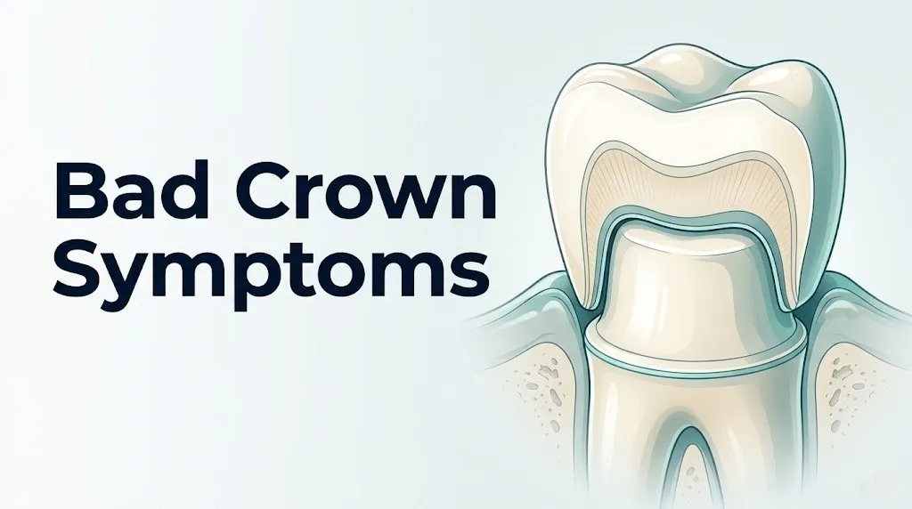
A well-functioning dental crown acts like a protective cap sealed over a prepared tooth. It distributes biting forces across its entire surface, prevents bacteria from reaching the dentinal tubules and dental pulp below, and sits flush against the gingival margin without irritating the surrounding gum tissue. When any of these functions breaks down, symptoms emerge.
The range of bad crown symptoms spans from minor and self-resolving to genuinely dangerous. Post-placement sensitivity that fades within two weeks is different from pain that worsens with heat, which signals possible irreversible pulpitis or pulp necrosis. A loose crown that comes off cleanly differs from a crown with an open margin that has allowed years of bacterial penetration. Understanding where your symptom sits on this spectrum is the essential first step.
Key bad crown symptoms, from least to most urgent:
- Mild temperature sensitivity lasting less than two weeks after placement: typically resolves as the tooth settles
- Bite that feels high immediately after placement: requires bite adjustment at the dental office
- Chronic temperature sensitivity beyond two weeks: suggests exposed dentinal tubules or early pulpal irritation
- Persistent pain when biting down: suggests occlusal trauma to the periodontal ligament or pulpitis
- Gum swelling or bleeding around the crown margin: suggests poor crown fit or gingival inflammation
- Bad taste or persistent bad breath from one area: suggests bacterial growth under the crown from cement breakdown
- Crown feels loose or rocks slightly: suggests cement washout and potential bacterial ingress
- Visible crack or chip in the crown material: requires prompt evaluation for structural integrity
- Fever, facial swelling, difficulty swallowing: dental emergency requiring immediate medical attention
People with pre-existing xerostomia (dry mouth) caused by medications such as antihistamines, tricyclic antidepressants, or antihypertensives face elevated risk of symptom progression because reduced saliva flow removes the natural protective buffering against bacterial acid beneath a poorly sealed crown margin.
What Makes a Dental Crown Go Bad
A dental crown goes bad through one or more of five primary failure mechanisms: cement breakdown, marginal gap formation, material fracture, occlusal overload, and biological failure of the tooth beneath the crown. Each mechanism produces a distinct symptom profile.
Think of a crown like a precisely fitted lid on a jar. If the seal degrades, moisture and bacteria get in. If the lid is the wrong size, it sits unevenly and lets things in around the edges. If the lid itself cracks, the protection is lost entirely. And if what is inside the jar spoils, the problem is not the lid at all.
Cement breakdown occurs when the dental cement bonding the crown to the prepared tooth dissolves or washes out over time. Older zinc phosphate and zinc polycarboxylate cements are more susceptible to dissolution by oral acids. When cement washes out, a microspace forms between the crown and the tooth, creating an ideal anaerobic environment for bacterial colonization.
Marginal gap formation happens when the crown edge does not seal precisely against the prepared tooth surface, often due to laboratory fabrication error, impression inaccuracy, or shrinkage of the crown material during firing. The American Dental Association (ADA) considers a marginal gap of greater than 120 micrometers to be clinically unacceptable, as gaps beyond this threshold allow bacterial microleakage to reach the underlying dentinal tubules.
Material fracture is particularly common in porcelain-fused-to-metal crowns, where the porcelain veneer layer is susceptible to chipping under lateral occlusal forces, and in all-ceramic crowns subjected to bruxism-related overload.
Occlusal overload from a crown placed too high in the bite produces excessive and uneven forces on the periodontal ligament (PDL), the fibrous connective tissue suspending the tooth root within the alveolar socket. Sustained PDL overload triggers a localized inflammatory response and can progress to bone resorption around the tooth root.
Biological failure refers to disease progression in the tooth itself: secondary caries developing under the crown margin, pulpal disease requiring endodontic treatment, or periodontal disease affecting the crown’s supporting structures.
According to a review published in the Journal of Prosthetic Dentistry, the most common reasons for crown replacement are caries (decay beneath the crown), endodontic failure (needing root canal therapy on an already-crowned tooth), and fracture of the crown or underlying tooth structure.
Older adults over age 65 face a higher rate of biological crown failure because both root caries susceptibility and gingival recession (which exposes unprotected root surface at the crown margin) increase with age, according to NIDCR epidemiological data.
Bad Fitting Crown Symptoms
A bad fitting crown produces symptoms including a bite that feels uneven, chronic gum irritation, food trapping between the crown and adjacent teeth, visible gaps at the gum line, and sensitivity at the crown margin. These symptoms stem from physical mismatch between the crown’s shape and the space it needs to occupy.
Crown fit has two dimensions: the internal fit (how well the crown sits over the prepared tooth structure) and the external fit (how the crown’s contour relates to adjacent teeth, opposing teeth, and the surrounding gum tissue). A problem with either dimension produces symptoms.
When the internal fit is imprecise, the crown sits too high or at an angle, creating occlusal prematurity. You’ll feel this as one tooth hitting before the others when you close your mouth. Biting on that tooth produces localized pain because the excess force is concentrated on a single point of the periodontal ligament rather than distributed across the full tooth surface. The PDL responds to this mechanical insult with localized edema and prostaglandin E2 release, producing an aching soreness that worsens with chewing and sometimes persists as a dull throb at rest.
When the external contour is incorrect, particularly when the crown overhangs the gum tissue (a condition called over-contoured emergence profile), the gingival sulcus, the natural crevice between tooth and gum, cannot be adequately cleaned. Plaque accumulates in this protected space, gingival inflammation follows, and the patient experiences consistent bleeding, redness, and soreness at the crown-gum junction.
Symptoms specific to a bad fitting crown:
- Bite feels noticeably higher on one side after crown placement
- Difficulty flossing because the contact point with the adjacent tooth is too tight or too loose
- Food consistently packing between the crown and the tooth next to it (open contact point)
- Gum tissue around the crown is chronically red, swollen, or bleeds with light brushing
- Visible ledge or step at the crown margin where it meets the tooth
According to the American College of Prosthodontists, fit assessment at crown delivery should include visual examination of the margin, probing of the gingival sulcus, floss resistance evaluation, and bite registration review. If your crown was placed without all of these checks, ask your dentist to perform a formal fit assessment at your next appointment.
Women who are pregnant experience increased gingival sensitivity due to elevated progesterone levels amplifying the gingival inflammatory response to plaque. A mildly over-contoured crown that would be asymptomatic in a non-pregnant patient can produce pronounced gum bleeding and soreness during pregnancy.
Dental Crown Pain After Placement
Dental crown pain after placement is expected for up to two weeks as mild soreness or sensitivity, but pain that worsens after the first week, spreads to adjacent teeth, intensifies with heat, or wakes you from sleep is not normal and requires evaluation. The type and timing of pain carry specific diagnostic meaning.
The tooth preparation process for crown placement removes enamel and sometimes dentin to create space for the crown. This preparation inevitably places the tooth’s dentinal tubules closer to the surface and may cause mild pulpal irritation through heat generated by drilling, desiccation of dentin, or chemical exposure from temporary cement. In the first one to two weeks, the dental pulp mounts a reversible inflammatory response, producing mild aching and temperature sensitivity that resolves as the pulp recovers.
Pain that worsens rather than improves beyond two weeks suggests the pulp did not recover from preparation trauma, which may indicate pre-existing pulpal disease that was subclinical before the crown was placed. This progression is classified as irreversible pulpitis: the point where the dental pulp’s inflammatory response becomes self-sustaining and the tissue proceeds toward necrosis rather than recovery. The American Association of Endodontists (AAE) describes irreversible pulpitis as characterized by lingering pain to cold lasting more than 30 seconds, spontaneous pain, and pain that intensifies with heat.
Pain spreading to adjacent teeth or to the jaw, ear, or temple may reflect referred pain through the trigeminal nerve pathway, where the inferior alveolar nerve (for mandibular teeth) or the superior alveolar nerves (for maxillary teeth) transmit pain signals that the brain localizes imprecisely.
To describe crown pain accurately to your dentist:
- Note when the pain started: immediately after placement or after a delay of days to weeks.
- Describe the character: sharp and brief versus dull and lingering versus spontaneous throbbing.
- Identify triggers: cold only, heat only, biting, or spontaneous with no trigger.
- Record duration: does it resolve within seconds, or does it persist for 30 or more seconds after the trigger is removed?
- Note whether it wakes you at night: nighttime pain suggests advanced pulpal disease rather than simple post-placement sensitivity.
People with a history of endodontic treatment on the crowned tooth may still develop pain if the prior root canal had untreated canals, a missed canal, or incomplete canal obturation, allowing residual bacteria to sustain periapical inflammation beneath the crown.
Key Takeaway: Crown pain that worsens after the first week, lingers more than 30 seconds after a cold stimulus, or appears spontaneously at rest is a clinical signal of irreversible pulpitis that requires endodontic evaluation, not watchful waiting.
Crown Sensitivity to Cold and Heat
Crown sensitivity to cold and heat signals that thermal stimuli are reaching the dental pulp through the dentinal tubules at a rate or intensity the pulp cannot tolerate without producing pain. The mechanism is well-established and depends on which thermal stimulus triggers it.
The hydrodynamic theory of dentin hypersensitivity, originally described by Brannstrom and confirmed by subsequent research, explains this sensation precisely. The dentinal tubules are microscopic fluid-filled channels running from the outer dentin surface to the dental pulp. Temperature changes cause the fluid within these tubules to contract (cold) or expand (heat), creating rapid fluid movement. This movement stimulates A-delta nerve fibers at the pulp-dentin border, producing a sharp, brief pain response that corresponds exactly to the timing of the thermal stimulus.
Cold sensitivity that resolves within one to five seconds after the cold is removed is typically a sign of reversible pulpitis or simple dentin hypersensitivity from an open crown margin or slight recession exposing the root surface at the crown edge. This type of sensitivity is painful but not structurally dangerous in the short term.
Heat sensitivity that lingers for 30 or more seconds after the heat stimulus is removed carries a more serious clinical implication. Lingering heat pain suggests the pulp’s inflammatory state has progressed to the point where vasodilation within the rigid pulp chamber creates sustained internal pressure. Because the pulp chamber is enclosed by dentin and cannot expand, any increase in internal pressure compresses the pulp’s own blood supply, accelerating tissue death. The AAE identifies lingering heat pain as a hallmark of irreversible pulpitis.
| Sensitivity Type | Duration After Stimulus | Clinical Implication | Recommended Action |
|---|---|---|---|
| Cold, resolves in 1 to 5 seconds | Brief | Reversible pulpitis or dentin hypersensitivity | Monitor; report to dentist at next visit |
| Cold, lingers 30+ seconds | Prolonged | Possible irreversible pulpitis | Dental appointment within 1 to 2 weeks |
| Heat, resolves quickly | Brief | Marginal gap or cement issue | Dentist evaluation for crown fit |
| Heat, lingers 30+ seconds | Prolonged | Irreversible pulpitis or partial pulp necrosis | Dental appointment within days |
| Spontaneous pain, no trigger | Constant | Advanced pulpitis or abscess | Same-day dental contact |
Metal crowns, particularly those made from gold or base metal alloys, conduct temperature more readily than porcelain or zirconia crowns. Patients with metal crowns or porcelain-fused-to-metal (PFM) crowns may experience temperature sensitivity that patients with all-ceramic or zirconia crowns do not, particularly in the early weeks after placement. This is not necessarily a sign of pathology but should be mentioned to the dentist if it persists beyond one month.
Pain When Biting Down on a Crown
Pain when biting down on a crown is produced by excessive occlusal force being transmitted through the crown to the periodontal ligament (PDL), which responds to mechanical overload with localized inflammation, edema, and pain via the trigeminal nerve. The most common cause is a crown that is positioned too high in the bite, creating an occlusal prematurity.
Each natural tooth in a functioning bite shares the load of chewing across the full dental arch. When one crown sits even a fraction of a millimeter too high, it receives the full impact of closing forces before any other tooth makes contact. The PDL, which normally absorbs and distributes these forces through its collagen fiber network, becomes overwhelmed by the concentrated load. Prostaglandin E2 and bradykinin accumulate in the ligament, the local capillaries dilate, and edema forms. The tooth feels sore to biting pressure, and may feel vaguely “longer” than surrounding teeth because the swollen PDL effectively elevates the tooth slightly within its socket.
This is one of the most common and most easily corrected bad crown symptoms. A single bite adjustment appointment, in which the dentist uses articulating paper to identify and selectively reduce the high point, typically resolves the pain within 24 to 72 hours.
Pain when biting that persists after a documented bite adjustment, or that develops weeks to months after crown placement when the bite initially felt fine, carries a different meaning. In these cases, the crown itself may not be the primary problem. The underlying tooth may have developed periapical periodontitis (inflammation at the tip of the root) from bacterial progression through a failed crown margin, producing percussion sensitivity that resembles bite pain but does not resolve with occlusal adjustment.
Quick Tip:
- Tap your upper and lower teeth together gently without food in your mouth. If one tooth hits noticeably before the others, this identifies the high point.
- Bite on a cotton roll or a folded piece of gauze placed over the crown. Pain with direct pressure that differs from the surrounding teeth suggests PDL inflammation, not just crown surface contact sensitivity.
- Older adults with posterior bone loss from long-term periodontal disease may have reduced PDL support, making bite pain from even minor occlusal discrepancies more intense than in younger patients with intact alveolar bone.
Gum Problems Caused by a Bad Crown
Gum problems caused by a bad crown include persistent gingival inflammation, bleeding on probing, gum recession specifically around the crown margin, and deepened periodontal pockets adjacent to the crown. Each sign reflects a different failure mode of the crown’s relationship with the surrounding periodontal tissues.
The gum tissue immediately around a crown is controlled by the crown’s emergence profile: the shape of the crown as it emerges from the gingival sulcus and transitions from the prepared tooth root to the visible crown surface. A well-designed emergence profile allows the patient to clean effectively with a toothbrush and floss, and does not press unnaturally against the gingival sulcus lining.
When the emergence profile is over-contoured (too bulky), the crown physically displaces gingival tissue, prevents floss from reaching the sulcus, and creates a stagnant zone where plaque accumulates without disruption. The gingival sulcus responds with chronic inflammation driven by bacterial lipopolysaccharides from gram-negative anaerobic species. This inflammation causes the classic signs of marginal gingivitis: redness, swelling, and bleeding on brushing or flossing localized specifically to the crown tooth rather than the whole mouth.
Long-term, persistent marginal inflammation can progress to periodontal pocket deepening around the crown, a sign that the supporting alveolar bone has begun to resorb in response to sustained bacterial challenge. A probing depth greater than 3 millimeters at the crown margin, particularly with bleeding on probing, indicates active periodontal disease that requires clinical management beyond crown replacement.
According to a 2019 study published in the Journal of Prosthodontics, crowns with open or overhanging margins showed statistically higher periodontal probing depths at five-year follow-up compared to crowns with clinically acceptable margins, confirming that crown fit quality directly influences long-term periodontal health.
People with type 2 diabetes mellitus who have a poorly fitting crown face compounded risk because hyperglycemia impairs neutrophil chemotaxis and phagocytosis, reducing the gingival tissue’s ability to defend against bacterial challenge. Gingival inflammation progresses faster, and pocket depths deepen more rapidly in poorly controlled diabetic patients compared to normoglycemic individuals.
Key Takeaway: Gum bleeding or swelling localized specifically to one crowned tooth rather than the whole mouth is a reliable early clinical signal of crown fit problems, not just poor oral hygiene, and deserves a dedicated dentist assessment of the crown’s marginal integrity.
Dark Line at the Gum Line Under a Crown
A dark line at the gum line under a dental crown is caused by the opaque metal substructure of a porcelain-fused-to-metal crown becoming visible as the gum tissue recedes away from the crown margin, exposing the grey metal collar at the base of the crown. This is primarily an aesthetic issue but can carry clinical implications depending on its cause.
Porcelain-fused-to-metal crowns are constructed with a metal coping, a thin thimble-shaped metal shell, over which porcelain is layered and fired. The porcelain covers the visible portion of the crown, but the base of the metal coping, called the metal collar or subgingival shoulder, is typically placed just at or slightly below the gum line when the crown is made. When the gum tissue recedes, this metal collar becomes partially visible as a dark or grey line against the tooth.
The clinical importance of the dark line depends on its cause. If it represents simple gingival recession from aging, previous periodontal disease, or aggressive toothbrushing technique, the crown itself may be structurally sound. The dark line is aesthetically unwanted, particularly in the front teeth, but does not indicate crown failure in isolation.
If the dark line represents a widening gap between the crown margin and the tooth surface due to cement dissolution or crown loosening, the clinical picture changes. The exposed margin becomes a direct pathway for bacterial ingress to the underlying dentin. This scenario requires dentist evaluation to assess whether the crown can be re-cemented or must be replaced.
Quick Tip:
- The dark line test at home: run a clean fingernail gently around the junction between the crown and the gum tissue. If you feel a distinct ledge or step where the crown edge meets the tooth rather than a smooth transition, that is a marginal discrepancy worth reporting to your dentist.
- All-ceramic and zirconia crowns do not produce a dark line because they have no metal substructure. If a dark line appears around a zirconia crown, it is more likely to represent actual tooth darkening from pulp necrosis beneath the crown rather than an aesthetic crown issue, which warrants urgent endodontic evaluation.
- Older adults and patients with a history of aggressive gum disease are more likely to experience gum recession that exposes crown margins, making regular dental monitoring of crown margins particularly relevant for these groups.
Bad Taste or Smell Under a Dental Crown
A persistent bad taste or smell from the area of a dental crown indicates bacterial growth in a space that should be sealed, most commonly caused by cement washout creating a gap between the crown and the prepared tooth, or by secondary caries developing under an imperfect crown margin. Neither situation resolves without dental intervention.
Dental cement is not a permanent adhesive in the biological sense. Over years of exposure to oral acids, thermal cycling, and mechanical stress from chewing, certain cement types dissolve gradually. Older glass ionomer and zinc phosphate cements are more vulnerable to this process than modern resin-based cements. When cement dissolves at the crown margin, a microscopic to macroscopic gap forms. Oral fluids carrying bacteria, food particles, and bacterial byproducts enter this space.
The anaerobic environment under a crown is ideal for gram-negative anaerobic bacteria such as Treponema denticola, Porphyromonas gingivalis, and Fusobacterium nucleatum, the same species responsible for periodontal disease. Their metabolic byproducts include volatile sulfur compounds (hydrogen sulfide and methyl mercaptan), which produce a distinctively unpleasant, rotten-egg or sulfurous taste and odor localized to the affected tooth. You may also notice the taste after pressing on the gum tissue around the crown or after using a water flosser on that area.
According to data reviewed by the American Dental Association, secondary caries beneath crown margins is among the most common reasons for crown failure and replacement, accounting for approximately 25 to 30 percent of crowns requiring replacement in long-term clinical studies.
Things to tell your dentist if you notice a bad taste under a crown:
- When you first noticed the taste or smell and whether it is constant or intermittent
- Whether the taste worsens after eating or drinking
- Whether you have had any recent dental X-rays of this tooth (within the past year)
- Whether you have had any pain, sensitivity, or swelling in the same area
- How long the crown has been in place and what type of cement was used (if known)
Patients who take proton pump inhibitors (PPIs) for acid reflux have chronically elevated gastric acid exposure in the mouth, which accelerates dental cement dissolution and increases secondary caries risk under crown margins.
Cracked Crown Symptoms
Cracked crown symptoms include sharp pain on release of biting pressure (a sign called cracked tooth syndrome when it occurs in the natural tooth), visible fracture lines or chips in the crown material, rough edges detectable with the tongue, and in severe fractures, crown mobility or loss of a crown segment. The symptom pattern depends on whether it is the crown material or the underlying tooth that is cracked.
A crown itself can fracture without the underlying tooth being damaged. This is most common with porcelain-fused-to-metal (PFM) crowns, where the porcelain veneer layer chips under lateral occlusal forces. The metal substructure remains intact and the tooth is still protected, but the fractured porcelain creates a rough surface that irritates the tongue and cheek, and the exposed metal is aesthetically unwanted. Small chips in posterior teeth may be monitorable with dental assessment. Large fractures that expose the metal substructure extensively, or any fracture in a front tooth, typically require crown replacement.
More serious is a crack that propagates through both the crown and the underlying tooth structure into the dentin or root. This type of fracture produces cracked tooth syndrome: a sharp, intense pain specifically on the release of biting pressure rather than on closure, caused by the cracked tooth segments briefly spreading apart under load and then springing back together, trapping fluid and stimulating pulpal nerve fibers with sudden pressure change. According to the AAE, cracked tooth syndrome is one of the most diagnostically challenging conditions in dentistry because the crack is often not visible on X-rays and requires clinical bite testing with a bite stick or similar device to localize.
Patients who grind their teeth (bruxism) face significantly elevated risk of crown fracture. The lateral grinding forces generated during bruxism can exceed 250 pounds per square inch, far above the 125 to 150 pounds per square inch range of normal chewing. Zirconia and gold crowns are generally more resistant to fracture under bruxism-related forces than porcelain or PFM crowns, and a nightguard (occlusal splint) is typically recommended for bruxism patients with crowns to reduce fracture risk.
| Crown Material | Fracture Risk Level | Most Common Fracture Pattern | Notes |
|---|---|---|---|
| Porcelain-fused-to-metal (PFM) | Moderate (porcelain layer) | Porcelain chipping, veneer delamination | Metal substructure usually intact |
| All-ceramic (lithium disilicate) | Low to moderate | Full crown fracture under excessive load | Strong in compression, vulnerable to lateral forces |
| Zirconia | Low | Rare fracture; more prone to opposing enamel wear | Preferred for bruxism patients |
| Gold alloy | Very low | Deformation rather than fracture | Strongest option for posterior crowns |
Loose or Fallen-Off Crown Symptoms
A loose or fallen-off dental crown produces symptoms including a sensation of the crown rocking or shifting on the tooth, a clicking sound when biting, sudden sensitivity of the prepared tooth when the crown fully detaches, and exposure of the underlying tooth to temperature, air, and bacteria. A fallen crown is not always a medical emergency but always requires prompt dental attention.
When a crown becomes loose but has not yet fallen off, you may feel it slightly shift when you bite in certain directions, detect a subtle click or movement with tongue pressure, or notice a new sensitivity that suggests the crown is no longer fully sealing the tooth. At this stage, bacteria are already entering the space between the crown and the prepared tooth. Every additional hour of delay increases the depth of bacterial penetration into the dentinal tubules and the risk of secondary caries.
A crown that has fully detached leaves the prepared tooth structure exposed. The prepared tooth has had its enamel removed and possibly some dentin reduced to accommodate the crown. Without the crown’s protection, the exposed dentin is acutely sensitive to temperature, sweet foods, and air contact through the same hydrodynamic mechanism that drives dentin hypersensitivity. The exposed dentin also has no acid-resistant protective layer, making it vulnerable to rapid surface caries development.
Steps to take if your crown falls off:
- Keep the crown. Do not throw it away. Your dentist may be able to re-cement it if the crown and underlying tooth are both intact.
- Clean the crown gently with a toothbrush and water. Do not use bleach or harsh chemicals.
- Do not attempt to super-glue or re-cement the crown yourself. Household adhesives are not biocompatible and can make professional re-cementation more difficult.
- If the exposed tooth is painful, over-the-counter dental cement (available at pharmacies) can be placed temporarily inside the crown to re-seat it until your appointment. This is a temporary measure, not a repair.
- Contact your dentist for an appointment within 24 hours. If pain is severe, sensitivity is acute, or any swelling develops, seek same-day dental care.
Older adults with bridges (crowns connected across multiple teeth) who lose one crown segment face a different situation. The remaining portion of the bridge may create lateral leverage forces on the abutment teeth, producing pain and possible tooth movement. Bridge complications should be evaluated by a general dentist or prosthodontist within 24 hours.
Key Takeaway: A fallen crown must be seen by a dentist within 24 hours because every hour the prepared tooth is exposed increases bacterial penetration into dentinal tubules and the likelihood that a simple re-cementation will need to be upgraded to crown replacement or root canal treatment.
Signs of Infection Under a Dental Crown
Signs of infection under a dental crown include persistent throbbing pain that is not clearly related to biting or temperature, swelling of the gum tissue adjacent to the crown, a small pimple-like bump on the gum (called a dental sinus tract or fistula), tooth discoloration beneath the crown, and in more advanced cases, facial swelling and fever. These signs indicate that bacteria have penetrated the dental pulp and that infection has extended beyond the crown-tooth interface.
The pathway from a failing crown to an infection under it follows a predictable sequence. A gap at the crown margin allows bacteria to enter. Bacteria penetrate the dentinal tubules and reach the pulp. The pulp’s immune response fails to clear the infection. Pulp tissue dies (pulp necrosis). The dead pulp becomes a substrate for continued anaerobic bacterial growth within the sealed pulp chamber. Bacteria and their toxins exit through the apical foramen (the opening at the root tip) into the surrounding periapical bone. The immune system mounts a response at the root tip, producing periapical periodontitis (early stage, typically visible on X-ray as a widening of the periodontal ligament space around the root apex) and then a periapical abscess (later stage, visible on X-ray as a dark round area at the root tip indicating bone destruction).
The dental sinus tract, a small raised bump or pimple on the gum, typically appears overlying the affected root tip. It represents a self-draining pathway for the abscess. The presence of a sinus tract means the abscess has enough internal pressure to erode through bone and gum tissue. It may intermittently drain a salty or unpleasant fluid. The drainage is actually a temporary pressure release and should not be interpreted as the problem resolving. The underlying infection is still active.
According to the American Association of Endodontists, periapical abscess is one of the most common causes of dental emergency visits in the United States, and a crowned tooth with years of marginal leakage is among the most common clinical scenarios leading to abscess development.
People who are immunosuppressed, including organ transplant recipients taking calcineurin inhibitors, patients receiving chemotherapy, and people with uncontrolled HIV infection, may not mount a typical inflammatory response to periapical infection. They may have minimal pain and no visible sinus tract even with an extensive periapical abscess, meaning the infection can spread silently. Any crown-related discomfort in immunosuppressed patients warrants expedited dental evaluation regardless of symptom intensity.
Tooth Abscess Under Crown Symptoms
A tooth abscess under a crown presents with severe throbbing pain that may pulse with the heartbeat, significant swelling of the face or jaw on the affected side, extreme tenderness of the tooth to even light touch, bad taste from abscess drainage, and in cases where the infection has begun spreading through fascial planes, fever, swelling of the neck, and difficulty opening the mouth fully. This symptom cluster represents a true dental emergency.
The periapical abscess forms when a collection of pus accumulates at the root tip of the infected tooth. Pus consists of dead white blood cells, bacterial debris, necrotic tissue, and inflammatory fluid produced by the immune system’s attempt to contain the infection. The enclosed space at the root tip cannot expand, so pressure builds rapidly, compressing the surrounding bone and producing intense, constant, throbbing pain that characteristically worsens when lying down because of increased blood pressure to the head in the supine position.
The pain of a true dental abscess under a crown is distinctly different from crown sensitivity or bite-related discomfort. It does not require a stimulus. It does not resolve with rest. It often keeps the patient awake at night, escalates over hours to days, and may feel as though the entire side of the face is affected even though the source is a single tooth.
As the abscess expands, it may penetrate through the cortical bone plate of the jaw and enter the surrounding soft tissues. The path it takes depends on the location of the tooth and the thickness of adjacent bone. Upper posterior teeth may drain toward the maxillary sinus, producing what feels like sinus pain and congestion on one side. Lower posterior teeth may drain downward into the submandibular space. When this happens, visible facial swelling below the jawline develops rapidly.
Symptoms of a dental abscess under a crown requiring same-day dental or emergency care:
- Throbbing, constant pain not relieved by over-the-counter analgesics
- Visible swelling of the face, jaw, or neck on the affected side
- Fever above 38°C (100.4°F) with dental pain
- Salty or foul-tasting drainage from the gum around the affected crown
- Pimple-like bump on the gum near the crowned tooth that may weep fluid
- Extreme sensitivity of the tooth to even light percussion with a fingertip
Patients with uncontrolled type 2 diabetes mellitus who develop a dental abscess face a bidirectional risk: the abscess drives blood glucose levels higher through the stress hormone cortisol response, and the hyperglycemia simultaneously impairs the immune response needed to contain the infection. A diabetic patient with a dental abscess should be seen the same day and may need emergency hospital-based dental care if the infection is spreading.
When Does a Bad Crown Become a Dental Emergency
A bad crown becomes a dental emergency when symptoms extend beyond the tooth itself to involve the surrounding facial structures, the airway, or systemic health, or when infection is spreading faster than the immune system can contain it. The critical threshold is facial swelling combined with fever, difficulty swallowing, or difficulty breathing.
Ludwig’s angina is the specific life-threatening complication of an untreated dental infection that spreads into the submandibular, sublingual, and submental fascial spaces simultaneously. It produces rapidly progressive bilateral swelling of the floor of the mouth, elevation of the tongue, drooling, inability to open the mouth fully (trismus), difficulty swallowing (dysphagia), muffled voice, and ultimately, progressive airway compromise if the swelling elevates the tongue enough to obstruct the oropharynx.
According to Mayo Clinic clinical documentation, Ludwig’s angina carries a mortality rate of approximately 8 to 10 percent even with modern antibiotic and surgical treatment, and historically was almost uniformly fatal before the antibiotic era. The condition can progress from an early fascial space infection to airway compromise within hours. It requires emergency room management with intravenous antibiotics, potential surgical drainage by an oral and maxillofacial surgeon, and possible emergent airway protection.
When Does a Bad Crown Become a Dental Emergency: Warning Symptoms
Certain symptoms associated with a bad dental crown require immediate emergency evaluation. Do not wait to see if these resolve on their own.
Call 911 or go to the nearest emergency room immediately if you experience:
- Swelling of the face, jaw, or neck that is growing visibly larger over hours: this indicates spreading fascial space infection, not simple dental inflammation
- Difficulty swallowing saliva, liquids, or food with a concurrent dental infection: this suggests the submandibular or parapharyngeal space is involved and the airway may be at risk
- Difficulty opening your mouth to less than two finger-widths (trismus) combined with fever and dental pain: this is a hallmark sign of masticatory space or parapharyngeal space infection
- Difficulty breathing or a sensation that your airway feels constricted along with jaw or neck swelling: this represents airway compromise and is a life-threatening emergency requiring 911 immediately
- Fever above 39°C (102.2°F) with dental pain and facial swelling that is not subsiding: this combination suggests the infection has exceeded local containment
These presentations can indicate Ludwig’s angina, descending cervical necrotizing fasciitis, or parapharyngeal space abscess, all of which require emergency medical assessment and management, not an urgent care appointment, a dentist’s office call, or watchful waiting at home.
When to See a Dentist for Bad Crown Symptoms
You should contact a general dentist within 24 to 48 hours for any crown symptom that is worsening rather than improving, any crown that has fallen off or feels loose, any new onset of pain or sensitivity that was not present before, or any visible damage to the crown material. Waiting longer than 72 hours for any of these presentations increases the risk of bacterial progression beneath the crown margin.
The right provider depends on the symptom:
- General dentist: First point of contact for all new crown symptoms. Can perform bite adjustment, re-cementation, X-ray evaluation, and basic clinical assessment of crown fit and margin integrity.
- Prosthodontist (board-certified specialist in dental restorations): Most appropriate for complex crown failures, crowns that have failed multiple times, assessment of whether an existing crown can be re-used, and full-mouth reconstruction cases.
- Endodontist (specialist in root canal and pulpal disease): Required for symptoms consistent with irreversible pulpitis (lingering heat sensitivity, spontaneous throbbing pain, nocturnal pain), for evaluation of periapical abscess, and for root canal therapy on crowned teeth.
- Oral and maxillofacial surgeon: Required for incision and drainage of a dental abscess, management of Ludwig’s angina, and evaluation of medication-related osteonecrosis of the jaw in patients taking bisphosphonates who have crown complications requiring extraction.
Preparing for your appointment:
- Bring the fallen crown if applicable, kept clean and dry in a small container.
- Write down all current medications including bisphosphonates, blood thinners, and immunosuppressants, as these affect treatment options.
- Note the approximate age of the crown and any previous procedures on the same tooth.
- Rate your pain on a 0-to-10 scale both at rest and with biting, and bring this information to the appointment.
- Ask specifically for a periapical radiograph and a clinical examination of the crown margin with a probe at your visit. These are the two essential diagnostic tools for crown assessment.
People taking anticoagulant medications (warfarin, apixaban, rivaroxaban) should inform their dentist before any procedure involving the crown or surrounding tissue, as these medications affect bleeding control during dental procedures. Anticoagulant therapy is not a reason to avoid dental care but requires coordinated management between the dentist and the prescribing physician.
Key Takeaway: Any crown symptom that is getting worse rather than better after 48 to 72 hours requires a dentist appointment, not continued home monitoring, because the window for simple management (bite adjustment, re-cementation, minor repair) closes as bacterial progression under the crown margin advances.
Frequently Asked Questions About Bad Crown Symptoms
How do I know if my dental crown is bad?
A dental crown is likely failing if you experience persistent pain beyond two weeks after placement, sensitivity to heat that lingers more than 30 seconds, a crown that feels loose or rocks when you bite, a bad taste localized to the crowned tooth, or visible damage to the crown material.
Any of these signs warrant a dentist appointment for crown margin assessment, periapical X-ray, and bite evaluation.
A crown that was previously comfortable but has developed new symptoms is a stronger warning sign than a crown that has always felt slightly off, because new symptoms often indicate progressive structural or biological failure.
How long should a dental crown last before it needs replacing?
According to the American College of Prosthodontists, dental crowns have an average clinical lifespan of 10 to 15 years, though many crowns last 20 or more years with good oral hygiene and no bruxism.
Crown longevity depends on the crown material, the quality of the original placement, oral hygiene habits, diet, bruxism status, and the biological health of the underlying tooth over time.
Regular dental checkups every six to twelve months allow your dentist to monitor crown margins for early signs of failure before symptoms develop.
Can a bad dental crown cause serious infection?
Yes, a failing dental crown can cause a dental abscess that, if untreated, can spread to the fascial spaces of the face and neck, potentially causing Ludwig’s angina, a life-threatening infection that can compromise the airway.
The pathway from a leaking crown margin to a periapical abscess involves bacterial microleakage through dentinal tubules, pulp necrosis, and infection spreading through the apical foramen into surrounding bone.
Any dental crown pain accompanied by facial swelling, fever, or difficulty swallowing requires emergency room evaluation, not a dental office appointment.
Why does my crown hurt when I bite down?
Crown pain on biting down is most commonly caused by a crown placed slightly too high in the bite, concentrating excessive occlusal force on the periodontal ligament of that tooth and producing localized inflammation.
This is corrected by a bite adjustment appointment in which the dentist uses articulating paper to identify and reduce the high point, typically resolving pain within 24 to 72 hours.
Bite pain that persists after a confirmed bite adjustment, or that develops weeks after placement when the bite initially felt normal, suggests a deeper problem such as periapical periodontitis and requires X-ray evaluation by a general dentist or endodontist.
What does an infected tooth under a crown feel like?
An infected tooth under a crown typically produces throbbing, constant pain that does not require a trigger and may worsen when lying down, along with swelling of the gum tissue near the crown and sometimes a small pimple-like bump on the gum that drains fluid.
In more advanced cases, the infection produces facial swelling, fever, and a persistent unpleasant taste, which are signs that the infection has spread beyond the tooth’s root tip into surrounding tissues.
These symptoms require same-day evaluation by a general dentist or endodontist, and any associated facial swelling with fever requires emergency room evaluation.
Can a dental crown fall off on its own and what should I do?
Yes, a dental crown can fall off on its own when the dental cement holding it in place dissolves or washes out over time, or when the underlying tooth structure changes due to decay or fracture.
If your crown falls off, keep it clean, do not use household glue to reattach it, and contact your dentist for an appointment within 24 hours to prevent bacterial penetration of the exposed prepared tooth.
Over-the-counter dental cement from a pharmacy can be used to temporarily reseat the crown until your appointment, but this is not a repair and the crown must still be professionally evaluated as soon as possible.
Closing
Bad crown symptoms tell a story about what is happening under the surface. Some stories are minor: a bite that needs adjusting, a cement joint that has aged, a porcelain chip that needs monitoring. Others are urgent: bacterial infiltration reaching the pulp, an abscess building pressure at the root tip, an infection beginning to spread toward structures far more important than a tooth.
The single most actionable thing you can take from this article: do not wait more than 48 to 72 hours for any crown symptom that is worsening rather than improving. Contact a general dentist for crown pain, looseness, a bad taste, or visible damage. Contact an endodontist for lingering heat sensitivity, spontaneous throbbing pain, or a sinus tract on the gum. And if you develop facial swelling with fever, difficulty swallowing, or any sense that your airway is affected, go to the emergency room immediately.
Your crown is a precision dental instrument sitting on a living tooth. Treating symptoms early keeps the solution simple. Waiting gives bacteria time to change a crown adjustment into a root canal, and a root canal into a hospital admission.