What Causes Double Chin? The Real Biology Behind It
What causes a double chin is a combination of submental fat accumulation, cervical skin laxity from collagen loss, specific hormonal patterns, genetic fat distribution tendencies, and in some cases, underlying medical conditions or medications that redistribute body fat. It is not simply a matter of being overweight, and understanding why requires a look at the specific anatomy and physiology of the submental region, not a generic list of lifestyle factors.
The National Institute of Diabetes and Digestive and Kidney Diseases (NIDDK) notes that body fat distribution is regulated by a complex interplay of genetics, sex hormones, and metabolic state, not uniform caloric storage. A double chin can develop in people with a healthy body mass index for reasons entirely distinct from the causes in someone with obesity, and the two situations require different approaches. Getting this distinction right is the starting point for understanding what is actually happening under your chin.
This article covers the specific anatomy of submental fat, the named biological mechanisms behind every major cause, how hormonal changes in menopause and stress create fat redistribution to the chin area, why some thin people develop a double chin while some heavier people do not, and exactly when submental fullness warrants evaluation by a physician rather than a cosmetic specialist.
What Causes Double Chin
A double chin is caused by fat accumulation in the submental fat compartment, loss of structural support from the platysma muscle and overlying skin, or both occurring simultaneously at different rates depending on a person’s age, genetics, hormonal profile, and body composition. These are distinct mechanisms that can operate independently or together.
The key clinical distinction is whether the double chin is primarily a fat-volume problem, a skin-laxity problem, or a combination. Someone in their 30s with a double chin from weight gain typically has predominantly fat accumulation with relatively intact skin elasticity. Someone in their 60s with a double chin that developed despite stable body weight is more likely dealing with platysma atrophy, collagen loss, and skin ptosis with minimal underlying fat increase. Treatment approaches differ completely based on this distinction.
A third and clinically important category: submental fullness that is not fat at all. Enlarged cervical lymph nodes, thyroid goiter projecting anteriorly, submandibular salivary gland enlargement, lipoma, and fluid from hypothyroidism can all produce a fullness under the chin that a person might dismiss as a cosmetic double chin. This is the category that most requires medical attention.
Causes of double chin by mechanism:
- Submental adipose accumulation from caloric surplus and adipogenesis
- Genetic predisposition to fat storage in the cervical and submental region
- Platysma muscle atrophy and cervical skin laxity from aging and collagen loss
- Hormonal changes (estrogen withdrawal at menopause, cortisol excess, hypothyroidism)
- Sustained forward head posture weakening submental muscle support structures
- Medication-induced fat redistribution (corticosteroids, antiretrovirals, atypical antipsychotics)
- Medical conditions producing submental swelling (lymphadenopathy, thyroid pathology, salivary gland disease)
People who are taking atypical antipsychotic medications such as olanzapine or quetiapine face a compounded double chin risk: these medications produce weight gain through histamine H1 receptor antagonism, driving appetite increase and adipogenesis, while simultaneously promoting insulin resistance that further amplifies fat storage efficiency.
What Is Submental Fat and Where It Forms
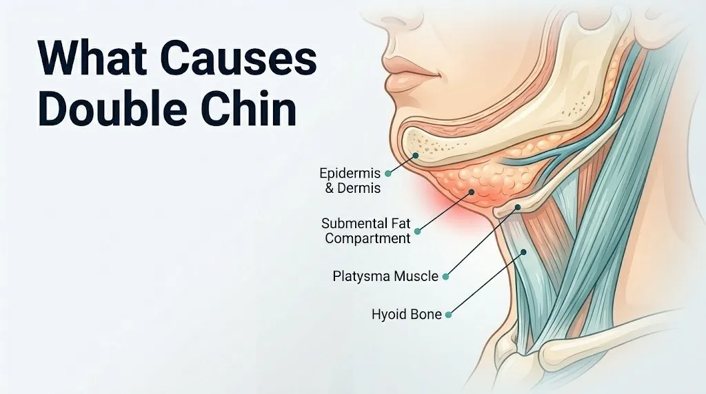
Submental fat is a discrete anatomical compartment of adipose tissue located beneath the skin of the chin and anterior neck, bounded by specific muscle layers that define its borders and clinical behavior. It is not simply a random accumulation of fat beneath the skin.
Think of the submental region like a precisely engineered sandwich. The top layer is the skin and thin layer of subcutaneous connective tissue. The middle layer is the submental fat compartment itself, a defined pocket of adipose tissue. The bottom structural layer is the platysma muscle, a broad, thin sheet of skeletal muscle that spans from the clavicle and upper chest up to the mandible and the skin of the lower face. Above the fat compartment and deeper still are the mylohyoid and geniohyoid muscles, which form the muscular floor of the mouth and provide the superior structural ceiling to the submental space. When any layer of this sandwich degrades or expands, the visual result is the same: chin fullness that the person sees as a double chin.
The superficial musculoaponeurotic system (SMAS) connects the platysma to the facial muscles laterally and plays a direct role in how fat and skin in the lower face and neck are mechanically supported. When the SMAS loses tension with age, the lower face and neck tissue descends, creating jowling and cervical skin laxity that contributes visually to submental fullness even without significant fat volume increase.
According to anatomy research reviewed in the Journal of Plastic and Reconstructive Surgery, the submental fat compartment is histologically distinct from adjacent cervical fat, with different adipocyte characteristics, lipolytic enzyme activity, and response to metabolic stimuli. This compartment-specificity is why the submental region often retains fat longer than other body areas during weight loss.
Older adults, particularly those over age 60, experience the greatest contribution from structural changes (platysma atrophy and SMAS laxity) relative to fat volume increase, meaning their double chin may look prominent even at a healthy weight.
Weight Gain and Double Chin
Weight gain causes a double chin by expanding the submental fat compartment through adipocyte hypertrophy and, at higher levels of adiposity, through adipogenesis (the creation of new fat cells) driven by sustained caloric surplus and hyperinsulinemia. This is the most straightforward mechanism but also the most commonly oversimplified.
The physiological pathway begins with caloric surplus. When caloric intake consistently exceeds energy expenditure, pancreatic beta cells secrete higher levels of insulin to manage elevated blood glucose. Hyperinsulinemia activates insulin receptor substrate-1 (IRS-1) signaling in preadipocytes throughout the body, including in the submental fat compartment, promoting their differentiation into mature, lipid-storing adipocytes. Existing adipocytes also undergo hypertrophy: they expand their intracellular lipid droplets as they store more triglycerides.
Fat does not distribute evenly across all body depots simultaneously. Individual genetics, sex hormone profiles, and depot-specific adrenergic receptor ratios determine which fat compartments fill fastest. The submental fat compartment in some individuals is particularly efficient at storing fat early in the weight gain process, before other regions show obvious change. This explains why someone can gain only 10 to 15 pounds and notice a dramatic change in their chin appearance before their waist circumference changes significantly.
According to data from the National Institute of Diabetes and Digestive and Kidney Diseases, even modest excess weight, a body mass index shift of just two to three units above a person’s metabolic set point, can produce visible changes in regional fat depots if those depots have a genetic predisposition to preferential filling.
People with type 2 diabetes mellitus or prediabetes (characterized by insulin resistance and compensatory hyperinsulinemia) face an amplified adipogenesis signal even without dramatic caloric excess, because the chronically elevated insulin environment persistently drives fat storage signals in preadipocytes throughout subcutaneous depots.
Key factors that amplify weight-related submental fat accumulation:
- Chronic hyperinsulinemia from insulin resistance or high-glycemic dietary patterns
- Low physical activity, reducing catecholamine-stimulated lipolysis throughout the day
- High dietary intake of refined carbohydrates, amplifying postprandial insulin spikes
- Sleep deprivation, which raises cortisol and ghrelin, promoting appetite and fat storage
- Alcohol consumption, which provides excess caloric load while impairing fatty acid oxidation
Key Takeaway: The submental fat compartment is a discrete anatomical space bounded by the platysma below and mylohyoid above, and its tendency to fill with fat during weight gain is determined largely by genetics and depot-specific adrenergic receptor ratios, not just overall body weight.
Genetics and Double Chin
Genetics causes a double chin by determining the distribution and density of fat-storing cells in the submental region, the structural integrity and thickness of the platysma muscle, and the baseline lipolytic efficiency of cervical fat depots, all of which are substantially heritable traits. This explains why some people develop a visible double chin at a healthy body weight while their relatives at higher weights do not.
The genetic architecture of regional fat distribution involves variants in multiple genes. The PLIN1 gene (perilipin-1), which encodes a protein coating the surface of lipid droplets in adipocytes and regulating their access to lipolytic enzymes, has variants associated with impaired fat mobilization from subcutaneous depots. A person inheriting lower-efficiency PLIN1 variants has adipocytes that release stored fat more slowly during caloric deficit or exercise, meaning fat accumulates in subcutaneous depots like the submental compartment and resists mobilization even with lifestyle changes.
Fat distribution genetics also includes variants near the FTO gene (fat mass and obesity associated) and several other loci identified in genome-wide association studies on body fat distribution, reviewed in a 2021 paper in Nature Genetics. These variants collectively explain a substantial portion of the variance in fat depot filling patterns, including why cervical fat accumulates prominently in some families across generations regardless of body weight.
Beyond fat cell distribution, genetics determines the structural characteristics of the platysma muscle. Platysma muscle thickness, fiber composition, and rate of age-related atrophy all have heritable components. A person who inherits a thinner platysma with a faster atrophy rate will show cervical skin laxity at a younger age and lower body weight than someone with a thicker, more durable platysma muscle.
According to the American Society of Plastic Surgeons, a family history of submental fullness is one of the most consistent predictors of an individual’s likelihood of developing a double chin, independent of body weight and age. This clinical observation aligns with the genetic research on fat distribution heritability.
People of East Asian ancestry have been documented in observational studies to accumulate more visible submental and cervical fat at lower overall body fat percentages compared to European-ancestry populations, attributed in part to differences in subcutaneous fat compartment distribution patterns and bone structure of the mandible and hyoid, which affects the cervicomental angle.
Aging and Skin Laxity Under the Chin
Aging causes a double chin through progressive loss of dermal collagen and elastin, age-related atrophy of the platysma muscle, and gradual descent of the superficial musculoaponeurotic system, all of which reduce the structural support that holds cervical skin and fat in a youthful position. This is why a visible double chin can develop or worsen even when body weight remains completely stable.
The molecular mechanism starts in the dermis. Dermal fibroblasts produce the collagen and elastin fibers that give skin its firmness and recoil. With advancing age, fibroblast activity declines: TGF-beta signaling (transforming growth factor beta), which normally drives fibroblasts to synthesize type I and type III collagen, becomes less effective. Simultaneously, matrix metalloproteinases (MMP-1 and MMP-3) produced by aging fibroblasts and keratinocytes increasingly degrade existing collagen and elastin at a rate that exceeds their replacement. The net result is a dermis with fewer and thinner collagen fibers and fragmented elastin, producing skin that is less firm, less elastic, and less capable of maintaining its position against gravity.
Ultraviolet radiation from sun exposure accelerates this process significantly through photoaging: UV-B radiation directly upregulates MMP-1 expression in skin fibroblasts and keratinocytes, accelerating collagen degradation beyond what chronological aging alone would produce. The anterior neck and submental region, frequently exposed to sun without sunscreen, accumulates significant photoaging-driven laxity over decades.
The American Academy of Dermatology reports that collagen production in the skin decreases by approximately 1 percent per year after age 20, with the rate of decline accelerating after age 40. By the time a person reaches age 60, they may have lost 30 to 40 percent of the dermal collagen density they had at age 20, a loss that is most visually apparent in areas of high gravitational load on lax skin, particularly the neck and jowl region.
Postmenopausal women experience an accelerated phase of collagen loss in the years immediately following menopause. Research published in the Journal of Investigative Dermatology found that skin collagen content decreases by approximately 2 percent per year during the first five years after menopause, compared to approximately 1 percent per year in premenopausal women, driven by the loss of estrogen’s collagen-stimulating effect on fibroblasts via estrogen receptor signaling.
Poor Posture and Double Chin
Poor posture, specifically sustained forward head posture, contributes to a double chin by chronically shortening and weakening the submental muscles that provide structural support to the floor of the mouth and submental tissues, while simultaneously overstretching and weakening the platysma, reducing its ability to maintain skin support in the anterior neck.
Forward head posture is the position in which the head sits in front of its ideal alignment over the shoulders rather than balanced directly above the cervical spine. For every inch the head moves forward from its neutral position, the effective gravitational load on the cervical spine and anterior neck structures increases. The mylohyoid, geniohyoid, and the anterior belly of the digastric muscle are all submental muscles that, in proper posture, contribute to holding the floor of the mouth and submental tissue architecture in a supported position.
When the head is chronically forward, these muscles adapt to a shortened resting length. Over time, they lose the capacity to generate meaningful tension in the anatomical position that would support submental tissue. The result: the submental tissue, including both fat and skin, has less muscular support from below and tends to hang in a lower position, making the cervicomental angle (the angle between the neck and chin) appear more obtuse and the double chin more visible.
Studies on head and neck posture reviewed in the Journal of Physical Therapy Science have found associations between sustained forward head posture and reduced activation of the suprahyoid muscle group (which includes the mylohyoid, geniohyoid, and digastric) during functional activities. This reduced functional activation further impairs the dynamic postural support these muscles provide to submental structures.
Quick Tip:
- Evaluate your resting head position: stand against a wall with your heels, buttocks, and upper back touching it. If the back of your head does not also touch the wall without effort, you have measurable forward head posture.
- People who spend six or more hours daily looking at a smartphone or laptop screen at a downward angle accumulate significantly greater forward head posture loading than those who work at eye-level screens, according to biomechanical analyses published in Surgical Technology International.
- Adolescents and young adults are now developing forward head posture at younger ages due to screen use patterns, making posture a relevant double chin contributor even in people under age 25.
Hormones and Double Chin
Hormones cause a double chin by regulating where the body stores fat, how efficiently it retrieves fat from specific depots, and how the structural integrity of skin and muscle in the cervical region is maintained over time. Three major hormonal axes drive submental fat changes: the sex hormone axis (estrogen and testosterone), the adrenal axis (cortisol), and the thyroid axis.
Insulin is the most fundamental hormonal driver of fat storage. When chronically elevated due to insulin resistance or a high-glycemic diet, insulin preferentially drives fat into subcutaneous depots by suppressing adipocyte lipolysis (via phosphodiesterase-3B inhibition) and promoting lipogenesis (via activation of fatty acid synthase). The submental fat compartment, like all subcutaneous fat depots, is subject to insulin’s lipogenic effect.
The sympathetic nervous system provides the counter-signal: catecholamines (epinephrine and norepinephrine) stimulate fat release from adipocytes via beta-adrenergic receptors. The problem specific to the submental compartment is its receptor composition. This depot has a relatively higher ratio of alpha-2 adrenergic receptors (which inhibit lipolysis) to beta-adrenergic receptors (which stimulate it) compared to abdominal fat, making it more resistant to catecholamine-driven fat mobilization during exercise and caloric deficit.
This receptor imbalance is why exercise and caloric restriction reliably reduce abdominal fat before visibly reducing submental fat in many individuals. The submental depot is physiologically slower to release its stored triglycerides, regardless of total body fat reduction.
According to research reviewed in the Journal of Clinical Endocrinology and Metabolism, regional fat depot differences in adrenergic receptor density are a primary reason why fat loss is not proportional across all body compartments during weight reduction interventions.
People taking beta-blocker medications (commonly prescribed for hypertension and cardiac conditions) may find that submental fat is particularly resistant to exercise-induced lipolysis, because beta-blockers directly antagonize the beta-adrenergic receptors that would normally drive fat mobilization from this depot during physical activity.
Key Takeaway: The submental fat compartment is physiologically resistant to fat mobilization compared to abdominal fat because of its higher alpha-2 to beta-adrenergic receptor ratio, which is why it often persists after weight loss that has already reduced fat elsewhere.
Menopause and Chin Fat
Menopause causes increased submental fat accumulation through two simultaneous mechanisms: the loss of estrogen’s role in regulating lipolysis in peripheral fat depots, and a shift in overall fat distribution from a gynecoid (hip and thigh predominant) to a more central and cervicodorsal pattern as estrogen-mediated fat storage preferences change with hormonal transition.
Estradiol, the primary estrogen produced by the ovaries before menopause, binds to estrogen receptor alpha (ERa) on adipocytes in peripheral fat depots, including the submental region. One of estrogen’s regulatory effects at this receptor is to maintain the sensitivity and density of beta-adrenergic receptors in these depots, supporting catecholamine-driven lipolysis. As estradiol levels fall during perimenopause and reach their postmenopausal nadir, this beta-adrenergic receptor support is withdrawn. The submental fat depot becomes even more resistant to catecholamine-stimulated lipolysis than it was before menopause.
Simultaneously, the decline in estrogen shifts the body’s fat distribution preference. Before menopause, estrogen promotes preferential storage of fat in peripheral depots (hips, thighs, breasts). After menopause, without this estrogenic preference, new fat storage shifts toward more central and upper body distribution, including the abdomen, upper back, and cervicodorsal region. The submental area is caught in both effects: it retains existing fat more stubbornly and receives a higher proportion of new fat deposition.
A 2018 study published in Menopause (the journal of the Menopause Society) found that postmenopausal women had measurably greater submental and cervical subcutaneous fat thickness compared to age-matched premenopausal women, independent of total body weight, confirming that the hormonal transition itself drives regional fat redistribution to the submental area.
Estrogen also has a collagen-stimulating effect on dermal fibroblasts via ERa. The loss of this effect after menopause accelerates the dermal collagen loss discussed in the aging section, compounding the visible effects of fat redistribution with skin laxity, producing a more pronounced double chin appearance than either mechanism alone would create.
Women on postmenopausal hormone replacement therapy (HRT) appear to have partially attenuated rates of submental fat accumulation and skin collagen loss compared to women not on HRT, based on observational data, though clinical guidelines from the American College of Obstetricians and Gynecologists (ACOG) do not endorse HRT for cosmetic fat redistribution specifically, as the benefits must be weighed against risks in individual patients.
Cortisol and Submental Fat Redistribution
Cortisol excess causes a distinctive pattern of fat redistribution that specifically targets the upper back, posterior neck, and submental region, producing a fullness under the chin that is mechanistically different from simple submental adipogenesis from caloric excess. This cortisol-driven redistribution can occur from endogenous overproduction (Cushing’s syndrome) or from exogenous corticosteroid medication use.
The mechanism involves glucocorticoid receptors (GR) on adipocytes. Glucocorticoid receptors are expressed in higher density in cervicodorsal and visceral fat depots compared to peripheral subcutaneous depots. When cortisol levels are chronically elevated, these high-density GR regions receive a proportionally stronger adipogenic signal. The GR, when activated by cortisol, upregulates lipoprotein lipase (LPL) activity in these adipocytes, increasing their capacity to extract circulating fatty acids from the bloodstream and store them as triglycerides. The cervical and submental fat compartments expand disproportionately as a result.
Cushing’s syndrome (ICD-10 E24), the clinical condition of pathological cortisol excess from a pituitary adenoma, adrenal tumor, or ectopic ACTH production, produces the characteristic “buffalo hump” (cervicodorsal fat pad) and “moon face” (facial rounding from temporal and mandibular fat accumulation) that visually includes a pronounced double chin. According to Mayo Clinic, Cushing’s syndrome affects approximately 10 to 15 people per million annually, making it uncommon but clinically important because its submental fat pattern mimics a purely cosmetic double chin while carrying systemic health implications requiring treatment.
Exogenous corticosteroid use (oral prednisone, prednisolone, and high-dose inhaled or topical steroids in some cases) produces the same glucocorticoid receptor-mediated redistribution. People on long-term corticosteroid therapy for conditions such as asthma, inflammatory bowel disease, lupus, or rheumatoid arthritis frequently develop submental fat accumulation as a medication side effect, not from dietary changes or weight gain.
Chronic psychological stress elevates cortisol through sustained hypothalamic-pituitary-adrenal (HPA) axis activation. While the cortisol elevation from psychological stress is typically lower in magnitude than in Cushing’s syndrome, prolonged HPA axis dysregulation still produces measurable shifts in fat distribution toward visceral and cervicodorsal depots over months to years, according to observational research published in the journal Obesity.
People on long-term corticosteroid therapy who develop submental fat accumulation should discuss the fat redistribution with their prescribing physician, as dosage adjustment, steroid-sparing agents, or therapy modification may be possible depending on the underlying condition being treated.
Thyroid Conditions and Double Chin Appearance
Thyroid conditions, particularly hypothyroidism (underactive thyroid), can produce submental fullness that visually resembles a double chin but is caused by soft tissue fluid accumulation (myxedema), reduced metabolic rate driving weight gain, or in some cases, a thyroid goiter projecting anteriorly into the anterior neck region. Distinguishing thyroid-related submental fullness from cosmetic fat accumulation requires clinical evaluation.
Hypothyroidism is caused by insufficient production of thyroid hormone (thyroxine, T4, and triiodothyronine, T3) from the thyroid gland, most commonly from autoimmune destruction (Hashimoto’s thyroiditis). Thyroid hormone regulates the basal metabolic rate through its action on mitochondria in virtually every cell in the body. When thyroid hormone levels fall, metabolic rate decreases, caloric expenditure falls, and body weight tends to increase even without any change in dietary intake. This metabolic suppression can drive fat accumulation throughout subcutaneous depots including the submental region.
Hypothyroidism also produces myxedema: a type of non-pitting edema caused by the accumulation of glycosaminoglycans (primarily hyaluronic acid and chondroitin sulfate) in the dermal and subcutaneous tissues. Unlike simple edema from fluid overload, myxedema is driven by the accumulation of hygroscopic polysaccharides that retain water in the extracellular matrix. This produces a characteristic puffy, doughy fullness of the face and anterior neck that can create or worsen the appearance of a double chin and does not respond to lipolytic treatments because it is not fat.
According to the American Thyroid Association, hypothyroidism affects approximately 4.6 percent of the U.S. population, with women five to eight times more likely to develop it than men. Women over age 60 have rates approaching 10 to 15 percent, making thyroid disease a clinically important consideration in this demographic when evaluating submental fullness.
A thyroid goiter (enlarged thyroid gland) projects anteriorly from its position at the base of the anterior neck and can create visible fullness in the lower anterior neck that is confused with submental fat. Goiters move upward when the patient swallows, which is a useful clinical distinguishing feature. Any visible or palpable swelling in the anterior neck that moves with swallowing warrants evaluation by a primary care physician and thyroid ultrasound, not cosmetic treatment.
Key Takeaway: Submental fullness that developed alongside unexplained weight gain, fatigue, cold intolerance, or dry skin warrants a TSH blood test to rule out hypothyroidism before any cosmetic evaluation, because myxedema-related cervical fullness requires thyroid hormone treatment, not fat reduction procedures.
Double Chin in Thin People
A double chin in a thin person who is not overweight is caused by genetic predisposition to preferential fat storage in the submental compartment, low cervicomental angle from mandibular or hyoid bone anatomy, platysma muscle thinness or laxity, or a combination of these factors that operate independently of total body fat percentage.
This is one of the most clinically misunderstood aspects of double chin causes, because most public health content implies that a double chin is simply a result of being overweight. The submental fat compartment can be disproportionately enlarged relative to total body fat if a person’s genetic fat distribution pattern assigns this compartment a high filling priority. A person with a body mass index of 21 (within the normal range) can have a visually prominent submental fat pad while having minimal abdominal or limb fat, if their genetically determined fat distribution pattern fills the cervical depots preferentially.
Bone structure is an underappreciated contributor. The cervicomental angle is the geometric angle formed between the line of the neck and the line of the chin. A more obtuse cervicomental angle (above approximately 105 to 120 degrees) produces a less defined jaw-neck transition, making even a small amount of submental soft tissue appear as a double chin. People with a naturally recessed mandible (retrognathia), a low-positioned hyoid bone, or a shorter neck relative to jaw length have a structurally obtuse cervicomental angle that makes a double chin visually apparent at very low submental fat volumes.
According to the American Society of Plastic Surgeons, a substantial proportion of patients seeking treatment for submental fullness are at or near a healthy body weight, with their double chin driven primarily by genetic fat distribution and cervicomental anatomy rather than overall adiposity. This population typically responds well to non-surgical or minimally invasive submental fat reduction rather than systemic weight management.
Adolescents and young adults who develop a double chin at a healthy weight frequently have this anatomy-and-genetics-driven pattern. Their submental fat compartment filled early in their development due to genetic distribution priorities, and it will not proportionally reduce with further weight maintenance or even with modest weight loss.
Double Chin in Men vs Women
The causes and patterns of double chin differ between men and women because of fundamental differences in sex hormone-regulated fat distribution, platysma muscle thickness, bone structure of the mandible and hyoid, and the timing and magnitude of age-related changes in submental tissue. These are not superficial differences but physiologically distinct processes.
Women have a higher percentage of total body fat than men at the same body mass index, driven by estrogen’s role in promoting peripheral subcutaneous fat storage for reproductive energy reserves. The submental fat compartment in women tends to fill earlier in the weight gain process and fill more readily at lower overall body fat percentages than in men, because estrogenic fat distribution preferences include peripheral subcutaneous depots like the submental region.
Men store a higher proportion of excess caloric intake as visceral (abdominal) fat, driven by androgen signaling. Testosterone promotes central fat deposition via androgen receptor-mediated effects on visceral adipocyte lipolytic activity, and it also promotes submental fat mobilization by maintaining a higher beta-adrenergic receptor density in cervical fat depots compared to the estrogenic environment. This means men at moderate degrees of excess weight tend to show abdominal obesity first and submental fat accumulation later in the weight gain trajectory compared to women.
However, men often have lower overall platysma muscle thickness than women and a heavier jaw structure that creates higher mechanical loading on submental skin over time. Men who develop submental fat accumulation after age 45 frequently also have concurrent platysma atrophy from age-related testosterone decline, producing a double chin that combines fat volume and structural laxity simultaneously.
| Factor | Women | Men |
|---|---|---|
| Primary fat driver | Estrogen-mediated peripheral depot filling | Caloric surplus driving visceral and then subcutaneous |
| Submental fat onset timing | Earlier in weight gain trajectory | Later, often after significant overall weight gain |
| Platysma laxity timing | Earlier (lower baseline estrogen from menopause accelerates) | Later, but accelerates with testosterone decline after 45 |
| Bone structure influence | Smaller mandible can reduce cervicomental angle definition | Stronger mandible typically provides clearer jaw line unless adiposity is high |
| Hormonal cause unique to group | Menopause, perimenopause, estrogen fluctuation | Hypogonadism (low testosterone) in older men |
| Medication-related cause | HRT changes, antipsychotic use | Anabolic steroid use, testosterone therapy changes |
Men with hypogonadism (clinically low testosterone) experience a shift toward an estrogen-dominant fat distribution pattern, increasing submental fat susceptibility and reducing the beta-adrenergic receptor support that normally aids cervical fat mobilization in men.
When a Double Chin Appears Suddenly
A double chin that appears suddenly over days to weeks, rather than developing gradually over months to years, is a clinical warning sign that requires medical evaluation, because this presentation pattern is inconsistent with the gradual mechanisms of adipogenesis, aging, and hormonal change and suggests a pathological cause of submental or cervical swelling.
Gradual double chin development from weight gain, aging, or hormonal change occurs over months to years. Fat cells accumulate slowly. Collagen loss occurs at approximately 1 percent per year. These are progressive, slow-onset processes. When submental fullness develops over days to weeks, the cause is almost certainly not fat accumulation. It is more likely to represent one of the following:
Cervical lymphadenopathy: Enlarged lymph nodes in the submandibular and submental lymph node chains (level I nodes) can produce visible and palpable swelling under the chin. Causes range from reactive enlargement from a local infection (dental abscess, pharyngitis, skin infection of the face or scalp) to lymphoma or metastatic malignancy. Reactive lymphadenopathy from infection typically resolves within two to four weeks. Lymph nodes that are hard, fixed, non-tender, or greater than 1 centimeter in diameter and persist beyond four weeks warrant evaluation by a primary care physician and potentially fine needle aspiration biopsy.
Submandibular salivary gland enlargement: The paired submandibular glands sit in the submandibular triangle and can enlarge rapidly from sialadenitis (salivary gland infection) or from salivary duct obstruction by a calculus (sialolithiasis). This produces a firm, often painful swelling that worsens at mealtimes when salivary flow is stimulated.
Angioedema: Acute allergic or hereditary angioedema (HAE) can produce rapid soft tissue swelling of the face, neck, and submental region within hours. This is a potential medical emergency requiring immediate attention.
Thyroid change: A rapidly enlarging thyroid (thyroiditis, thyroid cyst hemorrhage, or thyroid malignancy) can produce anterior neck swelling that appears suddenly.
Quick Tip:
- Press gently on the submental swelling. A double chin from fat feels soft, uniformly distributed, and non-tender. A swollen lymph node or salivary gland feels more discrete, has defined borders, and may be tender to pressure.
- Check your temperature. A sudden submental swelling with fever suggests an infectious cause (dental abscess drainage, lymphadenitis, sialadenitis).
- If the swelling came up in hours and you are having difficulty swallowing or breathing, this is a medical emergency. Call 911 or go to the nearest emergency room immediately.
When Does Neck Swelling Become a Medical Emergency
Sudden neck or submental swelling with certain associated symptoms requires immediate emergency evaluation. Do not wait to see if it resolves on its own.
Call 911 or go to the nearest emergency room immediately if you experience:
- Rapidly progressive swelling of the neck or under the chin combined with difficulty breathing or stridor: this can indicate angioedema, Ludwig’s angina, or a rapidly enlarging infectious process threatening the airway
- Difficulty swallowing saliva or liquids with associated neck swelling that developed over hours to days: this may indicate deep space neck infection or epiglottitis
- High fever above 38.5°C (101.3°F) with rapidly developing submental swelling and an inability to fully open your mouth: this combination can indicate Ludwig’s angina, a life-threatening bilateral submandibular space infection
- Sudden neck mass appearing over hours with throat tightening, tongue swelling, or hives: this suggests allergic or hereditary angioedema requiring immediate epinephrine and emergency management
These presentations can indicate airway-threatening infections or angioedema and require emergency medical assessment, not a dentist appointment or delayed clinic visit.
Medical and Non-Surgical Treatments for Double Chin
Medical and non-surgical treatments for a double chin include injectable deoxycholic acid (FDA-approved), cryolipolysis, radiofrequency skin tightening, and ultrasound-based treatment, each targeting a different component of the double chin anatomy, either the fat compartment volume or the overlying skin laxity. Surgical options include liposuction and neck-lift surgery.
Deoxycholic acid injection (brand name Kybella, FDA-approved in 2015) is the only FDA-approved injectable treatment for submental fat. Deoxycholic acid is a bile acid that disrupts adipocyte cell membranes when injected into the submental fat compartment, producing cytolysis of fat cells and a localized inflammatory response that is followed by fibrosis and tissue contraction. According to FDA approval data, clinical trials showed that 79 percent of treated patients had a measurable reduction in submental fat at six months. Multiple treatment sessions are typically required, spaced four to six weeks apart.
Cryolipolysis (commonly known as CoolSculpting): applies controlled cooling to the submental fat compartment, inducing apoptosis (programmed cell death) in adipocytes. The treated fat cells are cleared over approximately two to three months via the lymphatic system and macrophage phagocytosis. Research published in Dermatologic Surgery found submental fat reduction of approximately 20 to 25 percent at three months after a single cryolipolysis session.
Radiofrequency (RF) skin tightening devices target the dermis and SMAS layer with thermal energy, stimulating fibroblast collagen production and causing immediate collagen contraction. RF treatments address skin laxity rather than fat volume and are most effective in patients with mild to moderate submental laxity without substantial fat volume excess.
Ultrasound-based treatment (Ultherapy and similar devices) uses focused ultrasound energy to target the SMAS layer and deeper dermis, stimulating collagen neogenesis. These treatments work best as adjuncts to fat reduction in patients where platysma laxity and skin laxity are significant contributors to the double chin appearance.
For patients with prominent structural issues (severely lax platysma, significant cervicomental angle blunting), surgical platysmaplasty (neck-lift with platysma repair) combined with liposuction provides the most durable result, according to outcome data reviewed in the Journal of Plastic and Reconstructive Surgery.
Older adults with extensive skin laxity typically achieve better results from surgical options than from non-surgical treatments, because non-surgical modalities cannot replicate the structural platysma tightening that surgery achieves.
When to See a Doctor for a Double Chin
You should see a doctor for a double chin when it appeared suddenly rather than gradually, when it is accompanied by systemic symptoms like fatigue, weight gain, cold intolerance, or night sweats, when it is painful or tender to touch, when you can feel a distinct firm mass within the swelling, or when it has grown noticeably over a period of weeks to months.
The right provider depends on the clinical picture:
Primary care physician: First point of contact for any sudden onset submental swelling, any palpable neck mass, associated systemic symptoms, or concern about a medication side effect. A TSH blood test to evaluate thyroid function and a physical examination of the neck lymph nodes are appropriate first steps. Bring a list of all current medications to the appointment.
Board-certified endocrinologist: If a primary care physician identifies elevated TSH (hypothyroidism), low morning cortisol with clinical features suggesting adrenal insufficiency, or cortisol excess patterns suggesting Cushing’s syndrome, referral to an endocrinologist is appropriate for further evaluation with a 24-hour urinary free cortisol test, overnight dexamethasone suppression test, or adrenal imaging.
Board-certified dermatologist: For evaluation of non-surgical submental fat reduction options (deoxycholic acid injections, cryolipolysis, RF tightening) in patients where the double chin has been confirmed to be cosmetic in origin and no medical cause has been identified.
Board-certified plastic surgeon or facial plastic surgeon: For evaluation of surgical options including liposuction, platysmaplasty, or combined procedures in patients where non-surgical options are insufficient or where structural platysma laxity is the primary driver.
What to bring to your appointment:
- Timeline of when you first noticed the fullness and whether it appeared gradually or quickly
- Complete list of current medications, specifically corticosteroids, antiretrovirals, antipsychotics, and beta-blockers
- Family history of double chin in parents or siblings at similar ages and weights
- Any associated symptoms: fatigue, unexplained weight change, cold sensitivity, mood changes
- Recent weight history: have you gained, lost, or maintained weight over the past year?
People with a history of thyroid cancer or lymphoma who develop new anterior or submental neck swelling should contact their oncologist or primary care physician within days rather than treating the swelling as a new cosmetic concern.
Key Takeaway: Any submental fullness that appeared suddenly, is tender, feels firm or nodular rather than soft and diffuse, or came with systemic symptoms like fatigue or weight change needs a primary care physician evaluation and TSH blood test before any cosmetic assessment.
Frequently Asked Questions About What Causes Double Chin
What is the main cause of a double chin?
The most common cause of a double chin is submental fat accumulation driven by a combination of caloric surplus, genetic fat distribution patterns, and age-related platysma muscle laxity and skin collagen loss.
No single cause dominates in every person: in younger adults, fat accumulation and genetics tend to drive the presentation, while in older adults and postmenopausal women, skin laxity and hormonal fat redistribution become equally or more important.
Identifying which mechanism is primary in a given individual determines whether lifestyle changes, hormonal evaluation, or aesthetic treatment is the most appropriate path.
Can you get a double chin even if you are not overweight?
Yes, a double chin can develop in people with a completely healthy body weight due to genetic predisposition to submental fat storage, a structurally obtuse cervicomental angle from mandibular or hyoid bone anatomy, or platysma muscle laxity from aging.
According to the American Society of Plastic Surgeons, a substantial proportion of patients seeking submental fat treatment are at or near a healthy body weight, with their double chin driven by anatomy and genetics rather than overall body fat.
Non-surgical treatments like deoxycholic acid injections are clinically effective for this population because the target is submental fat specifically, not total body fat.
Does genetics determine if you get a double chin?
Genetics strongly influences double chin development by determining fat distribution priorities between body depots, platysma muscle thickness, and bone structure of the mandible and hyoid.
Variants in genes such as PLIN1 and FTO affect how efficiently fat is stored and mobilized from the submental compartment, explaining why some families show prominent double chins across generations at similar body weights.
If both of your parents developed a double chin at a relatively young age or healthy weight, your genetic risk for the same pattern is meaningfully elevated.
Can hormones cause a double chin?
Yes, hormones cause a double chin through several distinct mechanisms: estrogen withdrawal at menopause impairs lipolysis in the submental fat compartment, cortisol excess from chronic stress or corticosteroid medications drives cervicodorsal and submental fat redistribution, and hypothyroidism produces myxedema-related cervical tissue fullness.
These hormonal causes can produce submental fullness that looks identical to fat-driven double chin but requires different management, making clinical evaluation appropriate when the presentation is not explained by weight gain alone.
A TSH blood test and clinical assessment by a primary care physician are the starting points for evaluating a hormonally driven double chin.
How do I know if my double chin is fat or swollen glands?
Submental fat feels soft, diffuse, non-tender, and uniformly distributed under the chin, with no distinct firm borders.
Enlarged lymph nodes or a salivary gland feel more discrete, have defined edges, may be tender to gentle pressure, and are often accompanied by other signs of infection or illness such as sore throat, fever, or recent dental problems.
Any submental swelling that appeared within days, is tender, feels firm or nodular, or is accompanied by fever or difficulty swallowing warrants evaluation by a primary care physician, as these features are not consistent with simple fat accumulation.
Can a double chin go away on its own?
A double chin from submental fat accumulation can reduce with meaningful body weight loss, but the submental compartment’s high alpha-2 adrenergic receptor density makes it physiologically resistant to proportional fat loss compared to other body areas.
A double chin from age-related skin laxity and platysma atrophy does not resolve on its own, as collagen loss and muscle atrophy are progressive without intervention.
Submental fullness from hypothyroidism, cortisol excess, or medication side effects typically improves when the underlying hormonal or pharmacological cause is treated by the appropriate specialist.
Closing
A double chin is a single visual finding with genuinely different causes, and understanding which biology is driving yours changes everything about how to address it. Fat accumulation, skin laxity, hormonal redistribution, genetic anatomy, and posture all produce the same visual result through completely different mechanisms. The right approach depends entirely on which mechanism is at work in your specific situation.
If your double chin developed slowly alongside gradual weight change, genetics, aging, or posture are the most likely contributors. If it appeared over weeks, came with systemic symptoms, or does not match the pattern of gradual fat accumulation, a primary care physician evaluation including a TSH test and neck examination should precede any other assessment.
The most actionable step you can take is this: if the timeline, tenderness, firmness, or associated symptoms of your submental fullness do not match the gradual, soft, and asymptomatic profile of simple submental fat, bring it to a primary care physician before assuming it is cosmetic.