Burst Blood Vessel in Eye: Causes and Red Flags
The most common causes of a burst blood vessel in the eye include a sudden spike in venous pressure from coughing or straining, blunt trauma to the eye area, elevated arterial blood pressure, anticoagulant medications, and, in many adults, no identifiable triggering event at all. Most cases are harmless and resolve without treatment, but specific patterns of presentation can signal an underlying systemic condition that needs medical attention.
According to the National Eye Institute, a part of the National Institutes of Health, subconjunctival hemorrhage is one of the most common benign conditions seen in both ophthalmology and primary care settings. Research published in the British Journal of Ophthalmology has found that idiopathic cases (those with no identifiable cause) account for roughly 30 to 40 percent of all presentations, making isolated spontaneous rupture genuinely common in otherwise healthy adults.
This article covers every major and minor cause of a burst blood vessel in the eye, explains the specific vascular mechanism behind each one, identifies which presentations are self-limiting and which are not, and gives you clear guidance on when the red eye in the mirror warrants a phone call to your doctor or a trip to the emergency room.
Burst Blood Vessel in Eye: What It Is and Why It Happens
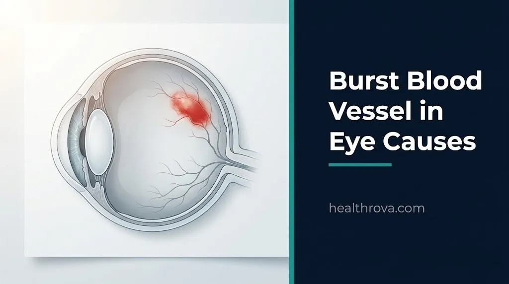
A burst blood vessel in the eye produces a bright red, flat patch on the white surface because blood has leaked out of a small conjunctival vessel and spread within the subconjunctival space between the transparent surface membrane and the underlying white coat of the eye.
The bulbar conjunctiva is the thin, transparent mucous membrane that covers the visible white surface of the eye (the sclera). It contains a dense network of small arterioles, venules, and capillaries. These vessels are tiny and relatively fragile by design. They serve the metabolic needs of the ocular surface, not high-pressure circulation.
When one of these vessels ruptures, blood collects between the conjunctival membrane and the sclera. Because this is a thin, sealed space, the blood spreads laterally rather than pooling. The result looks alarming because the color contrast against the white sclera is stark. However, the blood is entirely outside the eye’s internal structures and cannot affect vision in a typical presentation.
The condition is medically called subconjunctival hemorrhage. It is classified under ICD-10 code H11.3. The American Academy of Ophthalmology estimates that subconjunctival hemorrhage affects people of all ages, with incidence rising in adults over 50 due to age-related changes in conjunctival connective tissue and vascular wall elasticity.
Key facts about what makes this condition what it is:
- Blood is trapped in the subconjunctival space, not inside the eye
- The sclera beneath remains intact in typical cases
- The hemorrhage appears as a defined, flat red patch with sharp borders
- It does not cause pain in most presentations
- It does not produce discharge (distinguishing it from conjunctivitis)
- It typically resolves fully as the blood is resorbed by the body’s fibrinolytic system over 7 to 21 days
What Is a Subconjunctival Hemorrhage and How Does It Form
A subconjunctival hemorrhage forms when blood extravasates from a ruptured conjunctival microvessel into the potential space between the bulbar conjunctiva and the scleral surface, producing a visible red patch on the white of the eye.
The conjunctiva is nourished by a vascular network that includes contributions from the anterior ciliary arteries and branches of the episcleral venous plexus. These are very small caliber vessels. Their walls consist of an endothelial cell lining (the tunica intima), a thin smooth muscle and elastic tissue layer (the tunica media), and an outer connective tissue sheath (the tunica adventitia). In the conjunctiva, these walls are thinner than in most other tissues.
Think of these vessels like thin-walled garden hoses under variable water pressure. A momentary pressure spike, a small crack in the hose wall, or a weak section from repeated use can cause a localized rupture. The contents spill into the surrounding space and spread along the path of least resistance.
Once blood enters the subconjunctival space, the body’s fibrinolytic system begins breaking it down. Plasminogen, activated to plasmin by tissue plasminogen activator, cleaves fibrin and gradually resorbs the blood. This is why subconjunctival hemorrhage typically fades progressively over 1 to 3 weeks. The hemorrhage often appears to “migrate” toward the inner corner of the eye as resorption proceeds unevenly.
For people on anticoagulant medications or with low platelet counts, both the initial hemorrhage and the resorption period may be more prolonged because the fibrinolytic process depends on a functioning coagulation system for complete resolution.
| Feature | Subconjunctival Hemorrhage | Conjunctivitis |
|---|---|---|
| Color | Bright red, well-defined patch | Diffuse pink or red, no defined edge |
| Pain | Absent in most cases | Often present (irritation, burning) |
| Discharge | None | Often present (watery, mucoid, or purulent) |
| Vision change | Absent in typical cases | Absent unless corneal involvement |
| Cause | Vascular rupture | Infection or allergy |
| Resolution | 7 to 21 days | 7 to 14 days with appropriate treatment |
What Causes a Blood Vessel to Burst in Your Eye
A blood vessel bursts in the eye when intravascular pressure inside a conjunctival vessel exceeds the structural tolerance of that vessel’s wall, or when the vessel wall itself has been weakened by disease, mechanical wear, or medication effects.
Three distinct failure modes produce a subconjunctival hemorrhage. The first is a sudden pressure surge: venous or arterial pressure spikes abruptly beyond the vessel’s capacity, and the wall gives way at its weakest point. The second is wall weakness: the vessel wall has been structurally compromised by chronic disease, aging, inflammation, or drug effects, and normal or only slightly elevated pressure is sufficient to cause rupture. The third is direct mechanical disruption: trauma or friction physically tears the vessel wall.
Each named cause of burst blood vessel in the eye operates through one or more of these three mechanisms. Understanding which mechanism is at work helps predict whether the episode is a one-time pressure event or a sign of ongoing vascular vulnerability.
The tunica adventitia of conjunctival vessels loses tensile strength with age and with chronic oxidative stress from systemic conditions like diabetes and uncontrolled hypertension. Research published in JAMA Ophthalmology has noted that conjunctival vessel fragility increases measurably with advancing age, with more pronounced changes in adults over 60 years. This partially explains why subconjunctival hemorrhage is more frequent in older populations even without an obvious precipitating event.
The three core failure mechanisms:
- Pressure surge mechanism: Sudden transient rise in intravenous or arterial pressure overwhelms a structurally normal vessel wall
- Wall weakness mechanism: Chronic disease, medication, or aging thins the vessel wall, lowering the rupture threshold to near-normal pressure levels
- Mechanical disruption mechanism: Physical force directly tears the vessel wall regardless of pressure or wall integrity
Key Takeaway: A blood vessel bursts in the eye through one of three mechanisms: a sudden pressure surge, a weakened vessel wall, or direct mechanical trauma. Knowing which mechanism applies to your situation tells you a great deal about whether this is a one-time event or a pattern worth investigating.
What Causes Broken Blood Vessels in the Eye from Straining or Coughing
Straining, coughing, sneezing, vomiting, and heavy lifting cause a burst blood vessel in the eye by triggering the Valsalva maneuver, a physiological event in which forced expiration against a closed or partially closed airway sharply elevates intrathoracic pressure and subsequently raises venous pressure throughout the head and neck.
During a Valsalva maneuver, the diaphragm contracts forcefully against a closed glottis. Intrathoracic pressure rises dramatically. This increased pressure is transmitted backward through the superior vena cava into the venous drainage system of the head and neck, including the ophthalmic veins and the conjunctival venous network. The pressure spike is brief but intense. In a vessel with normal wall integrity, this may be enough to cause a localized rupture.
This is the physiological equivalent of briefly overpressurizing the thin-walled section of a balloon. The balloon may hold dozens of times, but if the pressure spike is large enough or the wall has a thin spot, it fails at the weakest point.
Coughing repeatedly, as during a bout of whooping cough (pertussis) or bronchitis, can cause multiple subconjunctival hemorrhages over a short period. Severe vomiting, particularly in cases of viral gastroenteritis, food poisoning, or bulimia nervosa, can produce the same mechanism. Childbirth is another high-pressure event: up to 33 percent of women develop subconjunctival hemorrhage during vaginal delivery, according to clinical observations reported in obstetric and ophthalmology literature.
For people with pre-existing hypertension or vascular fragility, the Valsalva pressure spike combines with an already elevated baseline venous pressure, substantially lowering the force needed to rupture a conjunctival vessel.
Common Valsalva triggers that cause burst blood vessels in the eye:
- Severe coughing fits (pertussis, bronchitis, chronic obstructive pulmonary disease exacerbations)
- Forceful sneezing
- Heavy lifting (weightlifting, manual labor, moving furniture)
- Bearing down during bowel movements (especially with constipation)
- Vomiting episodes
- Vaginal childbirth
- Vigorous crying in infants and young children
What Causes Burst Blood Vessel in Eye from High Blood Pressure
High blood pressure causes a burst blood vessel in the eye by chronically elevating arterial pressure against the walls of the conjunctival microvasculature, progressively damaging the endothelial lining and the tunica media until the wall loses structural integrity and spontaneously ruptures.
Arterial hypertension subjects small vessel walls to persistent mechanical stress. Over time, this stress drives pathological changes in the vascular wall: the endothelial cells become dysfunctional, the smooth muscle layer hypertrophies and then stiffens, and the elastic tissue within the tunica media degrades. The result is a vessel that is simultaneously under higher pressure and less capable of tolerating it.
Conjunctival vessels are part of the wider ophthalmic vascular territory and reflect systemic vascular health. A blood pressure reading above 140/90 mmHg (the threshold for stage 2 hypertension according to the American Heart Association) represents persistent excessive loading on vessels throughout the body, including the eye. The conjunctival vessels may be among the first to show signs of this loading because they are so thin-walled.
Hypertensive crisis (systolic blood pressure above 180 mmHg or diastolic above 120 mmHg) can cause sudden, severe subconjunctival hemorrhage as part of a broader pattern of acute hypertensive end-organ stress. In this situation, a burst blood vessel in the eye is not the primary concern. The simultaneous stress on the coronary arteries, cerebral vasculature, and kidneys is.
According to the Centers for Disease Control and Prevention, nearly 1 in 3 adults in the United States has hypertension, and approximately half of those do not have it adequately controlled. People in this group face a meaningfully higher background risk of spontaneous vascular rupture events, including subconjunctival hemorrhage.
For people with poorly controlled hypertension:
- A burst blood vessel in the eye without an obvious Valsalva trigger is a signal to check blood pressure immediately
- Repeated subconjunctival hemorrhages in a hypertensive individual warrant evaluation of blood pressure control strategy with a primary care physician or cardiologist
- If blood pressure at home or in a pharmacy exceeds 180/120 mmHg at the time of the hemorrhage, this meets the threshold for a hypertensive urgency or emergency requiring same-day medical evaluation
What Causes Blood Vessels to Burst in the Eye from Trauma
Trauma causes a burst blood vessel in the eye through direct mechanical disruption of conjunctival vessel walls, where physical force applied to the ocular surface or surrounding structures tears the vascular endothelium and the supporting wall layers, allowing blood to immediately extravasate into the subconjunctival space.
Blunt trauma is the most common traumatic cause. Being struck in or around the eye, impact during contact sports, a foreign body hitting the eye, or even a hard poke from a finger or object can produce this result. The magnitude of force needed is smaller than most people expect. The conjunctival vessels are superficial and protected only by the thin conjunctival membrane itself.
For any trauma-associated subconjunctival hemorrhage, it is essential to confirm that the injury is confined to the conjunctival surface and has not caused deeper damage. A hyphema (bleeding into the anterior chamber of the eye, between the cornea and the iris) looks similar on initial inspection but carries a very different risk profile, including the potential for secondary hemorrhage and acute elevated intraocular pressure. An ophthalmologist can distinguish these with a slit-lamp examination.
Trauma-associated hemorrhage can also accompany more serious ocular injuries:
| Condition | Clinical Feature | Urgency |
|---|---|---|
| Subconjunctival hemorrhage only | Flat red patch, no pain, no vision change | Monitor; evaluate if caused by significant force |
| Hyphema | Red blood visible in anterior chamber; may have blurred vision | Urgent: same-day ophthalmology evaluation |
| Orbital fracture | Periorbital bruising, diplopia, restricted eye movement | Emergency: go to ER |
| Ruptured globe | Irregular pupil, severe pain, visible wound, fluid from eye | Emergency: Call 911 immediately |
Age-specific note: older adults and children are at higher risk for significant ocular injury from seemingly minor trauma because of differences in periorbital tissue compliance and bony protection. Any trauma-associated hemorrhage in a child under 5 years that cannot be clearly explained by an accidental mechanism warrants evaluation by a pediatric emergency medicine physician to rule out non-accidental trauma.
What Causes a Popped Blood Vessel in the Eye from Blood Thinners
Anticoagulant and antiplatelet medications cause a burst blood vessel in the eye by disrupting the normal coagulation cascade or platelet aggregation pathway, which lowers the physiological threshold at which a conjunctival vessel rupture becomes clinically visible and slows the resorption of the resulting hemorrhage.
In people not taking these medications, minor microtrauma to conjunctival vessels happens constantly and is repaired quickly by the coagulation system before enough blood accumulates to be visible. Warfarin works by blocking the synthesis of vitamin K-dependent coagulation factors (factors II, VII, IX, and X). Apixaban and rivaroxaban directly inhibit Factor Xa. Aspirin irreversibly inhibits thromboxane A2 in platelets, reducing platelet aggregation. Clopidogrel blocks the ADP receptor on platelets, similarly impairing aggregation.
Any of these mechanisms means the body’s first-line response to a microrupture is blunted. What would normally be an invisible, spontaneously repaired microvascular event instead accumulates enough blood to produce a visible subconjunctival hemorrhage.
According to clinical guidance published by the American Academy of Ophthalmology, patients taking oral anticoagulants do experience subconjunctival hemorrhage at higher rates than the general population, and their hemorrhages tend to be larger in area and longer-lasting in duration.
What this means practically:
- A single subconjunctival hemorrhage in a patient on stable anticoagulant therapy with no recent dose change is generally not cause for alarm on its own.
- A hemorrhage that is dramatically large, bilateral, or accompanied by bleeding elsewhere (gums, easy bruising, nosebleed) may indicate over-anticoagulation and warrants checking the INR or contacting the prescribing physician.
- Patients on warfarin should have their international normalized ratio (INR) checked if a hemorrhage occurs outside their routine monitoring schedule, especially if the hemorrhage is unusually extensive.
- Any new subconjunctival hemorrhage in a patient who has recently started anticoagulant therapy or had a dose increase should prompt a call to the prescribing physician.
Key Takeaway: If you take a blood thinner (warfarin, apixaban, rivaroxaban, aspirin, or clopidogrel) and develop a burst blood vessel in your eye, a single small hemorrhage on stable therapy is usually a known side effect, but a large, bilateral, or recurrent hemorrhage with other bleeding signs warrants a same-day call to your prescribing physician to check your medication levels.
What Causes Broken Blood Vessels in Your Eye from Diabetes or Systemic Disease
Diabetes mellitus and other systemic diseases cause burst blood vessels in the eye by producing diabetic microangiopathy, a process in which chronically elevated blood glucose damages the structural proteins of small vessel walls throughout the body, including the conjunctival microvasculature, making them progressively more fragile and prone to spontaneous rupture.
In diabetes, persistent hyperglycemia drives glycosylation of the basement membrane proteins that form the scaffolding of vessel walls. The glycated proteins accumulate, the basement membrane thickens and loses flexibility, and the pericyte cells that normally reinforce capillary walls in the eye are selectively lost. This process makes conjunctival vessels simultaneously stiffer and weaker, lowering the pressure threshold at which they rupture.
Beyond diabetes, several other systemic conditions alter conjunctival vessel integrity:
| Systemic Condition | Mechanism of Increased Hemorrhage Risk |
|---|---|
| Diabetes mellitus | Glycation of basement membrane proteins; pericyte loss; microangiopathy |
| Thyroid disease (hypothyroidism) | Myxedematous deposits alter connective tissue, including around blood vessels |
| Chronic liver disease | Impaired production of coagulation factors (II, V, VII, IX, X, XI) |
| Systemic lupus erythematosus | Immune-mediated vascular inflammation and antiphospholipid antibodies affecting coagulation |
| Leukemia and lymphoma | Thrombocytopenia from marrow infiltration impairs platelet-based hemostasis |
| Severe anemia | Reduced blood viscosity and altered microvascular flow dynamics |
The National Institutes of Health notes that unexplained or recurrent subconjunctival hemorrhage in the absence of an obvious mechanical cause should prompt evaluation for systemic hematological or vascular conditions, particularly in adults over 40 with other undiagnosed symptoms such as fatigue, easy bruising, or unexplained weight loss.
For people with type 2 diabetes: subconjunctival hemorrhage in a poorly controlled diabetic patient is a visible sign of broader microvascular compromise. The same process damaging conjunctival vessels is operating in the retinal vessels, the renal glomerular capillaries, and the peripheral nerve microvasculature. A burst blood vessel in the eye in a diabetic patient is a reasonable prompt to schedule a diabetic eye exam with an ophthalmologist and a check of HbA1c with their primary care physician if overdue.
What Causes Blood Vessel in Eye to Burst from Eye Rubbing or Contact Lens Use
Mechanical friction from eye rubbing or contact lens use causes a burst blood vessel in the eye by applying direct shear force to the conjunctival surface, which physically disrupts the thin-walled vessels within the conjunctival epithelium and stroma.
Rubbing the eye seems harmless, but the mechanical force delivered to a small area of the conjunctival surface is substantially greater than it appears. The conjunctival vessels are located within approximately 0.1 millimeters of the surface. Firm rubbing presses and rolls these vessels against the harder sclera beneath, creating a pinching and shearing effect on the vessel wall.
Contact lens use introduces a different but related mechanism. Daily insertion and removal creates repeated mechanical contact with the superior and inferior conjunctival fornix. Lens wear for extended periods can cause localized conjunctival vessel dilation and, in some users, focal vessel wall fatigue. Research in the British Journal of Ophthalmology has documented higher rates of subconjunctival hemorrhage in long-term rigid gas permeable lens wearers compared to soft lens wearers, with the difference attributed to the greater mechanical interaction of rigid lenses with the conjunctival surface.
Quick Tip for contact lens wearers and those with eye rubbing habits:
- If a subconjunctival hemorrhage appears consistently on the same side of the eye or in the same location, consider whether a contact lens fit issue, lens edge design, or habitual rubbing pattern may be creating focal vessel stress
- An optometrist can assess lens fit under slit-lamp examination and identify whether lens edge lift or conjunctival papillae suggest chronic mechanical irritation
- For people who rub their eyes frequently due to allergic conjunctivitis: addressing the underlying allergic inflammation with an ophthalmologist or allergist reduces the itch-rub cycle that drives this mechanism
Older adults who rub their eyes are at higher risk because conjunctival tissue loses elasticity with age, reducing the vessel wall’s ability to absorb mechanical force without tearing.
Burst Blood Vessel in Eye Causes Related to Infection or Inflammation
Certain eye infections and inflammatory conditions cause a burst blood vessel in the eye by triggering local inflammatory cascades that increase vascular permeability, dilate conjunctival vessels to a degree that exceeds wall tolerance, and in some cases directly involve the vessel wall in an immune-mediated injury process.
Bacterial conjunctivitis caused by organisms such as Staphylococcus aureus or Streptococcus pneumoniae can produce enough conjunctival inflammation and vessel dilation that focal ruptures occur, particularly in the area of most intense inflammation. The associated redness from vasodilation and the redness from a true hemorrhage can coexist, making clinical distinction important.
Viral conjunctivitis, especially adenoviral conjunctivitis, can produce subconjunctival hemorrhage as a defining clinical feature. The specific variant called epidemic keratoconjunctivitis (EKC) and acute hemorrhagic conjunctivitis (AHC), caused by enterovirus 70 or coxsackievirus A24, produce subconjunctival hemorrhage as a primary presentation. Acute hemorrhagic conjunctivitis has appeared in outbreak patterns across Asia, Africa, and the Americas. The American Academy of Ophthalmology has documented these outbreaks, noting that the hemorrhagic form of viral conjunctivitis is accompanied by discharge, eye pain, and photophobia, distinguishing it from the painless, non-dischargey presentation of idiopathic subconjunctival hemorrhage.
Inflammatory conditions that can produce conjunctival hemorrhage:
- Severe allergic conjunctivitis with intense eye rubbing (combined mechanical and inflammatory mechanism)
- Episcleritis (inflammation of the episcleral tissue beneath the conjunctiva, which can involve the overlying conjunctival vessels)
- Scleritis (deeper inflammation of the sclera itself; associated hemorrhage here requires prompt ophthalmology evaluation as scleritis can indicate systemic autoimmune disease)
Key Takeaway: When a burst blood vessel in the eye is accompanied by discharge, eye pain, light sensitivity, or both eyes being affected simultaneously, infection or inflammation is a more likely cause than a simple pressure event, and evaluation by an optometrist or ophthalmologist within 24 to 48 hours is appropriate.
What Causes a Blood Vessel to Break in Your Eye Without Obvious Reason
A blood vessel can break in the eye without any obvious reason because in 30 to 40 percent of cases of subconjunctival hemorrhage, no specific precipitating event, medication, or underlying systemic condition is identified, a pattern clinicians call idiopathic subconjunctival hemorrhage.
The spontaneous rupture occurs because conjunctival vessels experience constant low-level mechanical stress from eye movements, blinking, and normal cardiovascular pulsations. Research in the British Journal of Ophthalmology has estimated that the average person makes approximately 15,000 to 20,000 blinks per day. Each blink applies a small frictional force across the conjunctival surface. Over decades, this cumulative mechanical stress contributes to focal areas of vessel wall fatigue that can ultimately rupture at normal pressure levels.
Idiopathic subconjunctival hemorrhage is a diagnosis of exclusion. It should only be considered the cause after obvious precipitating events (Valsalva, trauma) and contributing systemic factors (hypertension, coagulation disorder, medications) have been assessed. In a young, healthy adult who is not on any medications and has normal blood pressure, a single isolated episode is reasonably attributed to idiopathic causes. In an older adult or someone with risk factors, attributing it to idiopathic causes without at least checking blood pressure and medication history is clinically insufficient.
Factors that make idiopathic rupture more likely in a given person:
- Age over 50 (conjunctival connective tissue atrophy reduces vessel wall resilience)
- Dry eye disease (reduces the lubricating tear film that normally cushions conjunctival vessels from blinking friction)
- Hot, dry, or windy environments (promote conjunctival dryness and increased surface friction)
- Poor sleep with eye rubbing during sleep (unobserved mechanical trauma)
- Dehydration (alters blood viscosity and vascular wall hydration state)
What Causes Recurring Broken Blood Vessels in the Eye
Recurring broken blood vessels in the eye are caused by a persistent, inadequately addressed underlying factor such as poorly controlled hypertension, an undiagnosed coagulation disorder, an anticoagulant medication effect, chronic eye rubbing, poorly fitting contact lenses, or an underlying systemic disease affecting vascular wall integrity.
A single isolated subconjunctival hemorrhage in a healthy adult can be idiopathic. Two or more within a 30-day period, or three or more within a year, should not be attributed to coincidence. Recurrence is a pattern that demands explanation.
According to the Mayo Clinic, recurrent subconjunctival hemorrhage is one of the clinical presentations that should prompt laboratory evaluation for coagulation abnormalities and platelet disorders, particularly in the absence of an identified mechanical or pressure-related cause. The appropriate initial workup typically includes a complete blood count (CBC) to assess platelet count, a prothrombin time (PT/INR) and activated partial thromboplastin time (aPTT) to screen coagulation factor function, and a blood pressure measurement at minimum.
For recurrence in adults over 60 years: the risk of an underlying hematological condition, particularly a myelodysplastic syndrome or early leukemia affecting platelet production, increases with age. Fatigue, easy bruising, or unexplained weight loss alongside recurrent subconjunctival hemorrhage is a combination that warrants evaluation by a primary care physician within one to two weeks, not watchful waiting.
For recurrence in women of reproductive age: von Willebrand disease, the most common inherited coagulation disorder, is frequently diagnosed later in life and may first present with patterns of mucosal bleeding including subconjunctival hemorrhage, heavy menstrual bleeding, and easy bruising. A hematologist can confirm this diagnosis with von Willebrand factor antigen and activity assays.
When to seek evaluation for recurring hemorrhages:
- Two or more episodes within 30 days
- Three or more episodes within 12 months
- Any recurrence accompanied by bruising, nosebleeds, bleeding gums, or heavy menstrual bleeding
- Any recurrence in a person on anticoagulants with a recent dose change or compliance issue
- Any recurrence with no identifiable cause in a person who has not had blood pressure or coagulation status checked
Bilateral Burst Blood Vessel in Eye Causes
Bilateral subconjunctival hemorrhage, meaning the condition occurs in both eyes simultaneously or within a short period, is a clinically distinct presentation that strongly suggests a systemic cause rather than a local mechanical event, because a unilateral mechanical or pressure event almost never produces hemorrhage in both eyes at the same time.
A single cough, sneeze, or strain event can theoretically cause bilateral subconjunctival hemorrhage if the Valsalva pressure spike is severe enough, but this is uncommon. Much more often, bilateral presentation points to one of the following systemic mechanisms:
| Cause | Mechanism | Associated Features |
|---|---|---|
| Hypertensive crisis (>180/120 mmHg) | Severe arterial pressure overwhelms vessel walls in both eyes | Severe headache, confusion, chest pain, shortness of breath |
| Severe coagulopathy | Systemic failure of clotting allows bilateral microvascular hemorrhage | Bruising elsewhere, gum bleeding, petechiae |
| Thrombocytopenia | Platelet count too low for normal hemostasis | Easy bruising, petechiae on skin |
| Leukemia (acute) | Marrow infiltration suppresses platelet production | Fatigue, pallor, night sweats, lymph node swelling |
| Pertussis (whooping cough) | Prolonged, severe coughing creates repeated bilateral Valsalva events | Characteristic whoop, prolonged cough illness |
| Severe vomiting (cyclic vomiting, bulimia) | Repeated Valsalva during vomiting affects both eyes | History of repeated vomiting episodes |
Bilateral subconjunctival hemorrhage with any systemic symptoms such as severe headache, confusion, chest pain, fever, significant fatigue, or petechiae on the skin is a presentation that warrants same-day evaluation by a primary care physician or, in the presence of neurological symptoms or blood pressure above 180/120 mmHg, immediate emergency department assessment.
For children, bilateral subconjunctival hemorrhage warrants attention. In infants and young children, bilateral hemorrhage without a clear witnessed trauma or illness mechanism can be associated with non-accidental trauma (abusive head trauma). Pediatric emergency medicine evaluation is appropriate in any ambiguous case involving a child under 5 years with bilateral ocular hemorrhage.
Key Takeaway: A burst blood vessel in both eyes at the same time is rarely a coincidence. Bilateral presentation points toward a systemic cause, and any accompanying headache, neurological symptoms, or systemic bleeding signs requires same-day or emergency medical evaluation, not watchful waiting.
Burst Blood Vessel in Eye Causes with Vision Changes or Pain
A burst blood vessel in the eye that is accompanied by vision changes or eye pain is not a typical subconjunctival hemorrhage and requires immediate evaluation because the standard self-limiting presentation of this condition produces no visual disturbance and no pain.
True subconjunctival hemorrhage is a surface-level event. Blood in the subconjunctival space does not touch the cornea, does not enter the anterior or posterior chambers of the eye, does not affect the retina or optic nerve, and does not alter the refractive function of the lens. For these reasons, uncomplicated subconjunctival hemorrhage does not change visual acuity.
When vision changes occur alongside a burst blood vessel in the eye, the differential diagnosis expands to include conditions that are genuinely urgent:
- Hyphema: Blood in the anterior chamber of the eye (the space between the cornea and the iris). Produces a visible reddish layering in the lower part of the iris, often with blurred or hazy vision. Caused by trauma or spontaneously in people with certain eye conditions. Requires same-day ophthalmology evaluation.
- Vitreous hemorrhage: Bleeding into the vitreous cavity behind the lens, associated with diabetic retinopathy, posterior vitreous detachment, or retinal tear. Presents with sudden floaters, a “reddish haze,” or significant vision loss. Requires urgent ophthalmology evaluation.
- Retinal hemorrhage: Bleeding within the retinal layers from hypertensive retinopathy, diabetic retinopathy, or central retinal vein occlusion. Can accompany or occur independently of subconjunctival hemorrhage. Requires urgent ophthalmology evaluation.
- Acute angle-closure glaucoma: Presents with sudden severe eye pain, headache, nausea, and blurred vision with halos around lights. The eye may appear red. Requires immediate emergency evaluation. Call 911 or go to the nearest emergency room immediately.
Pain associated with a burst blood vessel in the eye is similarly a red flag. Uncomplicated subconjunctival hemorrhage may produce a very mild foreign body sensation but not true eye pain. True ocular pain (aching, stabbing, pressure) alongside a red eye requires same-day evaluation by an ophthalmologist or in an emergency setting.
How Long Does a Burst Blood Vessel in the Eye Last
A burst blood vessel in the eye typically resolves fully within 7 to 21 days as the body’s fibrinolytic system gradually resorbs the extravasated blood from the subconjunctival space.
The timeline depends on the size of the hemorrhage, the health of the individual’s fibrinolytic system, and whether any factors slow resorption. A small hemorrhage covering less than one-quarter of the visible scleral surface often clears within 7 to 10 days. A larger hemorrhage covering half or more of the visible white may take the full 14 to 21 days.
The healing process has a predictable appearance progression. The initial bright red color reflects oxygenated red blood cells. Over several days, the hemoglobin degrades, and the color shifts from red to orange to yellow-green, similar to the color progression of a bruise on the skin. This progression is normal and does not indicate worsening. Many patients find this color change alarming because they interpret the new color as a different problem when it is actually the expected resorption sequence.
No known topical treatment accelerates resorption. Artificial tears can relieve mild surface discomfort. A cold compress applied gently to the closed eye in the first few hours after a trauma-associated hemorrhage may reduce any associated local swelling, but it does not speed blood clearance from the subconjunctival space.
Timeline and resolution guidance:
| Phase | Approximate Timing | Appearance |
|---|---|---|
| Acute | Day 1 to 3 | Bright red, well-defined, may spread slightly |
| Early resolution | Day 4 to 7 | Darker red, edges beginning to fade |
| Mid resolution | Day 7 to 14 | Orange-red to orange, color change visible |
| Late resolution | Day 14 to 21 | Yellow-green to clear; fading from edges inward |
For people on anticoagulants, the timeline may extend to 28 to 35 days.
Any hemorrhage that has not shown clear improvement by day 21 in a person not on blood thinners warrants evaluation by an ophthalmologist or optometrist to confirm the diagnosis and assess for an underlying cause.
When to See a Doctor for a Burst Blood Vessel in the Eye
You should see a doctor for a burst blood vessel in the eye when the presentation does not match the typical self-limiting, painless, unilateral pattern of an isolated subconjunctival hemorrhage, or when specific clinical features suggest an underlying systemic or ocular condition requiring evaluation and treatment.
For the majority of healthy adults, a single unilateral subconjunctival hemorrhage with no pain, no vision change, no discharge, and a clear precipitating event (coughing, sneezing, heavy lifting, minor eye rubbing) can be monitored at home and does not require a doctor visit if it follows the expected resolution timeline.
Specific circumstances that warrant a doctor visit within 24 to 48 hours include:
- Any hemorrhage accompanied by eye pain beyond mild surface discomfort
- Any hemorrhage accompanied by even subtle vision change
- Bilateral hemorrhage without an obvious bilateral pressure event (such as severe prolonged coughing)
- A hemorrhage in a person whose blood pressure at the time reads above 160/100 mmHg
- A hemorrhage in a person taking anticoagulants who has not had recent INR monitoring
- A hemorrhage in a child under 5 without a clearly explained accidental cause
- Any hemorrhage associated with other bleeding signs (nosebleed, bruising, gum bleeding)
For these situations, the appropriate provider is an optometrist or ophthalmologist for isolated ocular evaluation, or a primary care physician for systemic evaluation including blood pressure assessment and basic blood work.
Step-by-step: what to do when you notice a burst blood vessel in your eye
- Look for the specific features that distinguish a typical subconjunctival hemorrhage from something more urgent: flat red patch with defined edges, no pain, no vision change, no discharge.
- Check your blood pressure if you have a home monitor or access to a pharmacy monitor. Record the reading.
- Review your current medications for anticoagulants, antiplatelet agents, or any recently changed doses.
- Consider whether a clear precipitating event (coughing, sneezing, straining, rubbing) occurred in the 12 to 24 hours prior.
- If all four of the above checks are reassuring (normal blood pressure, no relevant medications, clear precipitating event, no pain or vision change), monitor at home and expect resolution within 21 days.
- If any one of the above checks raises a concern, contact your primary care physician or optometrist within 24 to 48 hours.
- If pain, vision change, or bilateral hemorrhage with systemic symptoms is present, seek emergency evaluation the same day.
Emergency Symptoms: When to Call 911 or Go to the ER
Certain symptoms associated with a burst blood vessel in the eye require immediate emergency evaluation. Do not wait to see if these resolve on their own.
Call 911 or go to the nearest emergency room immediately if you experience:
- Sudden vision loss or significant vision change in one or both eyes alongside the red eye appearance
- Severe eye pain (not mild irritation, but true sharp, aching, or pressure-type pain)
- A burst blood vessel in the eye following significant blunt trauma to the head or eye, particularly if accompanied by double vision, restricted eye movement, or periorbital swelling
- Blood visible in the colored part of the eye (the iris area) or a visible wound on the eye surface (suggests hyphema or ruptured globe)
- A burst blood vessel in the eye accompanied by severe headache, confusion, chest pain, or shortness of breath (suggests hypertensive emergency with blood pressure likely exceeding 180/120 mmHg)
- Sudden onset of floaters, flashes of light, or a shadow or curtain across vision alongside the red appearance (suggests retinal hemorrhage or retinal detachment)
- A burst blood vessel in the eye in an infant or young child with no witnessed accidental cause, particularly if the child is also irritable, vomiting, or has altered responsiveness
These presentations can indicate hypertensive emergency, hyphema, ruptured globe, retinal hemorrhage, or acute angle-closure glaucoma, and require emergency medical assessment, not an urgent care appointment or a phone call.
Frequently Asked Questions About Burst Blood Vessel in Eye Causes
What is the most common cause of a burst blood vessel in the eye?
The most common identifiable cause of a burst blood vessel in the eye is a Valsalva maneuver, such as a coughing fit, forceful sneezing, vomiting, heavy lifting, or straining during a bowel movement, which temporarily spikes venous pressure in the head and neck beyond the tolerance of the small conjunctival vessels.
In roughly 30 to 40 percent of cases, according to research published in the British Journal of Ophthalmology, no specific cause is identified, and the hemorrhage is classified as idiopathic.
A single idiopathic episode in a healthy adult without hypertension, coagulation disorders, or relevant medications is generally benign and self-resolving within 7 to 21 days.
Can high blood pressure cause a blood vessel to burst in the eye?
Yes, high blood pressure can cause a blood vessel to burst in the eye by chronically stressing the walls of the conjunctival microvasculature until they lose structural integrity and spontaneously rupture.
A burst blood vessel in the eye without any obvious precipitating event in a person with known or undetected hypertension is a reasonable prompt to check blood pressure immediately.
If blood pressure exceeds 180/120 mmHg at the time of the hemorrhage, this is a hypertensive urgency or emergency requiring same-day medical evaluation, not home monitoring.
How long does a burst blood vessel in the eye take to heal?
A burst blood vessel in the eye typically resolves fully within 7 to 21 days as the body’s fibrinolytic system resorbs the blood from the subconjunctival space.
The color progression from red to orange to yellow-green is normal and represents the expected breakdown of hemoglobin during resorption, not a worsening of the condition.
People taking anticoagulant medications may experience a longer resolution period of 28 to 35 days due to impaired fibrin clearance.
Is a burst blood vessel in the eye serious?
A burst blood vessel in the eye is not serious in most healthy adults when it presents as a painless, unilateral, flat red patch with no vision change and a clear precipitating event.
The condition becomes clinically important when it is bilateral, recurrent, accompanied by pain or vision changes, or occurs in the context of poorly controlled hypertension, a coagulation disorder, or anticoagulant medication use.
Any presentation that includes eye pain, vision change, or symptoms elsewhere in the body alongside the ocular hemorrhage warrants same-day medical evaluation.
Can stress cause a blood vessel to pop in the eye?
Psychological stress does not directly rupture conjunctival blood vessels, but stress-related physiological responses may contribute indirectly by raising blood pressure and increasing the likelihood of Valsalva-type behaviors such as breath-holding, straining, or forceful coughing.
The current evidence for stress as an isolated direct cause is preliminary and based on observational associations rather than controlled physiological studies.
If a person consistently notices burst blood vessels in the eye during periods of intense psychological stress, checking blood pressure and evaluating whether stress-related behaviors are triggering Valsalva pressure spikes is a reasonable clinical first step with a primary care physician.
Should I be worried if I keep getting burst blood vessels in my eye?
Recurrent burst blood vessels in the eye, defined as two or more within 30 days or three or more within a year, should not be attributed to coincidence and warrant evaluation by a primary care physician.
The appropriate initial assessment typically includes a complete blood count (CBC) to check platelet count, a prothrombin time and INR to screen coagulation function, and a blood pressure measurement.
The Mayo Clinic notes that recurrent subconjunctival hemorrhage without an obvious mechanical explanation is one of the clinical patterns that can indicate an underlying coagulation disorder, platelet abnormality, or systemic condition such as leukemia that requires diagnosis and management.
A single burst blood vessel in the eye is an uncomfortable surprise, but for most healthy adults, it is a self-resolving event that needs nothing more than time and a blood pressure check. The window for concern opens when the presentation does not fit the typical pattern: when it happens repeatedly, when both eyes are affected, when pain or vision change accompanies the red patch, or when your medical history includes hypertension, coagulation disorders, or anticoagulant therapy.
The most useful thing you can do after noticing a subconjunctival hemorrhage is take a step back and assess those four factors honestly: Is it unilateral? Is there any pain or vision change? Is your blood pressure in range? Are you on any medications that affect clotting? If all four are reassuring, monitor and wait. If even one is not, contact your primary care physician or optometrist within 24 to 48 hours rather than waiting for a second episode to confirm the pattern.
Your eye’s surface has told you something about what is happening in your vasculature. That information, interpreted correctly, is worth paying attention to.