What Causes White Spots on Teeth? 8 Real Causes Explained
White spots on teeth form when the mineral structure of tooth enamel is altered during development or damaged after a tooth erupts, producing areas of subsurface porosity that scatter light differently from the surrounding healthy enamel. The result is an opaque, chalky, or stark white patch that can appear on one tooth or several, either symmetrically across the smile or in an irregular pattern depending on the underlying cause.
According to the National Institute of Dental and Craniofacial Research (NIDCR), dental conditions affecting enamel are among the most prevalent chronic health issues in the United States, and white spot lesions in particular are increasingly recognized as clinically important because they can signal either a stable developmental variation or an active early-stage cavity requiring timely treatment. The distinction matters far more than most general health content acknowledges.
This article covers every major cause of white spots on teeth with the actual physiological mechanism behind each, identifies which populations face elevated risk for specific causes, explains when a white spot is a cosmetic concern versus a dental emergency flag, and gives you specific, practical guidance on what to do next based on your situation.
What Causes White Spots on Teeth?
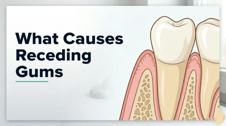
White spots on teeth are caused by disruptions to the normal mineral composition of dental enamel, the hard outer layer of the tooth crown made up of approximately 96% crystalline hydroxyapatite (a calcium phosphate mineral) organized into tightly packed rods called enamel prisms.
When that mineral matrix is either incompletely formed during tooth development or dissolved after the tooth has erupted, the affected enamel becomes porous. Porous enamel scatters light rather than transmitting it, making it appear white or opaque compared to the translucent glassy appearance of fully mineralized enamel. Think of it like frosted glass next to clear glass. The structure is similar, but the internal light behavior is completely different because of what’s missing at the microscopic level.
Two fundamentally different mechanisms produce this outcome. The first is developmental: something interferes with ameloblasts (the specialized cells that build enamel before a tooth erupts), causing incomplete or structurally abnormal mineral deposition. The second is post-eruptive: bacteria, acidic foods, or reduced salivary flow dissolve minerals out of erupted enamel, creating that same porous, light-scattering zone.
The cause matters because it determines clinical significance, reversibility, and the right course of action. Identifying which mechanism produced your white spots requires knowing when they appeared, where on the tooth they sit, how many teeth are affected, and your personal and dental history.
Common causes of white spots on teeth by mechanism type:
| Cause | Mechanism Type | Reversible? | Clinical Urgency |
|---|---|---|---|
| Dental fluorosis | Developmental (ameloblast disruption) | No | Low (cosmetic) |
| Enamel hypoplasia | Developmental (incomplete matrix formation) | No | Low to moderate |
| Molar incisor hypomineralization | Developmental (post-natal disruption) | No | Moderate |
| Demineralization from plaque | Post-eruptive (bacterial acid) | Partially | Moderate to high |
| Post-orthodontic white spots | Post-eruptive (acid under brackets) | Partially | Moderate |
| Xerostomia-related demineralization | Post-eruptive (reduced saliva buffering) | Partially | Moderate |
| Celiac-related enamel defect | Developmental (autoimmune disruption) | No | Moderate |
| Whitening artifact | Post-eruptive (temporary dehydration) | Yes (fully) | None |
Causes of White Spots on Teeth: A Clinical Overview
The causes of white spots on teeth fall into two categories: those that form before the tooth erupts (developmental) and those that develop on already erupted teeth (post-eruptive).
Developmental causes share a common feature: they affect specific teeth in a predictable way based on which teeth were forming during the period of disruption. Permanent incisors form their crowns between approximately ages 3 months and 5 years. Permanent first molars form between birth and age 3. Any insult to the body during those windows can affect enamel quality in those specific teeth, leaving other teeth unaffected because their crowns formed at a different time.
Post-eruptive causes are different. They affect teeth that are already fully formed and erupted, progressing in real time based on ongoing exposure to acidic environments, bacterial activity, or reduced salivary protection. Post-eruptive white spots can worsen over weeks to months if the underlying cause continues.
A 2021 review published in Caries Research confirmed that distinguishing between developmental and post-eruptive white enamel lesions is the first step in any clinical management decision, because the treatment approaches differ entirely. Developmental spots are managed cosmetically if at all. Post-eruptive spots, depending on depth, may need remineralizing treatment or restorative intervention to prevent cavity progression.
Key white spot causes at a glance:
- Dental fluorosis: Excess fluoride during enamel development
- Enamel hypoplasia: Incomplete enamel matrix formation from illness, nutrition, or birth complications
- Molar incisor hypomineralization: Post-natal systemic disruption during a critical developmental window
- Demineralization: Bacterial acid production from plaque buildup
- Post-orthodontic decalcification: Plaque accumulation around fixed brackets
- Dry mouth (xerostomia): Reduced salivary buffering capacity
- Celiac disease: Autoimmune disruption of ameloblast function
- Whitening dehydration artifact: Temporary and fully self-resolving
White Spots on Teeth From Fluorosis
Dental fluorosis forms when a child ingests too much fluoride during the period when permanent teeth are developing, disrupting ameloblast function in a specific and well-characterized way.
Normally, ameloblasts go through two stages. During the secretory stage, they deposit amelogenin and other protein scaffolding. During the maturation stage, they reabsorb those proteins and replace them with additional calcium and phosphate, completing enamel mineralization. Fluoride at elevated concentrations interferes with the maturation stage specifically, impairing the calcium signaling that drives protein reabsorption and final crystal growth. The result is enamel with a porous protein-rich interior layer that appears white or chalky under light.
According to the Centers for Disease Control and Prevention (CDC), the optimal fluoride level in community drinking water in the United States was set at 0.7 milligrams per liter in 2015 to balance cavity prevention against fluorosis risk. The CDC estimates that approximately 25% of U.S. adolescents show some form of dental fluorosis, the vast majority of which is mild (faint white lines or spots) with no effect on tooth function or health.
Severity of fluorosis correlates with fluoride dose and the developmental stage at exposure. The critical window for the upper front teeth, which are most cosmetically visible, is roughly 15 to 24 months of age. Children who regularly swallow fluoridated toothpaste during this period, or who are fed formula reconstituted with water containing naturally elevated fluoride, face the highest risk.
Fluorosis severity grading (Dean’s Index, used by the American Dental Association):
| Grade | Appearance | Tooth Function Impact |
|---|---|---|
| Questionable | Faint white flecks, barely visible | None |
| Very mild | Small opaque areas covering less than 25% of tooth surface | None |
| Mild | Opaque areas covering up to 50% of tooth surface | None |
| Moderate | All surfaces affected, brown staining possible | Minor |
| Severe | Pitting, brown staining, structural damage | Functional impact possible |
Children with kidney disease who receive dialysis face an elevated fluorosis risk because impaired renal clearance allows fluoride to accumulate at lower intake levels. A pediatric dentist should be consulted for fluoride management in this population.
White Spots on Teeth From Demineralization
Demineralization is the process by which mineral ions, primarily calcium and phosphate, are pulled out of the hydroxyapatite crystal lattice in enamel by acid, leaving behind a porous, weakened subsurface zone that appears white.
The acid comes primarily from Streptococcus mutans and Lactobacillus species, bacteria that colonize dental plaque and metabolize fermentable carbohydrates (sugars and refined starches) into lactic acid and other organic acids. When plaque pH drops below the critical threshold of 5.5, hydroxyapatite crystals begin to dissolve. The process is reversible in early stages: when pH rises again through saliva’s natural buffering action (driven by bicarbonate and phosphate), minerals can re-enter the enamel in a process called remineralization.
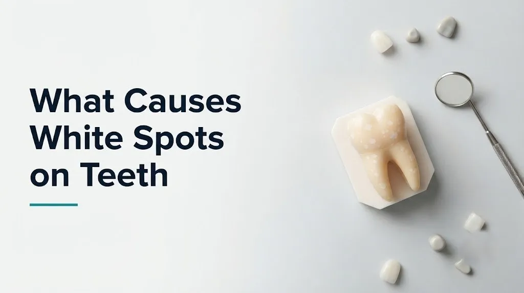
A white spot from demineralization is essentially an active early-stage cavity. The enamel surface may still be intact, but the interior has lost mineral density. Without intervention, continued acid exposure dissolves the surface layer too, creating a cavity that requires restorative treatment.
Saliva is the body’s primary defense. It buffers acid, delivers calcium and phosphate back to enamel, and contains antimicrobial proteins including lysozyme and lactoferrin. People who produce inadequate saliva, whether from dehydration, medication side effects, or salivary gland disease, lose this protection and face dramatically accelerated demineralization.
Risk factors that accelerate demineralization:
- Frequent consumption of sugary or acidic foods and beverages
- Poor oral hygiene allowing dense plaque biofilm accumulation
- Reduced salivary flow from any cause (see dry mouth section)
- High Streptococcus mutans counts in saliva (transmissible through shared utensils, particularly from caregiver to child)
- Gastroesophageal reflux disease (GERD), which exposes enamel to gastric acid
- Fixed orthodontic appliances creating plaque-retention sites
Adolescents wearing fixed orthodontic braces face particularly elevated demineralization risk at the bracket margins. A 2019 study published in the Journal of the American Dental Association found that white spot lesions were detectable around brackets in approximately 30% of orthodontic patients examined after at least 12 months of fixed appliance treatment.
Key Takeaway: Not all white spots are cosmetic. A white spot from active demineralization is an early cavity in progress, and catching it before surface breakdown occurs is the window for non-invasive remineralizing treatment.
White Spots on Teeth From Enamel Hypoplasia
Enamel hypoplasia refers to a deficiency in the quantity of enamel matrix laid down during tooth development, distinct from fluorosis, which affects mineralization quality rather than matrix quantity.
When ameloblasts are disrupted during the secretory phase of amelogenesis (rather than the maturation phase as in fluorosis), they produce less enamel than normal. The result can range from shallow surface pits or grooves to broad white opaque patches where enamel is abnormally thin. Because the underlying dentin may show through in some areas, affected teeth can also appear yellow-brown in spots adjacent to the white zones.
Causes of enamel hypoplasia are systemic. Any significant illness, high fever, severe nutritional deficiency (particularly vitamins A, C, and D, and calcium), or physiological stress during the period when a tooth’s crown is forming can disrupt ameloblast function. Preterm birth is one of the strongest risk factors. Research published in the European Archives of Paediatric Dentistry has found that preterm infants (born before 37 weeks gestation) show enamel hypoplasia rates approximately two to three times higher than term-born children, likely because neonatal illness, hypocalcemia, and metabolic instability during the period of active crown formation disrupt secretory-stage ameloblasts.
The pattern of affected teeth provides a developmental timeline. If only the incisors and first molars show hypoplasia, the disruption occurred in early childhood. If all teeth show defects, either a prenatal or early infant insult or a genetic cause (such as amelogenesis imperfecta) may be responsible.
Conditions associated with enamel hypoplasia:
- Preterm birth or low birth weight
- Severe febrile illness in infancy
- Vitamin D deficiency (rickets)
- Lead exposure during early childhood
- Antibiotic exposure during enamel formation (particularly tetracycline for different discoloration; ampicillin and amoxicillin linked to MIH)
- Celiac disease (covered separately below)
- HIV infection and antiretroviral treatment during infancy
Children who spent time in neonatal intensive care units should have a dental evaluation by a pediatric dentist by age 12 months regardless of visible tooth abnormalities, because early identification of hypoplasia allows protective sealants or remineralizing treatments to be applied before the teeth are exposed to a full caries risk environment.
Molar Incisor Hypomineralization
Molar incisor hypomineralization (MIH) is a distinct developmental enamel defect affecting the first permanent molars and often the permanent incisors, characterized by white, cream, yellow, or brown demarcated opacities that are sharply defined at their borders.
MIH differs from generalized enamel hypoplasia in its specificity. It affects the first permanent molars in all or most cases, and at least some of the permanent incisors in approximately two-thirds of cases, because those teeth all begin their crown mineralization during the same developmental window: the last trimester of pregnancy through approximately the first year of life. A review published in Community Dentistry and Oral Epidemiology in 2021 estimated the global prevalence of MIH at approximately 14%, making it one of the most common developmental dental conditions in children worldwide.
The exact cause of MIH remains under investigation as of 2026. Proposed factors include prenatal illness, early childhood respiratory infections (particularly repeated otitis media and upper respiratory illness in the first year), antibiotic use (specifically amoxicillin), perinatal hypoxia, and dioxin exposure, all of which could disrupt ameloblast function during that narrow post-natal window. No single causal agent has been definitively confirmed by large controlled human trials, but the clinical association between early childhood illness and MIH is supported by consistent epidemiological data.
What makes MIH clinically important beyond its appearance is that the hypomineralized enamel is structurally fragile. It breaks down more easily under chewing stress, making affected teeth disproportionately susceptible to rapid caries progression and post-eruptive breakdown. Children with MIH-affected first molars may experience pain from temperature sensitivity even before cavitation is visible. Because MIH enamel is porous and difficult to bond to, restorations in these teeth are technically demanding and show higher failure rates than restorations in normal enamel.
Distinguishing MIH from dental fluorosis:
| Feature | MIH | Dental Fluorosis |
|---|---|---|
| Teeth affected | First molars and incisors specifically | All teeth forming during fluoride exposure |
| Border of opacity | Sharp, well-demarcated | Diffuse, blends into surrounding enamel |
| Color range | White, cream, yellow, brown | White to brown (severity-dependent) |
| Post-eruptive breakdown | Common in moderate/severe cases | Rare except in severe fluorosis |
| Geographic symmetry | Often asymmetric | Usually symmetric across arch |
| Cause | Post-natal systemic illness window | Excess fluoride during development |
Children with MIH who experience molar pain or sensitivity should be evaluated by a pediatric dentist promptly. Delayed treatment in MIH-affected molars is associated with rapid enamel breakdown and increased restorative complexity.
Key Takeaway: Molar incisor hypomineralization is not the same as fluorosis. If your child has white or chalky patches on first molars and front teeth with sharply defined borders, and those teeth are sensitive, a pediatric dentist evaluation is warranted within two to four weeks, not at the next annual visit.
White Spots on Teeth After Braces
White spots appearing on teeth after orthodontic braces are removed are one of the most common and clinically avoidable causes of post-eruptive enamel damage in adolescents and young adults.
Fixed orthodontic brackets create protected zones where plaque biofilm accumulates in areas that standard brushing cannot effectively reach. Specifically, plaque collects at the margins where the bracket adhesive meets the tooth surface. In these microenvironments, cariogenic bacteria continuously produce acid from dietary sugars, and the area receives less saliva contact and less mechanical cleaning than exposed enamel surfaces. Over months of treatment, this sustained acid exposure progressively dissolves mineral from the subsurface enamel, creating white spot lesions that become visible immediately after the brackets are removed.
The lesions follow a characteristic pattern: they appear as white halos or rings around the bracket attachment sites, most prominently on the upper front teeth, canines, and first premolars, which are the teeth with the highest sugar exposure and the most plaque accumulation in most patients. The pattern is so distinctive that it essentially confirms orthodontic decalcification as the cause without requiring additional investigation.
According to the American Journal of Orthodontics and Dentofacial Orthopedics, white spot lesions are detectable in approximately 50 to 75% of orthodontic patients at bracket removal when they are assessed with sensitive tools such as quantitative light-induced fluorescence (QLF). Many of these lesions are subclinical at removal but can progress to frank cavitation if diet and hygiene do not improve after treatment ends.
Prevention during active treatment relies on fluoride varnish applications at every orthodontic appointment, intensive oral hygiene instruction reinforced at each visit, and, in high-risk patients, prescription-strength fluoride rinses. Patients with poor hygiene compliance during treatment who then receive inadequate follow-up monitoring are at the highest risk for continued lesion progression after brackets are removed.
Steps to minimize white spot formation during orthodontic treatment:
- Brush each tooth surface twice daily using a soft-bristle brush angled at 45 degrees toward the bracket-tooth junction.
- Use a proxabrush (interdental brush) to clean beneath the archwire and around each bracket base at least once per day.
- Rinse with an ADA-accepted fluoride mouthwash before bedtime, after brushing, so fluoride remains in contact with enamel overnight.
- Limit fermentable carbohydrates, particularly sticky or acidic foods, to mealtimes rather than consuming them throughout the day.
- Attend every scheduled orthodontic appointment to receive fluoride varnish application and clinical hygiene assessment.
- Request a post-treatment white spot evaluation at the appointment when brackets are removed, and ask your orthodontist or general dentist to assess lesion activity before planning any cosmetic treatment.
White Spots on Teeth and Dry Mouth
Xerostomia (dry mouth) dramatically elevates the risk of white spot lesion formation because saliva is the primary remineralization mechanism protecting erupted tooth enamel.
Saliva does far more than keep the mouth comfortable. It buffers plaque acid through its bicarbonate content, delivers calcium and phosphate ions directly to the enamel surface for remineralization, reduces bacterial counts with antimicrobial proteins, and mechanically washes food debris and acid from tooth surfaces. A normal adult produces approximately 0.5 to 1.5 liters of saliva daily. When that output drops significantly, the demineralization-remineralization balance shifts decisively in the direction of mineral loss.
Multiple common causes reduce salivary flow. Anticholinergic medications, which include many antihistamines, antidepressants (particularly tricyclics), antipsychotics, bladder medications (oxybutynin), and certain antihypertensives, block muscarinic receptors in salivary gland acini, directly reducing secretion. Radiation therapy targeting head and neck tumors can destroy salivary gland parenchyma permanently, with parotid glands being particularly vulnerable. Sjogren syndrome, a chronic autoimmune condition affecting predominantly middle-aged women, causes lymphocytic infiltration of salivary and lacrimal glands, progressively destroying their secretory capacity.
The National Institutes of Health estimates that over 400 commonly prescribed medications list dry mouth as a side effect, making medication-induced xerostomia one of the most widespread but underrecognized contributors to dental demineralization in adults.
White spots developing in an adult with adequate oral hygiene and no recent orthodontic treatment should prompt a review of current medications and a discussion with the prescribing provider and a general dentist about salivary stimulation strategies or prescription fluoride supplementation.
Conditions and medications that significantly reduce salivary flow:
- Tricyclic antidepressants (amitriptyline, nortriptyline)
- First-generation antihistamines (diphenhydramine, chlorpheniramine)
- Anticholinergic bladder medications (oxybutynin, tolterodine)
- Antipsychotics (quetiapine, olanzapine, clozapine)
- Radiation therapy to the head and neck
- Sjogren syndrome
- Dehydration from any cause
- Mouth breathing during sleep (reduces salivary protection of front tooth surfaces)
Key Takeaway: If white spots are forming on your teeth as an adult and you’re not wearing braces, reviewing your medication list for anticholinergic drugs with your dentist or primary care physician is the most clinically productive first step.
White Spots on Teeth From Celiac Disease
Celiac disease causes white spots and other enamel defects through a mechanism entirely distinct from fluoride exposure or bacterial acid: an autoimmune process that disrupts ameloblast function during tooth development.
Celiac disease is a chronic autoimmune condition triggered by gluten ingestion in genetically susceptible individuals. The primary immune response targets the small intestinal lining, producing anti-tissue transglutaminase antibodies (tTG-IgA) and anti-endomysial antibodies that drive intestinal villous atrophy. However, the immune dysregulation is systemic, and research has consistently found that these antibodies, or related immune mechanisms, can also affect ameloblast function during enamel formation.
The resulting enamel defects, called celiac-associated dental enamel defects, follow a characteristic pattern. They are symmetric (appearing in the same locations on corresponding teeth on both sides of the arch), demarcated, and distributed in a chronological pattern corresponding to the period when active gut inflammation impaired nutrient absorption or immune-mediated enamel disruption occurred. They affect all four quadrants of dentition rather than just specific arch segments, which differentiates them from post-eruptive demineralization.
A study published in the Journal of Clinical Gastroenterology found that enamel defects meeting diagnostic criteria for celiac-associated dental changes were present in approximately 85% of celiac disease patients in one clinical cohort, and that dental enamel defects were sometimes the presenting finding that led to celiac diagnosis in patients with otherwise subclinical gut involvement.
This matters clinically because dentists are often the first clinicians to observe a pattern of symmetric, chronologically distributed enamel defects in otherwise healthy-appearing patients. Recognizing celiac-associated enamel defects as a potential systemic sign rather than a purely dental finding can prompt referral for serologic testing (tTG-IgA antibody panel) and gastrointestinal evaluation.
Distinguishing celiac-associated enamel defects from fluorosis:
| Feature | Celiac-Associated Defects | Fluorosis |
|---|---|---|
| Symmetry | Yes, bilateral | Yes, bilateral |
| Teeth affected | All four quadrants | Teeth forming during fluoride exposure |
| Associated GI symptoms | Possible (diarrhea, bloating, failure to thrive) | None |
| Responds to gluten-free diet? | Stops new defects; existing ones persist | N/A |
| Antibody testing | tTG-IgA positive | Negative |
White enamel defects in a child who also has chronic gastrointestinal symptoms, unexplained iron-deficiency anemia, or growth delay should prompt a conversation between the dentist and the child’s pediatrician, because the dental findings may reflect an underlying systemic condition.
White Spots on Baby Teeth
White spots on primary (baby) teeth can occur through the same mechanisms as permanent teeth, but two specific patterns deserve particular attention in infants and toddlers.
The first is early childhood caries (ECC), formerly called baby bottle tooth decay. This is aggressive demineralization of primary upper front teeth produced by prolonged exposure to fermentable carbohydrates, most commonly from formula, breast milk, or juice when fed to an infant in a bottle at naptime or bedtime. The infant falls asleep with liquid pooled around the front teeth, the pH in the oral environment drops due to bacterial fermentation, and with minimal salivary clearance during sleep, the acid exposure is prolonged and severe. White spots appear first at the gumline of the upper front teeth, the zone with the longest liquid contact, before progressing to yellow-brown discoloration and frank cavitation.
The American Academy of Pediatric Dentistry (AAPD) recommends that all infants have their first dental visit by age 1 or within six months of the first tooth erupting, specifically to identify early caries risk and counsel caregivers on feeding practices that prevent ECC.
The second pattern is enamel hypoplasia on primary teeth, which forms from prenatal or perinatal disruptions to ameloblast function. Primary tooth enamel begins forming in utero (primary central incisors begin mineralizing around week 14 of fetal development), so maternal illness, severe nutritional deficiency during pregnancy, or perinatal hypoxia can disrupt primary tooth enamel quality before the child is born.
Signs of white spots on baby teeth that warrant prompt evaluation by a pediatric dentist:
- White or chalky patches at the gumline of upper front teeth, particularly if the child drinks from a bottle at night
- White spots on multiple primary teeth without an obvious dietary pattern
- White spots accompanied by visible brown or dark discoloration
- Teeth that appear to be breaking down or chipping without trauma
- Any dental finding accompanied by a child’s refusal to eat hard or cold foods
Premature infants and children with a history of neonatal intensive care unit admission have higher rates of enamel hypoplasia in primary teeth and should receive a pediatric dental evaluation earlier than routine guidelines suggest, ideally at or before 12 months of age.
Key Takeaway: White spots at the gumline of a toddler’s upper front teeth are not cosmetic. They are the earliest sign of a highly aggressive caries pattern that can destroy primary teeth quickly, and a pediatric dentist evaluation should happen within two weeks of the parents noticing them.
White Spots on Teeth in Adults
White spots on teeth in adults follow a different clinical logic than childhood-onset white spots, and the differential diagnosis shifts substantially depending on whether the spots are new or have been present since childhood.
Stable white spots that have existed since childhood, are symmetric, and have not changed over years almost certainly represent a developmental cause: fluorosis, enamel hypoplasia, or MIH. They don’t indicate active disease, they don’t pose a cavity risk independently, and they require cosmetic evaluation rather than urgent clinical management.
New white spots developing in an adult are a different matter. An adult who notices new white spots appearing on teeth where no spots existed before needs to consider: active demineralization from plaque (often near the gumline), recession-related root surface exposure (root cementum is less acid-resistant than enamel), xerostomia from new medications, acid exposure from GERD or frequent vomiting, or, rarely, a systemic condition affecting salivary or immune function.
Adults with the following characteristics face higher risk for developing new post-eruptive white spots:
- Individuals taking multiple medications with anticholinergic activity
- People with Sjogren syndrome or other autoimmune conditions affecting salivary glands
- Anyone who has undergone radiation therapy to the head and neck region
- People with poorly controlled GERD causing nighttime acid reflux exposure
- Adults in active orthodontic treatment with clear aligners or fixed appliances
- Individuals with high sugar or acidic beverage intake combined with reduced salivary flow
Adults over 65 are at particular risk because polypharmacy (taking five or more medications) is common in this age group, many of which have anticholinergic effects. Research cited by the American Dental Association has found that dry mouth affects approximately 30% of adults over age 65, contributing to substantially elevated late-life caries rates.
Postmenopausal women experience an additional risk factor: declining estrogen levels affect salivary gland secretory capacity in some individuals, and the hormonal shift also coincides with increased use of medications (antidepressants, antihypertensives, bladder medications) that independently reduce salivary flow.
White Spots on Teeth From Whitening
White spots appearing immediately after professional or at-home tooth whitening are almost always a temporary dehydration artifact and represent the single most benign entry on this cause list.
Whitening agents, whether hydrogen peroxide (professional concentration typically 25 to 40%) or carbamide peroxide (at-home concentration typically 10 to 22%), penetrate enamel and release free oxygen radicals that oxidize chromogen molecules embedded in the enamel matrix, which is the mechanism behind the lightening effect. During and immediately after this process, the whitening agent draws water out of enamel. This temporary dehydration makes all enamel appear more opaque, and any preexisting subtle hypomineralized zones that were previously invisible against slightly translucent normal enamel suddenly become starkly visible as the surrounding enamel loses its translucency.
In individuals with preexisting subclinical fluorosis or small developmental white spots, whitening can dramatically increase the visual contrast between the white spots and the surrounding tooth. This is not the whitening causing new damage. It’s revealing a structural variation that was always present but previously masked by the optical properties of normally hydrated enamel.
Within 24 to 72 hours of stopping whitening treatment, as enamel rehydrates, the appearance of many of these spots diminishes or disappears entirely. The general guideline is to avoid assessing the cosmetic result of whitening for at least 48 to 72 hours after the last treatment session for this reason.
However, in a small number of cases, whitening can genuinely increase the visibility of preexisting enamel defects in ways that don’t fully resolve. If prominent white spots persist beyond 72 hours post-treatment, a consultation with a general dentist or prosthodontist is appropriate to discuss whether resin infiltration or microabrasion might improve uniformity.
Distinguishing temporary whitening artifact from a new structural problem:
- Temporary artifact: spots appear within hours of whitening, affect multiple teeth diffusely, reduce visibly over 24 to 72 hours after treatment ends
- Persistent structural defect: spots were present before whitening but now appear more pronounced; limited to specific teeth; no reduction in visibility after 72-hour rehydration period
Individuals with known fluorosis or developmental white spots should discuss this risk with their dentist before beginning whitening treatment, because the contrast amplification effect can be significant enough to produce a cosmetic outcome the patient did not anticipate.
Key Takeaway: White spots immediately after whitening are almost always temporary rehydration effects. Wait 72 hours before drawing any conclusions about their significance, and discuss preexisting developmental white spots with your dentist before any whitening treatment.
Are White Spots on Teeth Reversible?
Whether white spots on teeth are reversible depends entirely on their cause and depth.
Developmental white spots from fluorosis, enamel hypoplasia, or MIH are not reversible in the conventional sense because the underlying enamel structure was formed abnormally and cannot be reformed. The mineral that was deposited incorrectly or incompletely cannot be reorganized. However, cosmetic treatments can alter their appearance dramatically. Techniques include microabrasion (removing a thin outer enamel layer to reduce the surface expression of a subsurface defect), resin infiltration (the Icon resin system, which draws a low-viscosity resin into the porous enamel to fill the light-scattering voids and restore optical uniformity), professional bleaching to reduce contrast between the white spot and surrounding enamel, or composite bonding to mask larger defects. These approaches don’t repair the enamel but they can effectively eliminate the cosmetic concern.
Post-eruptive white spot lesions from active demineralization are a different story. In their earliest stages, when the enamel surface is still intact and the subsurface mineral loss is less than 30% of normal density, remineralization is genuinely possible. Fluoride (at therapeutic concentrations from fluoride varnish, prescription fluoride toothpaste, or professional treatments) promotes hydroxyapatite crystal growth back into the porous zone. Casein phosphopeptide-amorphous calcium phosphate (CPP-ACP), a dairy-derived remineralizing agent available in products such as MI Paste, supplies bioavailable calcium and phosphate directly to the enamel surface under conditions that facilitate crystal regrowth.
A 2022 Cochrane systematic review on remineralization of white spot lesions found strong evidence that fluoride varnish application and CPP-ACP use are both effective at reducing the visual severity and measured mineral deficit of early white spot lesions, particularly those identified within the first six months after orthodontic bracket removal.
Once the enamel surface breaks down into a frank cavity, the lesion is no longer reversible without restorative treatment.
Reversibility summary by cause:
| Cause | Reversible? | How Managed |
|---|---|---|
| Fluorosis (mild-moderate) | No (structure) but cosmetically manageable | Resin infiltration, microabrasion, bleaching |
| MIH | No | Protective sealants, restorations if breakdown occurs |
| Early demineralization | Partially (with active remineralization treatment) | Fluoride varnish, CPP-ACP, improved hygiene |
| Post-orthodontic white spots | Partially (if treated within 6 months) | Fluoride varnish, resin infiltration |
| Whitening dehydration | Fully (within 72 hours) | Wait for rehydration |
| Enamel hypoplasia | No | Cosmetic restoration, sealants for protection |
White Spot Lesions and Early Cavities
White spot lesions occupy a specific and clinically important position in the caries spectrum: they are the earliest detectable stage of dental caries (tooth decay), representing subsurface demineralization that has not yet progressed to surface cavitation.
Understanding this distinction changes the urgency calculus completely. A white spot lesion is not yet a cavity in the conventional sense. The enamel surface is still intact. The tooth does not yet have a hole. This is the window for non-invasive treatment, and it is a short one. The International Caries Detection and Assessment System (ICDAS), which is the clinical tool used by dentists worldwide to code caries severity, classifies white spot lesions as ICDAS code 1 (visible only after prolonged air drying) or code 2 (visible without air drying on a wet surface). Both codes represent active lesions that require monitoring and preventive treatment rather than restorative drilling.
The rate of progression from white spot lesion to frank cavitation varies based on diet, oral hygiene, salivary flow, and fluoride exposure. In high-risk individuals (poor hygiene, high sugar intake, reduced salivary flow), progression can occur within weeks to a few months. In lower-risk individuals with good diet control and fluoride use, lesions can remain stable for years or even partially remineralize.
The clinical significance is this: white spots in the right pattern (near the gumline, at bracket sites, on smooth surfaces without developmental explanation) are a signal that active caries disease is present in the mouth, and that signal warrants a caries risk assessment and preventive treatment plan from a general dentist, not just reassurance and a plan to monitor.
What to tell your dentist if you’ve noticed new white spots:
- When you first noticed the white spots (months, weeks, days ago).
- Whether any spots are on teeth that previously looked normal, versus spots you’ve had since childhood.
- Your current dental hygiene routine in specific detail: brushing frequency, type of toothpaste (fluoride concentration), flossing, mouthwash use.
- Any changes to your medications, diet, or health status in the past year.
- Whether you’ve been wearing any orthodontic appliances (clear aligners, retainers with brackets).
- Whether you experience dry mouth, and at what times of day it’s most pronounced.
White Spots on Teeth: Prevention
Preventing white spots on teeth requires strategies tailored to their cause, because the prevention of developmental white spots differs entirely from the prevention of post-eruptive demineralization.
For fluorosis prevention, the American Academy of Pediatric Dentistry (AAPD) recommends that children under 3 use only a rice-grain-sized smear of fluoride toothpaste to minimize ingestion risk, and that children ages 3 to 6 use a pea-sized amount. Parents should check the fluoride concentration in their home drinking water (testing kits are available; municipal water reports are public) and discuss fluoride supplementation versus restriction with a pediatric dentist if using formula with fluoridated water as the primary infant food source.
For demineralization prevention, the primary targets are reducing the frequency of fermentable carbohydrate exposure (how often, not just how much sugar), optimizing mechanical plaque removal, and maximizing fluoride contact with enamel surfaces. The CDC recommends brushing with fluoride toothpaste twice daily as the foundational caries prevention measure for all ages.
For orthodontic patients, professional fluoride varnish applied at every orthodontic appointment reduces white spot lesion formation rates, and some clinical centers also use fluoride-releasing bonding cements to provide continuous low-level fluoride delivery at the bracket margin throughout treatment.
Practical prevention measures by cause:
- Fluorosis: Use age-appropriate toothpaste amounts; supervise brushing through age 8; test home water fluoride levels if on well water
- Early childhood caries: No bottle at naptime or bedtime after the first tooth erupts; wipe infant gums with a damp cloth after feeding; first dental visit by age 1
- Post-orthodontic demineralization: Fluoride varnish at every appointment; electric toothbrush; proxabrush for daily bracket cleaning; avoid sugary drinks during treatment
- Dry mouth-related demineralization: Review medications with dentist; use xylitol-containing gums to stimulate salivary flow; prescription high-fluoride toothpaste (5,000 ppm) where indicated by the dentist
- General demineralization: Limit acidic beverages to mealtimes; use a straw for acidic drinks; wait 30 minutes after eating acidic foods before brushing (to avoid brushing softened enamel); professional fluoride varnish application twice yearly for moderate-to-high-risk patients
When to See a Dentist for White Spots
The right timing for a dental evaluation depends on whether the white spots are stable and long-standing or new and potentially active.
See a general dentist within two to four weeks if:
- New white spots have appeared on teeth that previously looked normal
- White spots are visible at the gumline of multiple teeth, particularly the upper front teeth
- White spots appeared after orthodontic bracket removal and are persisting beyond four to six weeks
- White spots are accompanied by tooth sensitivity to cold or sweet foods
- White spots appear chalky or pitted rather than smooth
- A child’s teeth show white or chalky areas on the first permanent molars or front teeth with sharply defined borders
See a pediatric dentist at the first tooth eruption or by age 1 if:
- You have any questions about your infant’s dental development
- Your infant was born prematurely or had significant illness in the newborn period
- Your child has been on long-term antibiotic therapy or experienced repeated serious illness in the first two years of life
- You are using formula with fluoridated water as the primary dietary fluid
Discuss with your general dentist at the next routine visit if:
- You have stable white spots that have been present since childhood and have not changed
- White spots appeared immediately after whitening and are fading over 72 hours
- You’re curious about cosmetic options for preexisting developmental white spots
A dentist may use the following tools to assess white spots:
| Diagnostic Tool | What It Detects | Clinical Application |
|---|---|---|
| Visual examination under air drying | Surface texture, opacity contrast | Distinguishes surface from subsurface lesion |
| DIAGNOdent laser fluorescence | Mineral density change | Quantifies depth of demineralization |
| Quantitative light-induced fluorescence (QLF) | Subsurface mineral loss | Monitors lesion size over time |
| Bitewing radiographs | Proximal caries, dentin involvement | Rules out deeper lesion extension |
| Transillumination (FOTI/DIFOTI) | Lesion shadow vs. opacity | Differentiates enamel from dentin lesion |
No white spot on a tooth has ever been correctly diagnosed from a photograph or a written symptom description alone. A trained dentist examining the actual tooth, under proper lighting, with air drying, and ideally with adjunctive detection tools, is the only way to accurately determine whether a white spot represents active disease, a stable developmental finding, or a cosmetic concern. If you’re uncertain about a spot, the answer is always to have it examined, not to wait until the next scheduled appointment if that appointment is months away.
Frequently Asked Questions About White Spots on Teeth
What causes white spots on teeth suddenly?
White spots appearing suddenly on teeth are almost always caused by active demineralization from bacterial acid production in dental plaque, or by temporary enamel dehydration after whitening treatment.
New white spots that appear without whitening and persist beyond a few days represent early-stage enamel mineral loss and should be evaluated by a general dentist within two to four weeks.
Sudden white spots across multiple teeth in someone taking new medications may also reflect reduced salivary flow from anticholinergic drug effects.
Are white spots on teeth a sign of cavities?
White spots on teeth can be an early sign of a cavity, specifically the earliest stage of dental caries when subsurface enamel has lost mineral density but the surface is still intact.
Not every white spot is a cavity: developmental white spots from fluorosis or enamel hypoplasia indicate a structural variation, not active decay.
A dentist using clinical tools including air drying, laser fluorescence, and radiographs can determine whether a white spot is an active lesion or a stable developmental finding.
Can white spots on teeth go away on their own?
Developmental white spots from fluorosis or enamel hypoplasia do not go away on their own because the underlying enamel structure cannot reform.
Early demineralization white spots can partially or fully reverse if remineralization is promoted through fluoride application and improved oral hygiene before the enamel surface breaks down.
Whitening-related white spots typically resolve within 24 to 72 hours as enamel rehydrates, and no treatment is needed.
What causes white spots on teeth after braces?
White spots after braces are caused by demineralization: plaque accumulates around bracket bases and adhesive margins where brushing cannot reach, and bacteria in that plaque produce acid that dissolves mineral from the underlying enamel.
The process occurs gradually over months of orthodontic treatment, and the white spots become visible when the brackets are removed and expose the decalcified enamel.
Fluoride varnish applied by the orthodontist throughout treatment and careful daily cleaning around brackets are the most evidence-supported prevention strategies.
Do white spots on teeth mean too much fluoride?
White spots can indicate too much fluoride during tooth development, a condition called dental fluorosis, but fluorosis is only one of several possible causes.
Fluorosis-related white spots are symmetric, affect multiple teeth in the same developmental group, and have diffuse rather than sharply defined edges.
White spots from demineralization, enamel hypoplasia, molar incisor hypomineralization, or celiac disease can look similar to fluorosis, and accurate identification requires a dental examination.
What doctor do I see for white spots on teeth?
For white spots on adult teeth, a general dentist is the appropriate first provider for evaluation, diagnosis, and management.
For white spots on a child’s teeth, a pediatric dentist (pedodontist) provides both diagnostic evaluation and age-appropriate preventive and restorative care.
If a dentist identifies a pattern suggesting a systemic cause such as celiac disease, they may recommend referral to the patient’s primary care physician or a gastroenterologist for serologic testing and evaluation.
White spots on teeth have specific, identifiable causes, and knowing which one applies to your situation determines everything about whether action is needed and what kind. Developmental spots from fluorosis or hypoplasia are stable findings without ongoing disease risk. Active white spot lesions from demineralization are a different clinical reality: early-stage caries that can progress to cavitation if the underlying cause continues unchecked.
The practical step is straightforward. Stable spots that have existed for years and haven’t changed: mention them at your next routine dental visit and ask about cosmetic options if they bother you. New spots, spots in a child, spots after braces, or spots accompanied by sensitivity: schedule an evaluation with a general dentist or pediatric dentist within two to four weeks, describe specifically when you noticed them and on which teeth, and bring your complete medication list to the appointment.
You now know more about the cellular mechanism behind enamel white spots than most general health content will ever tell you. Use that understanding to ask the right questions when you sit in the dental chair.