What Causes Ear Bleeding: 16 Causes and Warning Signs
Ear bleeding, known clinically as otorrhagia, is caused by disruption of blood vessels in the external auditory canal, the tympanic membrane (eardrum), or the middle ear cavity, and identifying the correct source determines whether the cause is minor or a medical emergency. The causes of ear bleeding range from a small scratch from aggressive ear cleaning to a ruptured eardrum, a severe infection, or bleeding caused by a basilar skull fracture following head trauma.
According to the National Institute on Deafness and Other Communication Disorders (NIDCD), injuries to the ear canal and eardrum are among the most common reasons patients seek otolaryngology evaluation in the United States. Blood from the ear is a symptom that most people find alarming, and rightly so: while many causes are self-limiting, several require same-day or emergency evaluation.
This article covers all 16 medically recognized causes of ear bleeding, the specific physiological mechanism behind each, how to tell minor from serious, and the exact clinical threshold at which ear bleeding requires you to call 911 or go to an emergency room.
What Causes Ear Bleeding
Ear bleeding is caused by damage to blood vessels anywhere along the ear’s anatomy, from the outer canal surface to the eardrum to the middle ear structures, and the cause determines both the clinical picture and the appropriate response.
The human ear is divided into three functional zones. The external auditory canal runs about 2.5 centimeters from the outer opening to the eardrum. It is lined with thin squamous epithelium overlying a rich submucosal vascular network supplied by branches of the external carotid artery. That thin skin bleeds easily when scratched or infected. The tympanic membrane itself contains a fibrous middle layer with a network of small vessels that rupture when pressure across the membrane exceeds their structural limit. Behind the eardrum, the middle ear cavity contains the ossicular chain (malleus, incus, and stapes) and is lined with a vascular mucosa.
Blood visible at the outer ear opening can originate from any of these three zones, and each zone has a distinct set of causes. A surface cut in the canal bleeds bright red and usually stops within minutes. Hemotympanum, blood pooled behind an intact eardrum, appears as a dark blue or red discoloration seen through the drum and does not drip from the canal unless the drum is also ruptured. Blood mixed with clear fluid can indicate cerebrospinal fluid leakage from a basilar skull fracture, which is a neurological emergency.
| Origin Zone | What It Looks Like | Most Likely Causes | Urgency Level |
|---|---|---|---|
| External auditory canal | Bright red, stops quickly | Canal abrasion, infection, foreign body | Low to moderate |
| Tympanic membrane (ruptured) | Moderate flow, may have discharge | Barotrauma, infection, direct trauma | Moderate to high |
| Middle ear (hemotympanum) | Dark discoloration behind drum | Head trauma, severe infection, tumor | High |
| Mixed blood and clear fluid | Thin, watery blood from canal | Basilar skull fracture | Emergency |
Older adults with thinned canal skin may notice bleeding from minor friction that would not affect a younger person’s canal epithelium. Children’s external auditory canals sit at a more horizontal angle and are narrower, making them more vulnerable to foreign body injury and more difficult to examine without specialist equipment.
Ear Canal Injuries and Abrasions
Ear canal abrasions are the most common cause of ear bleeding and occur when the delicate squamous epithelium lining the external auditory canal is physically disrupted by direct mechanical trauma.
The canal’s outer one-third is fibrocartilaginous and contains hair follicles, ceruminous glands, and sebaceous glands. The inner two-thirds overlies bone and is covered by an extremely thin skin layer, averaging only 0.2 millimeters thick, with no subcutaneous fat layer beneath it. When this skin tears, the submucosal capillary network bleeds into the canal. Even modest shear force from a fingernail, a hairpin, or the corner of a tissue produces a visible bleed.
Most canal abrasions are self-limiting. The bleeding typically stops within 5 to 10 minutes because the canal is narrow and generates natural tamponade pressure. Research published in the American Journal of Otolaryngology has documented that surface canal abrasions account for the majority of ear bleeding presentations in primary care and urgent care settings.
Individuals taking aspirin or clopidogrel (antiplatelet medications) may experience prolonged bleeding from canal abrasions that would otherwise stop quickly in someone with intact platelet function. Elderly patients are disproportionately affected because aging skin loses collagen density and vascular support, making the canal epithelium significantly more fragile.
Signs that a canal abrasion does not need emergency care:
- Bleeding is bright red and slows or stops within 10 to 15 minutes
- No preceding head impact or forceful blow
- Hearing feels normal in the affected ear
- No vertigo, facial weakness, or severe pain
- The injury followed known contact (a fingernail, a hairpin, a cotton swab)
If bleeding from a suspected canal abrasion does not stop within 20 minutes, or if there is any uncertainty about the cause, evaluation by a primary care physician the same day is appropriate.
Ruptured Eardrum Causes and Mechanism
A ruptured eardrum, clinically called a tympanic membrane perforation, causes ear bleeding when the tearing of the membrane’s fibrous and vascular layers releases blood into the canal.
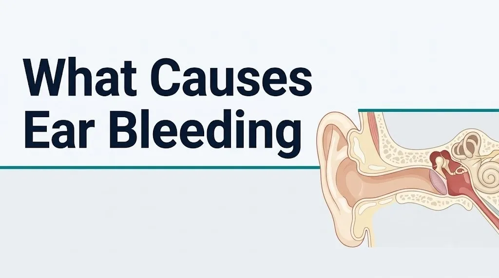
The tympanic membrane is structured like a three-layer sandwich: an outer squamous epithelial layer continuous with the canal skin, a fibrous middle layer containing radial and circular collagen fibers and a submucosal blood vessel network, and an inner mucosal layer continuous with the middle ear mucosa. When the membrane tears, all three layers are disrupted. The fibrous middle layer’s vasculature bleeds into the canal, producing the characteristic bloody discharge associated with perforation.
Four primary mechanisms produce tympanic membrane rupture. Direct impact from a slap to the ear or an explosion wave compresses air in the canal faster than the membrane’s elastic limit. Infection-related perforation occurs when middle ear pressure from acute otitis media builds until the membrane’s weakest point, typically the pars tensa (the central larger portion), gives way. Barotrauma creates a sudden pressure differential across the membrane. Iatrogenic injury from improper ear cleaning or ear candling also accounts for a subset of perforations.
According to the American Academy of Otolaryngology Head and Neck Surgery (AAO-HNS), the majority of traumatic tympanic membrane perforations in adults heal spontaneously within 8 to 12 weeks without surgical intervention, provided the middle ear is kept dry and free of infection. However, perforations exceeding 50% of the membrane’s total surface area or those complicated by ossicular chain disruption may require tympanoplasty (surgical reconstruction).
Children under age 12 are more prone to infection-related perforations because the pediatric Eustachian tube sits at a more horizontal angle, impairing natural middle ear drainage and increasing the frequency of acute otitis media. Adults with a prior history of chronic ear infections may have scarred, less elastic tympanic membrane tissue that tears more readily under modest pressure.
Ear Infections Causing Ear Bleeding
Ear infections cause bleeding through two distinct mechanisms depending on whether the infection originates in the outer ear canal or the middle ear, and the two presentations carry different urgency levels.
Acute otitis externa (infection of the external auditory canal, commonly called swimmer’s ear) causes bleeding when bacterial or fungal infection produces enough inflammatory hyperemia and mucosal breakdown to disrupt the thin canal skin. The dominant organisms are Pseudomonas aeruginosa and Staphylococcus aureus. Prostaglandins and histamine released during the inflammatory response increase local capillary permeability and friability. Scratching the intensely itchy, infected canal adds mechanical disruption on top of inflammatory fragility, producing a serosanguineous (blood-tinged) discharge that is often mistaken for a more serious bleed.
Acute otitis media (middle ear infection) produces bleeding through a different pathway. As bacterial or viral infection causes mucosal inflammation inside the middle ear cavity, pressure builds behind the eardrum. When middle ear pressure rises high enough, it ruptures the tympanic membrane’s pars tensa. This perforation releases not just the accumulated purulent fluid but also blood from the torn membrane’s submucosal vessels. The Centers for Disease Control and Prevention (CDC) reports that acute otitis media is one of the most common childhood infections, with most children experiencing at least one episode before age 5, making infection-related tympanic perforations a clinically frequent cause of ear bleeding in pediatric practice.
People with diabetes mellitus face a heightened risk of severe otitis externa progressing to a destructive form called malignant otitis externa because diabetic impairment of neutrophil chemotaxis and phagocytosis allows infection to spread beyond the canal into cartilage and bone.
Quick Tip:
- Ear bleeding from infection is almost always accompanied by pain, itching, or a sense of fullness in the ear
- Clear or yellow discharge mixed with blood from an ear infection suggests tympanic membrane rupture and warrants same-day evaluation by a primary care physician
- Fever above 38.5 degrees Celsius (101.3 degrees Fahrenheit) with ear bleeding and pain requires same-day evaluation, not watchful waiting
Key Takeaway: Ear bleeding from an infection, whether from otitis externa or a ruptured eardrum from otitis media, is nearly always accompanied by pain or discharge; painless ear bleeding after head trauma is a separate and more urgent clinical category entirely.
Barotrauma and Pressure-Related Ear Bleeding
Barotrauma causes ear bleeding when a rapid change in ambient air or water pressure creates a pressure differential across the tympanic membrane that exceeds the membrane’s structural tolerance, rupturing its fibrous layers and the submucosal vessels within them.
Think of the tympanic membrane like a drum skin stretched tightly across a frame. Under normal conditions, the Eustachian tube equalizes pressure between the middle ear cavity and the environment by opening briefly during swallowing or yawning. When ambient pressure changes faster than the Eustachian tube can compensate, the pressure difference across the membrane builds. If that differential reaches approximately 500 to 600 millimeters of mercury, the membrane ruptures, according to data referenced in the otolaryngology literature.
This commonly occurs during airplane descent, scuba diving ascent or descent, an explosion blast wave, or a hyperbaric oxygen treatment session. Divers who hold their breath during ascent and pilots during rapid cabin depressurization are at particular risk. The resulting perforation produces sharp pain, a sudden decrease in tinnitus or pressure, and bleeding into the canal.
A related barotrauma mechanism involves the round window membrane, a thin membrane separating the middle ear from the cochlea. Forceful Valsalva maneuvers (bearing down against a closed airway, as in heavy lifting, forceful nose-blowing, or childbirth) can rupture the round window membrane. This produces a perilymph fistula: leakage of inner ear fluid into the middle ear. While perilymph fistula does not always produce frank blood, it can produce serosanguineous discharge when accompanied by middle ear mucosal injury.
Individuals with a history of Eustachian tube dysfunction, chronic sinusitis, or prior ear surgery have impaired pressure equalization capacity and are at elevated risk for barotrauma at pressure differentials that would not affect a person with normal Eustachian tube function.
Head Trauma and Hemotympanum
Head trauma causes ear bleeding through two distinct and clinically separate pathways, and correctly distinguishing them is the difference between a manageable injury and a life-threatening emergency.
The first pathway is direct trauma to the external auditory canal or tympanic membrane from a blow to the side of the head. A direct impact can abrade the canal skin or rupture the eardrum through the same compression mechanism described under barotrauma. This type of injury produces bright red canal bleeding and is managed similarly to other traumatic tympanic perforations.
The second and far more serious pathway is hemotympanum from a basilar skull fracture. The base of the skull includes the petrous portion of the temporal bone, which houses the middle ear structures. When a basilar skull fracture occurs, blood from fractured bone and torn meningeal vessels accumulates in the middle ear cavity behind an intact tympanic membrane. This produces a characteristic dark blue or purple discoloration seen through the drum, called hemotympanum. Blood may also leak from the canal if the tympanic membrane is co-fractured.
The clinical hallmarks that distinguish basilar skull fracture from minor head trauma are named and specific. Battle’s sign is bruising over the mastoid process (the bony prominence behind the ear) that appears 24 to 48 hours after the injury. Raccoon eyes (periorbital ecchymosis) appear when blood tracks forward along fascial planes. Cerebrospinal fluid otorrhea, the leakage of clear brain fluid from the ear, may occur alongside or instead of bloody discharge.
A 2022 review published in Annals of Emergency Medicine confirmed that hemotympanum is present in approximately 50% of confirmed basilar skull fractures and that the combination of hemotympanum with Battle’s sign has a positive predictive value exceeding 85% for basilar fracture on CT imaging.
Any ear bleeding following a head impact must be treated as a potential basilar skull fracture until CT imaging of the temporal bone and head has ruled it out.
Key Takeaway: Ear bleeding after any head impact requires immediate emergency room evaluation because hemotympanum from basilar skull fracture is clinically indistinguishable from minor canal trauma without CT imaging of the temporal bone.
Malignant Otitis Externa and Temporal Bone Involvement
Malignant otitis externa (also called necrotizing otitis externa) is a rare but life-threatening infection that begins in the external auditory canal and progressively destroys the cartilage, soft tissue, and bone of the temporal bone, producing ear bleeding as one of its clinical signs.
Despite the term “malignant,” this condition is infectious, not cancerous. The name refers to its aggressive, destructive behavior. Pseudomonas aeruginosa is the causative organism in over 90% of cases, according to data published in JAMA Otolaryngology Head and Neck Surgery. The bacteria produce several proteolytic enzymes, including exotoxin A, alkaline protease, and elastase, that digest perichondrium and soft tissue faster than the immune system can respond.
The infection begins as severe otitis externa, then penetrates the cartilaginous-bony junction of the canal floor (an area called the fissures of Santorini), enters the skull base, and can progress to osteomyelitis of the temporal bone, facial nerve palsy (when it reaches the stylomastoid foramen where cranial nerve VII exits), and intracranial spread to the meninges or sigmoid sinus.
Bleeding in this context comes from erosion of the granulation tissue and submucosal vasculature of the canal as the infection destroys the surrounding architecture. The bleeding is often modest but persistent, accompanied by severe ear pain disproportionate to what a typical ear infection produces, otorrhea, and tenderness over the mastoid.
People with diabetes mellitus account for approximately 90% of malignant otitis externa cases, according to data cited in the AAO-HNS clinical literature, because the combination of small vessel disease and impaired neutrophil function creates the conditions for unchecked bacterial invasion. Immunocompromised individuals, including those on chemotherapy, long-term corticosteroid therapy, or living with HIV, make up most of the remaining cases.
Any diabetic or immunocompromised patient with ear pain, canal bleeding, and granulation tissue visible in the canal requires urgent evaluation by a board-certified otolaryngologist, not watchful waiting, because delayed treatment correlates directly with temporal bone osteomyelitis and cranial nerve involvement.
Ear Canal Tumors and Growths Causing Bleeding
Both benign and malignant tumors of the ear canal and middle ear can cause ear bleeding by eroding through overlying tissue and disrupting the vascular supply of the structures they invade.
The most clinically important vascular tumor of the middle ear is the glomus tympanicum, a paraganglioma arising from chemoreceptor cells along the tympanic branch of the glossopharyngeal nerve on the medial wall of the middle ear. Glomus tympanicum tumors are among the most highly vascularized tumors in the body. They appear as a red or blue pulsatile mass visible through the tympanic membrane on otoscopy. Bleeding occurs when tumor neovascularization extends to the tympanic membrane surface and the membrane ruptures or is disrupted. The bleeding is typically pulsatile because the tumor’s vascular network follows the arterial pulse.
Cholesteatoma is a distinct cause of ear bleeding involving abnormal accumulation of keratinized squamous epithelium in the middle ear or mastoid, forming an expanding cyst-like mass. As a cholesteatoma grows, it produces collagenase enzymes that erode the ossicular chain, mastoid bone, and adjacent structures. Erosion into the mucosal lining vessels produces chronic, low-volume, foul-smelling bloody discharge from the ear. The Mayo Clinic identifies cholesteatoma as one of the most underdiagnosed causes of chronic ear bleeding because its early presentation overlaps with simple chronic otitis media.
Squamous cell carcinoma of the external auditory canal is rare, accounting for approximately 1 in 1 million population per year based on population registry data, but it presents with persistent ear bleeding, otalgia, and hearing loss that does not respond to standard infection treatment. Any patient with recurrent ear bleeding and no identified benign cause requires evaluation by a board-certified otolaryngologist to exclude malignancy.
Foreign Objects and Ear Bleeding in Children
Foreign objects lodged in the external auditory canal cause ear bleeding when the object’s edges lacerate the canal skin during insertion or during a child’s attempt to remove it.
Children between ages 2 and 8 are the predominant demographic affected by ear foreign bodies. The pediatric external auditory canal differs anatomically from the adult canal in two key ways: it is narrower relative to the object being inserted, and it sits at a more horizontal orientation. This horizontal angle means that when a child inserts a small bead, bean, button battery, or piece of a toy and attempts to remove it by pushing or pulling, the canal’s natural curves act as friction points that tear the thin overlying epithelium.
The objects that cause the most severe bleeding are those with sharp edges (broken toy pieces, small metal clips) and those that expand when moistened (beans, seeds, erasers). Button batteries deserve separate attention: they generate an electrical current against the moist canal skin that produces liquefactive necrosis within as little as two hours. This is a pediatric emergency requiring same-day otolaryngology evaluation regardless of whether bleeding is present.
According to the American Academy of Pediatrics, foreign body removal from the ear should not be attempted at home because home removal attempts using tweezers, cotton swabs, or water irrigation frequently push the object deeper, worsen the laceration, or rupture the tympanic membrane.
If a parent suspects a foreign body in a child’s ear with associated bleeding, same-day evaluation by a pediatrician or a board-certified otolaryngologist is appropriate. If the child is in significant distress, if there is any possibility of a button battery, or if the object is not visible, emergency room evaluation is warranted rather than waiting for a scheduled appointment.
Key Takeaway: A button battery lodged in a child’s ear requires emergency evaluation the same day it is suspected, regardless of whether bleeding is visible, because electrical current from the battery can destroy the canal and eardrum within hours.
Ear Bleeding After Cleaning or Cotton Swab Use
Cotton swab use is one of the most common causes of ear bleeding in adults and represents a clear mismatch between what people believe cotton swabs do and what they actually do anatomically.
The external auditory canal is a self-cleaning structure. The canal epithelium migrates outward at approximately the same rate fingernails grow, carrying cerumen (earwax) and debris naturally to the outer ear opening. When a cotton swab is inserted, it does not clean the canal. It compresses earwax against the bony canal wall and drags the cotton tip along the skin of the inner canal, which is the area covered by the thinnest, most fragile epithelium in the entire ear anatomy.
The bony-cartilaginous junction of the canal, located roughly 1 centimeter from the eardrum, is where swab-related lacerations most commonly occur. At this junction, the canal curves slightly, the skin is thinner, and there is no underlying subcutaneous fat. A cotton swab pushed past the canal’s natural resistance at this junction tears epithelium easily, and the resulting bleed can look alarming despite being superficial.
The American Academy of Otolaryngology Head and Neck Surgery formally recommends against the insertion of any object, including cotton swabs, into the ear canal. This recommendation applies regardless of the purpose: wax removal, moisture absorption after swimming, or itch relief.
Adults with dry skin conditions affecting the canal, including eczema or psoriasis, are at significantly elevated risk for swab-related canal bleeding because chronic inflammation has already compromised the structural integrity of the canal epithelium. Older adults face the same elevated risk due to age-related collagen loss in canal skin.
What Causes Spontaneous Ear Bleeding
Spontaneous ear bleeding, meaning bleeding that occurs without identifiable external trauma, infection, or pressure event, points toward a smaller set of causes and warrants prompt evaluation because several of those causes are not self-limiting.
The term “spontaneous” in a clinical context means the patient is unaware of a precipitating event. However, most apparently spontaneous ear bleeding has an identifiable cause on examination: a minor canal abrasion the patient did not notice, a cholesteatoma eroding through mucosa, a vascular tumor like a glomus tympanicum, or a systemic bleeding disorder causing canal mucosa to bleed from normal friction.
True vascular causes of spontaneous ear bleeding include hereditary hemorrhagic telangiectasia (HHT), a genetic disorder affecting the vascular endothelium through mutations in genes encoding endoglin or activin receptor-like kinase 1. HHT produces abnormal vessel formations (telangiectasias and arteriovenous malformations) throughout the body, including in the external auditory canal mucosa. Patients with HHT frequently report spontaneous bleeding from multiple body sites, including the nose and mouth, which provides a clinical clue.
Systemic coagulopathies, including thrombocytopenia (low platelet count) from any cause, von Willebrand disease (the most common inherited bleeding disorder in the United States, affecting approximately 1 in 100 people according to the National Institutes of Health), and severe liver disease impairing clotting factor synthesis, can all produce spontaneous mucosal bleeding including from the ear canal.
Any patient with recurrent, apparently spontaneous ear bleeding without a history of trauma or infection should be evaluated by a primary care physician with a complete blood count and coagulation panel as the initial diagnostic workup, before assuming the bleeding is benign.
Anticoagulant Medications and Ear Bleeding Risk
Anticoagulant and antiplatelet medications do not independently cause ear bleeding, but they significantly amplify the severity and duration of bleeding from any of the underlying causes already described.
Warfarin inhibits the synthesis of clotting factors II, VII, IX, and X by blocking vitamin K epoxide reductase in the liver. Direct oral anticoagulants including apixaban and rivaroxaban inhibit Factor Xa directly, while dabigatran inhibits thrombin. Antiplatelet agents including aspirin (which irreversibly acetylates cyclooxygenase-1 in platelets) and clopidogrel (which blocks the ADP receptor P2Y12 on platelet surfaces) reduce the platelet aggregation response that normally forms the primary hemostatic plug.
When a person on any of these medications sustains even a minor canal abrasion or develops mild otitis externa, what would normally be a self-limiting 5-minute bleed can persist for 30 minutes or longer, and what would otherwise be a modest perforation bleed can produce enough volume to cause genuine concern.
A 2021 study published in JAMA Otolaryngology Head and Neck Surgery found that anticoagulated patients presenting to otolaryngology clinics with ear bleeding were significantly more likely to require procedural intervention for hemostasis compared to non-anticoagulated patients with the same underlying injury, even when the initial tissue injury appeared identical on examination.
Patients on anticoagulant or antiplatelet therapy should inform their primary care physician or otolaryngologist about any ear bleeding promptly rather than waiting for it to resolve, because the normal self-limiting timeline does not apply to them. They should not stop their anticoagulant medication without explicit physician guidance, as the risks of thrombosis from abrupt discontinuation typically outweigh the risk of minor ear bleeding.
Key Takeaway: Anticoagulated patients cannot rely on the normal “it stopped in 10 minutes” rule for ear bleeding; any ear bleed that persists beyond 20 minutes or recurs warrants a call to their prescribing physician the same day.
Ear Bleeding With No Pain vs. Ear Bleeding With Pain
The presence or absence of pain alongside ear bleeding is one of the most clinically informative features for narrowing the differential diagnosis, and the distinction carries direct implications for urgency.
Painful ear bleeding is the more common presentation. It accompanies canal abrasions (the torn epithelium has pain-sensing nerve endings supplied by branches of the auriculotemporal nerve, a branch of cranial nerve V3, and the auricular branch of the vagus nerve), otitis externa (prostaglandin-mediated nociceptor sensitization), acute tympanic membrane perforation from barotrauma or infection (the sudden tear is acutely painful), and malignant otitis externa (severe, deep, constant otalgia is a diagnostic hallmark).
Painless ear bleeding is a smaller and more clinically concerning category. A glomus tympanicum or glomus jugulare tumor produces pulsatile, painless bleeding because the tumor grows slowly and the surrounding structures accommodate its expansion without generating acute nociceptive signals. Cholesteatoma produces chronic painless bloody discharge because it erodes slowly and the squamous epithelium lining the cholesteatoma sac lacks pain-sensing innervation. Squamous cell carcinoma of the ear canal is often painless in its early stages.
The most clinically important painless presentation is blood from the ear following head trauma in a patient who has experienced a period of altered consciousness. The altered consciousness itself may suppress the pain response, making the painless nature of the ear bleeding a misleading reassurance rather than a true indicator of a minor injury.
| Presentation | Likely Cause | Urgency |
|---|---|---|
| Painful + bright red + recent cleaning | Canal abrasion | Low: monitor |
| Painful + discharge + fever | Otitis media or externa with perforation | Moderate: same-day evaluation |
| Painless + pulsatile + gradual onset | Glomus tumor | Moderate: scheduled ENT |
| Painless + chronic + foul odor | Cholesteatoma | Moderate: scheduled ENT |
| Painless or mild pain + after head impact | Basilar skull fracture / hemotympanum | Emergency: 911 or ER |
Ear Discharge Mixed With Blood Versus Pure Blood
The character of the fluid coming from the ear is as diagnostically informative as the presence of blood itself, and distinguishing different types of discharge requires attention to color, consistency, and smell.
Pure bright red blood that flows steadily from the canal points to active surface bleeding from a laceration in the external auditory canal or a freshly ruptured tympanic membrane. It behaves like blood from any fresh wound: it may clot at the canal opening, it is viscous, and it is bright red unless it has pooled for several hours.
Serosanguineous discharge (a mixture of blood and serous fluid, appearing pink or light red and thinner than pure blood) typically indicates otitis externa with mucosal breakdown, healing stage of a tympanic membrane perforation, or a vascular tumor with a serous component. This type of discharge is common in the resolving phase of an ear infection where the ruptured membrane is beginning to heal.
Watery, blood-tinged discharge following significant head trauma is the presentation that demands the most urgent attention. Cerebrospinal fluid (CSF) is thin, clear, and slightly yellowish when not mixed with blood. When a basilar skull fracture tears the meninges, CSF escapes into the middle ear and out through the canal if the tympanic membrane is disrupted. When mixed with blood, it appears as thin, pink, or blood-streaked watery fluid. A clinical bedside test for suspected CSF otorrhea is the halo test: a drop of the fluid placed on white gauze or filter paper produces a central blood spot surrounded by a clear halo ring if CSF is present.
Foul-smelling, thick, brown or grey-tinged bloody discharge from a chronically affected ear points strongly toward cholesteatoma, where keratin debris accumulates and becomes infected. This pattern is distinct enough that experienced otolaryngologists recognize it on the history alone before examining the ear.
Emergency Symptoms: When to Call 911 or Go to the ER
Certain presentations of ear bleeding require immediate emergency evaluation. Do not wait to see if these resolve on their own.
Call 911 or go to the nearest emergency room immediately if ear bleeding is accompanied by any of the following:
- Loss of consciousness, confusion, or disorientation following any head impact, because these indicate possible intracranial injury requiring immediate CT imaging
- Severe or worsening headache after a head injury with ear bleeding, because this pattern suggests rising intracranial pressure from an epidural or subdural hematoma
- Blood or watery blood-tinged fluid from the ear after a head impact with no clear canal laceration, because this may represent CSF otorrhea from a basilar skull fracture
- Bruising behind the ear (over the mastoid bone), especially appearing 12 to 48 hours after a head injury, because Battle’s sign indicates basilar temporal bone fracture
- Purple or blue discoloration around both eyes after a head injury with ear bleeding, because raccoon eyes indicate anterior fossa skull fracture
- Weakness or drooping of one side of the face alongside ear bleeding, because facial nerve (cranial nerve VII) palsy indicates temporal bone involvement by fracture or severe infection
- Sudden hearing loss, severe vertigo, or inability to walk straight combined with ear bleeding, because these suggest inner ear structure damage from trauma or intracranial hemorrhage
- Ear bleeding in a child following possible button battery insertion, because the electrochemical current destroys tissue within hours
These presentations can indicate basilar skull fracture, epidural or subdural hematoma, temporal bone fracture with facial nerve injury, or meningeal hemorrhage, and every one of these requires emergency medical assessment in a hospital setting with CT imaging capability, not an urgent care appointment.
How Ear Bleeding Is Diagnosed
Diagnosing the cause of ear bleeding begins with a structured clinical history and a direct examination of the ear canal and eardrum, then proceeds to imaging and laboratory testing based on what the examination reveals.
The first tool is otoscopy: direct visualization of the external auditory canal and tympanic membrane using a handheld otoscope with a light source. A primary care physician can perform basic otoscopy. A board-certified otolaryngologist uses a binocular operating microscope for higher-magnification, three-dimensional visualization of the canal and drum, allowing detection of subtle perforations, early cholesteatoma, vascular tumors, and canal carcinoma that basic otoscopy would miss.
Pneumatic otoscopy adds a bulb that gently pressurizes the canal to assess tympanic membrane mobility. A perforation shows no mobility response. A middle ear effusion shows restricted mobility. Normal mobility effectively rules out many middle ear pathologies.
When head trauma is involved, computed tomography (CT) of the temporal bone is the definitive imaging study. CT clearly shows basilar skull fractures, hemotympanum, mastoid air cell opacification, and ossicular disruption. It does not show soft tissue detail well, so magnetic resonance imaging (MRI) of the brain and temporal bone is added when intracranial extension, meningeal involvement, or soft tissue tumor staging is needed.
For suspected glomus tumors, CT angiography or digital subtraction angiography maps the tumor’s vascular supply before any surgical or embolization intervention. For systemic bleeding causes, a complete blood count (CBC), coagulation panel (prothrombin time, INR, activated partial thromboplastin time), and von Willebrand factor antigen and activity levels form the initial laboratory workup.
To describe symptoms effectively to your physician:
- State exactly when the bleeding started and whether it began suddenly or gradually
- Describe any preceding event: head impact, ear cleaning, flight, diving, recent illness, or nothing identifiable
- Note whether the discharge is bright red, dark, watery, thick, or has an odor
- State whether you have pain, hearing change, dizziness, or facial weakness on the same side
- List all current medications including aspirin, blood thinners, and any supplements
- Note any prior ear surgeries, chronic ear disease, or diabetes diagnosis
This history gives an otolaryngologist or emergency physician the clinical information needed to prioritize imaging and examination steps and avoid delays in diagnosis.
Frequently Asked Questions About Ear Bleeding
Is ear bleeding always serious?
Ear bleeding is not always serious, but it always warrants attention because the cause cannot be confirmed without examination.
Minor canal abrasions from ear cleaning are the most common cause and are typically self-limiting, stopping within 10 to 15 minutes without treatment.
Ear bleeding after any head impact, accompanied by hearing loss, facial weakness, or watery discharge, is a potential emergency requiring evaluation in the nearest emergency room the same day.
What does it mean when blood comes out of your ear after a head injury?
Blood from the ear after a head injury may indicate a basilar skull fracture with hemotympanum or a ruptured tympanic membrane from the impact force.
If the blood is thin and watery or the fluid appears clear with blood streaks, it may contain cerebrospinal fluid leaking from a meningeal tear, which is a neurological emergency.
Call 911 or go to the nearest emergency room immediately after any head injury accompanied by ear bleeding, regardless of how mild the impact seemed.
Can an ear infection cause your ear to bleed?
Yes, ear infections cause ear bleeding through two mechanisms: otitis externa inflaming and breaking down canal skin, and acute otitis media building enough middle ear pressure to rupture the tympanic membrane.
Infection-related ear bleeding is almost always accompanied by pain, ear fullness, and sometimes fever.
A primary care physician should evaluate any ear infection with associated bleeding the same day, because a ruptured eardrum from infection requires treatment to prevent further complications.
Why is my ear bleeding but I have no pain?
Painless ear bleeding is a clinically distinct pattern that points toward a smaller set of causes, including a vascular tumor like glomus tympanicum, a cholesteatoma, early squamous cell carcinoma of the ear canal, or a systemic bleeding disorder.
Head trauma with ear bleeding that produces no pain may indicate a brief period of altered consciousness that suppressed the pain response, making the absence of pain particularly dangerous in this context.
Any painless ear bleeding that is not immediately explained by an obvious minor canal scratch warrants scheduled evaluation by a board-certified otolaryngologist within a few days.
What should I do if my child’s ear is bleeding?
Keep the child calm and do not attempt to remove any object or flush the ear with water, because home removal attempts frequently worsen the injury.
If a button battery insertion is possible, go to the nearest emergency room immediately regardless of whether the child is in pain or the ear is visibly bleeding.
For any other child with ear bleeding without a clear cause, same-day evaluation by a pediatrician or otolaryngologist is appropriate; if the child is in significant distress or there was a head impact, go to the emergency room.
Can earwax removal cause bleeding?
Earwax removal can cause ear bleeding when instruments or cotton swabs contact the thin skin of the bony inner portion of the external auditory canal, which has no subcutaneous fat layer and tears easily.
Professional earwax removal using irrigation, microsuction, or curette by a primary care physician or otolaryngologist carries a much lower injury risk than home removal attempts.
The American Academy of Otolaryngology Head and Neck Surgery formally recommends against inserting cotton swabs or any object into the ear canal for any reason.
Ear bleeding covers a wide clinical spectrum, from a superficial canal scratch that resolves in minutes to a basilar skull fracture requiring emergency neurosurgical evaluation. The single most practical point from everything covered here is this: the cause of ear bleeding cannot be confirmed without direct visualization of the canal and tympanic membrane, and the urgency level depends entirely on whether there was a head impact, what the discharge looks like, and what other symptoms accompany it.
If ear bleeding followed a head injury, do not wait. Go to the nearest emergency room. If it followed ear cleaning and stopped in 10 minutes with no hearing change, monitoring briefly at home is reasonable, but a recurrence warrants a call to your primary care physician. If ear bleeding is painless, recurrent, or foul-smelling, schedule an appointment with a board-certified otolaryngologist this week, not next month.
What you know now about the anatomy, the mechanisms, and the red flags gives you the clinical frame to make that decision clearly.