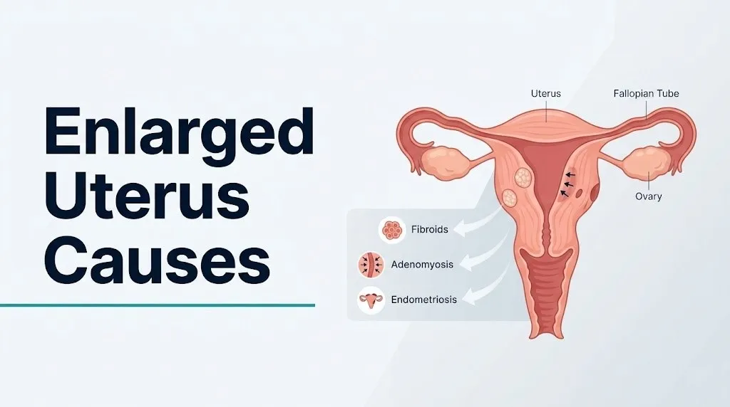
Enlarged Uterus Causes: What’s Really Behind It 2026
The most common enlarged uterus causes are uterine fibroids and adenomyosis, but the full list includes endometrial hyperplasia, endometrial cancer, hematometra from cervical stenosis, uterine sarcoma, and normal pregnancy, each producing uterine enlargement through a completely different physiological mechanism. Knowing which cause is most likely in your situation depends on your age, reproductive status, symptom pattern, and whether the enlargement is diffuse or focal.
The American College of Obstetricians and Gynecologists (ACOG) reports that uterine fibroids affect between 20 and 80 percent of women by age 50, making them the dominant driver of uterine enlargement in reproductive-age women. What most general health articles skip is why this happens at the tissue level, and that mechanism determines everything from how the enlargement feels on exam to which treatment approach applies.
This article covers every clinically recognized cause of uterine enlargement with the specific hormonal and cellular mechanism behind each one, identifies which causes are benign and manageable versus which require urgent evaluation, and tells you exactly what type of provider to contact based on your symptom pattern and age.
Enlarged Uterus Causes: The Full Clinical Picture
Enlarged uterus causes span a wide spectrum from entirely benign and common conditions to rare but potentially serious pathology, and the cause cannot be determined from symptoms alone.
The eight primary causes of uterine enlargement are:
- Uterine leiomyomas (fibroids): benign smooth muscle tumors, the most common cause in reproductive-age women
- Adenomyosis: ectopic endometrial gland invasion into the myometrial wall, producing diffuse wall thickening
- Normal pregnancy: the most physiologically dramatic cause, with the uterus expanding from approximately 70 grams at baseline to over 1,100 grams at term
- Endometrial hyperplasia: abnormal proliferation of the endometrial glandular lining under unopposed estrogen stimulation
- Endometrial carcinoma: malignant transformation of endometrial epithelium, the most common gynecologic cancer in the United States
- Hematometra: blood accumulation within the uterine cavity from outflow obstruction, distending the uterine body
- Uterine sarcoma (including leiomyosarcoma): rare malignant tumors arising from uterine smooth muscle or connective tissue stroma
- Large endometrial polyps: pedunculated overgrowths of the endometrial lining, less commonly producing appreciable global uterine enlargement
The clinical distinction that matters most is whether the enlargement is diffuse and symmetrical (suggesting adenomyosis or endometrial pathology) or focal and asymmetric (suggesting discrete fibroid nodules). A board-certified obstetrician-gynecologist can often distinguish these patterns on pelvic examination before imaging is performed.
| Cause | Type of Enlargement | Most Common Age Group | Requires Urgent Evaluation? |
|---|---|---|---|
| Uterine fibroids | Focal, asymmetric | Reproductive age, 30s-50s | Only if hemorrhage or rapid growth |
| Adenomyosis | Diffuse, symmetric, boggy | Late 30s-50s | No, but OB-GYN workup needed |
| Pregnancy | Global, progressive | Reproductive age | No (normal) |
| Endometrial hyperplasia | Mild, diffuse | Perimenopausal | Within weeks |
| Endometrial carcinoma | Variable, often asymmetric | Postmenopausal | Yes, within days |
| Hematometra | Global distension | Any age with obstruction | Yes, if acute and painful |
| Uterine sarcoma | Rapid, irregular | Reproductive and post-menopausal | Yes, immediately |
What Is an Enlarged Uterus and How Big Is Too Big
An enlarged uterus is one that exceeds the normal anatomical dimensions of the non-pregnant uterus, which measures approximately 7.5 cm in length, 4.5 cm in width, and 3.0 cm in depth, with a normal weight of 60 to 90 grams in a woman of reproductive age.
The Cleveland Clinic uses a practical reference point: a normal uterus is roughly the size of a pear. Clinicians and radiologists describe uterine enlargement in terms of gestational-size equivalents when fibroids or other pathology dramatically increases uterine volume. A “12-week size” uterus, for example, means the uterus has enlarged to approximately the dimensions of a uterus at 12 weeks of pregnancy, with a fundal height reaching near the umbilicus on abdominal examination.
Uterine size increases measurably with parity. Women who have carried pregnancies to term typically have a slightly larger uterus than nulliparous women, with normal post-pregnancy measurements running approximately 8.5 cm in length. This is physiological and not pathological.
The threshold for clinical concern is not a single number. A 9-cm uterus in a postmenopausal woman who has never had pregnancies warrants prompt evaluation, while a 10-cm uterus in a 40-year-old woman with known fibroids and stable symptoms may be monitored without immediate intervention.
Quick Tip:
- Uterine size on ultrasound is measured in three dimensions, and the total volume is calculated using the prolate ellipsoid formula (length x width x depth x 0.523). A volume above 120 to 160 mL is generally considered enlarged for a non-pregnant adult woman.
- Women with a uterus described as larger than “12-week size” on clinical examination should have a transvaginal ultrasound to characterize the cause before any management decision is made.
- Postmenopausal women whose uterus should be atrophying after estrogen withdrawal should have any measured enlargement evaluated regardless of absolute size.
Uterine Fibroids: The Most Common Cause of Enlarged Uterus
Uterine fibroids (uterine leiomyomas) are the most common cause of an enlarged uterus in reproductive-age women, occurring in an estimated 20 to 80 percent of women by age 50, with the highest clinical burden falling between ages 30 and 50.
Fibroids are benign monoclonal tumors arising from a single myometrial smooth muscle cell that undergoes genetic mutation. The mutation activates growth programs that would normally be suppressed. Estrogen drives the initial and sustained growth of fibroid tissue through upregulation of estrogen receptor alpha (ERalpha) in the fibroid cells, making these cells far more sensitive to circulating estradiol than normal myometrial cells. Progesterone, acting through progesterone receptor B (PRB), maintains fibroid volume by suppressing apoptosis (programmed cell death) within the tumor. This is why fibroids typically grow during the reproductive years, often accelerate in the hormone-rich perimenopausal transition, and shrink after menopause when ovarian estrogen production falls.
Fibroids also produce their own estrogen locally. The enzyme aromatase (CYP19A1) is expressed within fibroid tissue, converting circulating androgens into estradiol within the tumor itself. This local estrogen amplification helps explain why fibroids can grow even in women with normal systemic estrogen levels.
The location of a fibroid within the uterus determines how it contributes to enlargement. Intramural fibroids grow within the myometrial wall and produce the most pronounced global uterine enlargement. Subserosal fibroids project outward from the uterine outer surface and may create an asymmetric lumpy contour without dramatically increasing overall uterine volume. Submucosal fibroids project inward into the uterine cavity and are the most likely to cause heavy bleeding, even when small.
According to the NICHD Uterine Fibroid Research Program, Black women in the United States develop fibroids at two to three times the rate of white women, with earlier onset and larger fibroid burden at diagnosis. The mechanism behind this disparity involves both genetic factors and differences in vitamin D levels, which has an inhibitory effect on fibroid growth pathways.
Adenomyosis: When the Uterine Wall Itself Expands
Adenomyosis causes an enlarged uterus through a fundamentally different mechanism than fibroids: ectopic endometrial glands and stromal cells invade directly into the myometrium, triggering a reactive expansion of the smooth muscle surrounding each invasive focus.
Think of it this way: fibroids are like knots tied into a rope (discrete nodules within the muscle wall). Adenomyosis is like soaking the entire rope in water until it swells uniformly throughout. The uterus in adenomyosis typically feels diffusely enlarged, tender, and soft or “boggy” on pelvic palpation, rather than the firm, nodular, asymmetric texture characteristic of fibroids.
The mechanism of invasion in adenomyosis is not fully established, but the current evidence points to disruption of the endometrial-myometrial junction (the junctional zone, visible on MRI as a distinct hypointense band). When this boundary breaks down, endometrial glands migrate inward. Once embedded in the myometrium, these ectopic glands respond to the monthly cycle of estrogen and progesterone stimulation exactly as normal endometrium does: they proliferate under estrogen, differentiate under progesterone, and attempt to shed during menstruation. Because the shed tissue has nowhere to go, it causes localized micro-hemorrhage and inflammation within the myometrial wall. The surrounding smooth muscle hypertrophies in response, thickening the myometrial wall globally.
Research published in the American Journal of Obstetrics and Gynecology has documented that adenomyosis produces elevated levels of prostaglandin E2 and prostaglandin F2alpha within the affected myometrium. These prostaglandins drive the severe cramping dysmenorrhea that distinguishes adenomyosis from other causes of uterine enlargement.
Adenomyosis is most commonly diagnosed in women aged 35 to 50. Parous women and women who have had prior uterine surgery, including cesarean section, are at higher risk, as uterine wall disruption may facilitate endometrial cell migration into the myometrial layer.
Key Takeaway: Adenomyosis produces a diffuse, boggy, symmetrically enlarged uterus caused by endometrial tissue invading the uterine muscle wall, while fibroids produce a firm, asymmetric, nodular enlargement from discrete smooth muscle tumors, and a pelvic examination by a board-certified OB-GYN can often distinguish these two patterns before imaging is ordered.
Causes of Enlarged Uterus During Pregnancy
Normal pregnancy is the most physiologically expected cause of uterine enlargement, and the uterus undergoes one of the most dramatic size changes of any organ in the human body over a 40-week period.
A non-pregnant uterus weighs approximately 60 to 90 grams. By 40 weeks of gestation, the uterus weighs approximately 1,100 grams, an increase of more than tenfold. The fundal height rises from below the pubic symphysis in early pregnancy to approximately 36 to 37 cm above the pubic symphysis at term, reaching the level of the xiphoid process.
This growth happens through two distinct cellular processes in the myometrium. In early pregnancy, myometrial smooth muscle cells increase in size through hypertrophy driven by rising estrogen and progesterone from the developing placenta. After approximately 20 weeks, smooth muscle cell division (hyperplasia) contributes to further wall expansion to accommodate the growing fetus. The decidualization of the endometrium, in which the glandular lining transforms into supportive tissue for the implanted embryo, also adds to overall uterine volume.
Beyond term gestation, abnormal pregnancy-related conditions can cause pathological uterine enlargement. Gestational trophoblastic disease (hydatidiform mole), in which abnormal placental trophoblast tissue grows rapidly within the uterine cavity, can produce a uterus that is large for gestational dates and is often accompanied by markedly elevated beta-human chorionic gonadotropin (beta-hCG) levels.
Women of advanced maternal age (35 years or older) and women pregnant for the first time at older ages have a higher statistical risk of gestational trophoblastic disease than younger women, according to epidemiological data published in the Journal of Reproductive Medicine. Any uterus that measures significantly larger than expected for gestational dates warrants ultrasound evaluation to exclude molar pregnancy.
Endometrial Hyperplasia and Uterine Enlargement
Endometrial hyperplasia causes uterine enlargement through abnormal proliferation of the glandular epithelium lining the uterine cavity, driven almost exclusively by prolonged exposure to estrogen without adequate progesterone opposition.
Under normal menstrual cycle physiology, estrogen stimulates endometrial proliferation during the follicular phase, and progesterone from the corpus luteum converts this proliferative lining to a secretory lining after ovulation. When ovulation does not occur (anovulatory cycles, common in perimenopause and PCOS), progesterone is not produced. Estrogen stimulation of the endometrium continues unchecked, producing abnormal glandular thickening, cystic dilation of endometrial glands, and eventual overcrowding of glandular architecture.
The ACOG Practice Bulletin on Endometrial Hyperplasia classifies this condition into two types based on histological architecture and the presence or absence of cellular atypia: hyperplasia without atypia (which carries a low risk of malignant transformation, estimated at approximately 1 to 3 percent over 20 years) and atypical hyperplasia (which carries a substantially higher risk of progression to endometrial carcinoma, estimated at approximately 8 to 29 percent if untreated, per ACOG data).
The uterine enlargement in endometrial hyperplasia is typically mild to moderate and symmetric, reflecting the thickened endometrial cavity rather than a myometrial mass effect. The endometrial stripe measurement on transvaginal ultrasound is the key imaging marker: a stripe above 4 to 5 mm in a postmenopausal woman or above 12 to 14 mm in a premenopausal woman warrants endometrial biopsy.
Women with obesity face a disproportionately elevated risk of endometrial hyperplasia. Adipose tissue is a significant site of peripheral estrogen synthesis through aromatase activity, converting androgens to estrone (a weaker estrogen). In women with high adipose tissue mass, this peripheral estrogen production creates a state of chronic, low-grade, unopposed estrogen exposure that continuously stimulates endometrial growth.
Endometrial Cancer as a Cause of Enlarged Uterus
Endometrial carcinoma is the most common gynecologic malignancy in the United States and causes uterine enlargement through invasion and expansion of malignant endometrial cells into the uterine cavity and eventually into the myometrial wall.
The Society of Gynecologic Oncology (SGO) reports that endometrial cancer is diagnosed in approximately 66,000 women in the United States annually. It is predominantly a disease of postmenopausal women, with a median age of diagnosis of 63 years. The most common histological subtype, endometrial endometrioid adenocarcinoma, arises from endometrial hyperplasia through progressive accumulation of genetic mutations that disable tumor suppressor pathways (most commonly PTEN loss and microsatellite instability).
Early-stage endometrial carcinoma may produce only a subtle increase in uterine size, primarily through enlargement of the endometrial cavity with tumor tissue. As the disease advances, myometrial invasion deepens and the uterus enlarges more noticeably. At more advanced stages, the cervix and parametrial tissues may be involved, and the overall uterine mass can become substantial.
The cardinal warning symptom is postmenopausal vaginal bleeding. Approximately 90 percent of women with endometrial cancer experience abnormal vaginal bleeding before diagnosis, according to Mayo Clinic clinical data. Any postmenopausal bleeding, including spotting, must be evaluated with endometrial biopsy and transvaginal ultrasound. Waiting to see if bleeding resolves is not appropriate clinical management.
Women on tamoxifen therapy for breast cancer have an elevated risk of endometrial cancer. Tamoxifen acts as an estrogen agonist in the uterus while blocking estrogen in breast tissue, and long-term use produces endometrial stimulation similar to unopposed estrogen. Any woman on tamoxifen who develops abnormal uterine bleeding or a thickened endometrial stripe on surveillance imaging should be evaluated by a board-certified gynecologic oncologist without delay.
Key Takeaway: Any postmenopausal woman with an enlarged uterus and vaginal bleeding, even light spotting, requires endometrial biopsy and transvaginal ultrasound evaluation by a board-certified obstetrician-gynecologist within days, not weeks, because endometrial carcinoma is the most common gynecologic cancer in the United States and is highly treatable when identified early.
Hematometra and Cervical Stenosis Causing Uterine Swelling
Hematometra is uterine enlargement caused by the accumulation of blood within the uterine cavity, occurring when the internal or external cervical os is obstructed and menstrual blood cannot drain normally.
The uterus in this condition is not enlarged because of a mass or wall thickening; it is enlarged because the cavity is distended with retained blood, functioning like a balloon slowly filled with fluid that has no outlet. As pressure within the uterine cavity increases, the thin uterine walls are pushed outward, producing a globally distended, often tender uterus that can be palpated abdominally when the hematometra is substantial.
Cervical stenosis is the most common underlying cause of hematometra. The cervical canal narrows through several mechanisms: post-procedural scarring after cervical LEEP (loop electrosurgical excision procedure), cone biopsy, or laser ablation for cervical dysplasia; post-radiation cervical fibrosis following pelvic radiation therapy for cervical or endometrial cancer; or post-menopausal cervical atrophy in women whose estrogen levels have declined and whose cervical tissue has contracted. Uterine or endocervical cancer obstructing the cervical canal is an additional cause that must always be excluded.
Women who have undergone prior cervical procedures should be aware that cervical stenosis can develop months to years after the procedure. The presenting symptom is often cyclic pelvic pain without associated menstrual flow (cryptomenorrhea), or simply absent menstruation with progressive lower abdominal distension in a woman who was previously menstruating regularly.
Quick Tip:
- Hematometra producing acute severe pelvic pain and hemodynamic symptoms (lightheadedness, rapid pulse, pallor) from rapid uterine distension or secondary infection (pyometra) warrants immediate emergency evaluation.
- Women who are postmenopausal and develop hematometra should have cervical obstruction evaluated to exclude endocervical or endometrial malignancy as the obstructing cause.
- Adolescents with primary amenorrhea and cyclic pelvic pain should be evaluated for imperforate hymen or transverse vaginal septum, which produces hematometra through outflow obstruction at the vaginal rather than cervical level.
Uterine Sarcoma: A Rare but Serious Cause
Uterine sarcoma is a malignant tumor arising from the smooth muscle or connective tissue of the uterine wall and represents one of the most clinically serious causes of uterine enlargement, though it accounts for only 3 to 7 percent of all uterine malignancies.
The most common subtype, uterine leiomyosarcoma, arises from the smooth muscle of the myometrium and is genetically and biologically distinct from uterine leiomyomas (fibroids). It does not arise from a pre-existing fibroid that “turns malignant,” despite the popular misconception. Leiomyosarcomas develop de novo from myometrial cells that have acquired chromosomal instability, most commonly involving deletions in chromosomes 10q and 13q affecting tumor suppressor gene loci.
The clinical challenge is that uterine sarcoma can appear similar to large fibroids on imaging. The features that raise suspicion for sarcoma rather than fibroid include: rapid uterine growth (particularly after menopause when fibroid growth should have slowed or stopped), heterogeneous internal echogenicity on ultrasound suggesting necrosis or hemorrhage within the mass, and elevated lactate dehydrogenase (LDH) on serum testing. No imaging modality reliably differentiates sarcoma from fibroid preoperatively, which is why the finding of unexpected leiomyosarcoma in surgical fibroid specimens occurs in an estimated 1 in 400 to 1 in 500 cases, according to research published in the Journal of the American Medical Association (JAMA).
Postmenopausal women with any rapidly enlarging uterine mass, particularly one accompanied by pain and postmenopausal bleeding, should be evaluated by a board-certified gynecologic oncologist. Uterine sarcoma carries a substantially worse prognosis than endometrial carcinoma, with five-year survival rates for stage III and IV leiomyosarcoma below 30 percent, making early surgical evaluation essential.
What Causes an Enlarged Uterus After Menopause
An enlarged uterus after menopause is a finding that requires a different and more urgent clinical interpretation than the same finding in a reproductive-age woman, because the postmenopausal uterus should be atrophying rather than growing.
After the final menstrual period, ovarian estrogen production falls sharply. The endometrium thins progressively, the uterus contracts in size, and the myometrium loses the hormonal stimulation that drove fibroid growth and myometrial hypertrophy during the reproductive years. In most postmenopausal women who are not on hormone therapy, uterine volume decreases measurably within two to three years of menopause and continues to decline gradually.
Any uterine enlargement in this context is therefore an active process, not a residual one. The possible causes include:
- Endometrial carcinoma: the most common malignant cause in postmenopausal women; produces endometrial cavity distension and eventual myometrial invasion
- Uterine sarcoma: produces rapid, often painful uterine enlargement in postmenopausal women
- Fibroid persistence under exogenous estrogen: women on systemic hormone therapy (estrogen-containing menopausal hormone therapy) may have fibroids that persist or rarely grow because exogenous estrogen continues to stimulate ERalpha in fibroid tissue
- Cervical stenosis with hematometra: cervical atrophy and fibrosis in postmenopausal women can obstruct the cervical os, causing blood or fluid accumulation in the uterine cavity
- Pyometra: pus accumulation within the uterine cavity (usually from cervical obstruction and secondary infection) producing a tender, distended uterus in an often elderly woman with systemic signs of infection
According to ACOG clinical practice guidance, any postmenopausal woman with vaginal bleeding, pelvic pain, or a uterus found to be larger than expected on imaging should have endometrial pathology excluded with endometrial sampling before any other management is considered. The default clinical posture in postmenopausal uterine enlargement is to rule out malignancy first.
Key Takeaway: A uterus that is enlarging after menopause is an active pathological process until proven otherwise, and any postmenopausal woman with unexplained uterine enlargement should be evaluated by a board-certified obstetrician-gynecologist with endometrial sampling and transvaginal ultrasound within days to one week of the finding, not at a routine annual appointment scheduled months away.
Swollen Uterus Causes: Risk Factors and Who Is Most Affected
The risk factors for a swollen uterus vary considerably by the specific underlying cause, and understanding which factors apply to you helps predict which cause is most likely before diagnostic testing is complete.
Risk factors for uterine fibroids:
- Black or African American ancestry: two to three times higher risk than white women, per NICHD data, with fibroids typically presenting earlier and in larger numbers
- Family history of fibroids in a first-degree relative: estimated 2.5-fold increased risk, pointing to a heritable genetic component
- Early menarche (onset of menstruation before age 10): increases lifetime cumulative estrogen exposure
- Nulliparity: pregnancy is associated with fibroid regression in some women, so women who have not been pregnant may have higher cumulative fibroid burden
- Obesity: elevated adipose tissue aromatase activity increases peripheral estrogen production
- Vitamin D deficiency: research published in Fertility and Sterility has found an association between low serum 25-hydroxyvitamin D levels and higher fibroid prevalence, particularly in Black women
Risk factors for adenomyosis:
- Parity: counterintuitively, adenomyosis is more common in women who have had pregnancies, possibly because uterine wall trauma during childbirth facilitates endometrial-myometrial junction disruption
- Prior uterine surgery: cesarean section, myomectomy, or uterine curettage
- Late 30s to 40s age group: adenomyosis prevalence peaks in this reproductive decade
Risk factors for endometrial hyperplasia and carcinoma:
- Postmenopausal status: most endometrial cancers occur after age 55
- Obesity (body mass index above 30): adipose-derived estrogen creates chronic unopposed endometrial stimulation
- Unopposed exogenous estrogen therapy without progestogen
- Tamoxifen use
- Lynch syndrome (hereditary non-polyposis colorectal cancer): carries a 40 to 60 percent lifetime risk of endometrial cancer per the National Cancer Institute
- Diabetes mellitus and metabolic syndrome: associated with elevated insulin and IGF-1 levels, which independently stimulate endometrial cell proliferation
Symptoms Produced by an Enlarged Uterus
The symptoms of an enlarged uterus depend more on the specific cause and the direction of uterine expansion than on uterine size alone, and some women with substantially enlarged uteri remain asymptomatic.
The most clinically consistent symptoms across causes of uterine enlargement include:
- Menorrhagia (heavy menstrual bleeding): most pronounced with submucosal fibroids, endometrial hyperplasia, or endometrial carcinoma; caused by increased endometrial surface area, abnormal vascular architecture, and impaired uterine contractility
- Dysmenorrhea (painful menstruation): most severe in adenomyosis, where prostaglandin E2 and F2alpha from ectopic glands drive intense myometrial cramping
- Pelvic pressure and heaviness: produced by the gravitational effect of an enlarged uterus on the pelvic floor and surrounding structures
- Urinary frequency and urgency: caused by anterior uterine enlargement compressing the posterior wall of the bladder, reducing functional bladder capacity
- Lower back pain and sciatica-like symptoms: caused by the weight of the enlarged uterus creating tension on the uterosacral ligaments and compressing sacral nerve roots when the uterus is retroverted
- Abdominal distension: in women with very large uteri (greater than 20-week size), the uterus displaces abdominal contents upward and produces visible abdominal protrusion
Women with iron deficiency anemia from chronic menorrhagia may present with fatigue, pallor, exertional dyspnea, and heart palpitations as secondary symptoms. A complete blood count (CBC) with serum ferritin is a standard part of the initial workup when heavy bleeding accompanies an enlarged uterus.
Postmenopausal women with an enlarged uterus may be entirely asymptomatic or may note only mild pelvic pressure, which is why the finding of uterine enlargement on imaging done for an unrelated reason in a postmenopausal woman must not be dismissed as incidental without proper endometrial evaluation.
Pelvic Pain, Heavy Bleeding, and Bloating from Uterine Enlargement
Pelvic pain, heavy menstrual bleeding, and bloating are the three most functionally disabling symptom clusters produced by an enlarged uterus, and each points to a somewhat different underlying mechanism and cause.
Pelvic pain from uterine enlargement takes two distinct forms. Chronic pelvic pressure is the dull, aching, constant discomfort produced by a heavy, enlarged uterus resting on pelvic floor musculature, the bladder, and the rectum. It is characteristic of large fibroids and globally enlarged adenomyotic uteri. Acute cyclic pelvic pain (dysmenorrhea) is the cramping pain that occurs during or around menstruation, most severe in adenomyosis because of prostaglandin-driven myometrial contractions against the resistance of fibrotic, infiltrated myometrial tissue.
Heavy menstrual bleeding (menorrhagia) from uterine enlargement is a physiological consequence of increased endometrial surface area and altered uterine vascular architecture. A fibroid-distorted uterus has a greater total endometrial surface area from which blood vessels must be shed during menstruation, and submucosal fibroids disrupt the normal contractile mechanism the myometrium uses to compress and close uterine blood vessels after menstruation begins. The ACOG defines clinically significant menorrhagia as blood loss exceeding 80 mL per menstrual cycle, a threshold most women with symptomatic fibroids or adenomyosis exceed substantially.
Bloating from uterine enlargement is not intestinal bloating from gas. It is the sensation of lower abdominal fullness produced by upward displacement of bowel loops by a large uterus and simultaneous rectal compression reducing normal bowel transit. Women with very large fibroids (greater than 10 to 12 week uterine size) sometimes report that their abdomen “looks pregnant,” which is an accurate anatomical description of what is occurring.
Women with both fibroids and hypothyroidism experience disproportionately heavy bleeding because thyroid hormone deficiency impairs platelet aggregation and coagulation factor synthesis, compounding the bleeding already produced by fibroid-related endometrial changes. A thyroid-stimulating hormone (TSH) test is clinically warranted in women with severe menorrhagia to exclude hypothyroidism as a contributing factor.
How an Enlarged Uterus Is Diagnosed
An enlarged uterus is first detected during a pelvic examination, then characterized in detail by imaging, and further evaluated with tissue sampling when endometrial pathology is suspected.
The diagnostic process for an enlarged uterus typically proceeds in this order:
- Bimanual pelvic examination: the clinician places one or two fingers inside the vagina and the other hand on the lower abdomen to assess uterine size, shape, contour, mobility, and tenderness. A normal uterus occupies the space between the examiner’s hands comfortably. An enlarged uterus is palpable between the hands with a size estimation in “weeks” or centimeters. A boggy, tender, symmetrically enlarged uterus suggests adenomyosis. A firm, irregular, asymmetrically enlarged uterus suggests fibroids.
- Transvaginal ultrasound (TVUS): the primary imaging tool for uterine assessment. The transducer placed inside the vagina allows high-resolution imaging of the uterus, endometrial lining, myometrial architecture, and ovaries. TVUS measures uterine dimensions, identifies and maps individual fibroid locations and sizes, measures endometrial stripe thickness, and can identify adenomyosis through characteristic features including myometrial heterogeneity, cystic spaces within the myometrium, and asymmetric wall thickening.
- Pelvic MRI: ordered when the uterus is too large for complete transvaginal ultrasound characterization, when sarcoma is suspected, when the number and location of fibroids must be mapped precisely before myomectomy, or when adenomyosis must be confirmed. MRI visualizes the junctional zone (normally less than 12 mm thick; greater than 12 mm is diagnostic for adenomyosis by the criteria used in published clinical research) with higher specificity than ultrasound.
- Endometrial biopsy (EMB): performed using a thin flexible cannula (Pipelle) inserted through the cervical os to sample endometrial tissue. Required whenever endometrial hyperplasia or carcinoma is suspected based on symptoms, age, or ultrasound findings. Performed in the office without anesthesia in most women, though women with cervical stenosis may require cervical dilation first.
- CBC with serum ferritin: ordered to quantify the degree of iron deficiency anemia from chronic blood loss.
- Hysteroscopy: a thin lighted camera inserted through the cervical os under sedation, used to directly visualize the endometrial cavity and take directed biopsies when endometrial biopsy results are inconclusive or when submucosal fibroids or polyps must be evaluated and treated in the same procedure.
Emergency Symptoms: When to Call 911 or Go to the ER
Certain presentations associated with an enlarged uterus represent acute medical emergencies. Do not wait to see if these symptoms resolve on their own.
Call 911 or go to the nearest emergency room immediately if you experience:
- Sudden severe pelvic pain that is significantly worse than your usual menstrual cramps, particularly if accompanied by lightheadedness, rapid heart rate, or pallor (these signs suggest acute hemorrhage or torsion of a subserosal fibroid)
- Heavy vaginal bleeding that soaks through a menstrual pad in less than one hour for two or more consecutive hours (suggests acute hemorrhage from fibroid-related vascular rupture or severe menorrhagia requiring immediate hemostatic intervention)
- Severe pelvic pain with fever above 38.5 degrees Celsius (101.3 degrees Fahrenheit) and vaginal discharge in a woman with known uterine enlargement (suggests pyometra, an infected uterine cavity requiring emergency drainage)
- Postmenopausal vaginal bleeding accompanied by rapidly worsening pelvic pain and a palpable abdominal mass (the combination raises urgent concern for uterine sarcoma or advanced endometrial carcinoma requiring immediate oncological evaluation)
- Any abdominal rigidity, inability to walk upright, or loss of consciousness associated with pelvic symptoms (suggests peritoneal involvement from a ruptured uterine pathology requiring surgical emergency assessment)
These presentations require emergency medical assessment at a hospital emergency department, not an urgent care appointment or a next-day phone call. Delayed diagnosis of uterine hemorrhage or pelvic sepsis carries life-threatening risk.
What Causes a Enlarged Uterus: Distinguishing Benign from Serious Causes
Distinguishing a benign from a serious cause of an enlarged uterus requires integrating your age, menopausal status, symptom character, rate of uterine growth, and imaging findings together, not any single factor in isolation.
Benign causes (fibroids, adenomyosis, normal pregnancy) dominate in reproductive-age women. These conditions cause real symptoms and require treatment, but they do not carry inherent malignant potential. Fibroids do not transform into uterine sarcomas. Adenomyosis does not become cancer. The key feature distinguishing these benign causes from serious ones is the clinical trajectory: benign causes tend to be stable or slow-growing over months and years, while serious causes tend to produce rapid change in symptoms, bleeding patterns, or uterine size.
The features that consistently point toward a serious cause requiring prompt evaluation:
- New uterine enlargement after menopause
- Postmenopausal vaginal bleeding of any volume
- Rapid increase in uterine size over weeks to months rather than years
- Uterine mass with heterogeneous internal structure on ultrasound (suggesting necrosis or hemorrhage within the mass, features more consistent with sarcoma than fibroid)
- Constitutional symptoms (unexplained weight loss, night sweats, fatigue) alongside uterine enlargement
Benign causes associated with slower clinical timelines and premenopausal reproductive-age presentation:
- Gradual uterine enlargement over years with cyclic, predictable heavy periods pointing toward fibroids
- Symmetric uterine enlargement with severe dysmenorrhea and a history of prior uterine surgery pointing toward adenomyosis
- Uterine enlargement with rising beta-hCG in a reproductive-age woman pointing toward pregnancy or gestational trophoblastic disease
According to Mayo Clinic, the distinction between benign and serious uterine enlargement causes cannot be made from symptoms alone and always requires a combination of pelvic examination, transvaginal ultrasound, and in many cases endometrial tissue sampling. Any woman who has received information about an enlarged uterus from a healthcare provider and has not had a clear diagnostic explanation should ask her obstetrician-gynecologist specifically: “Has endometrial sampling been done, and what were the results?”
| Cause | Benign or Serious | Urgency of Evaluation | Provider Type |
|---|---|---|---|
| Uterine fibroids | Benign | Scheduled (within weeks) | Board-certified OB-GYN |
| Adenomyosis | Benign | Scheduled (within weeks) | Board-certified OB-GYN |
| Normal pregnancy | Physiological | Routine obstetric care | Obstetrician |
| Endometrial hyperplasia without atypia | Benign but monitor | Within 2 to 4 weeks | Board-certified OB-GYN |
| Atypical endometrial hyperplasia | Premalignant | Within 1 to 2 weeks | Board-certified OB-GYN or gynecologic oncologist |
| Endometrial carcinoma | Malignant | Within days | Board-certified gynecologic oncologist |
| Hematometra | Variable by cause | Within days if acute | Board-certified OB-GYN or ER |
| Uterine sarcoma | Malignant | Immediately | Board-certified gynecologic oncologist |
Key Takeaway: Benign causes of an enlarged uterus (fibroids and adenomyosis) are distinguished from serious causes by the clinical trajectory, reproductive status, and imaging findings together, and no woman who has been told she has an enlarged uterus should accept a management plan without knowing whether endometrial tissue sampling has been performed and what it showed.
Frequently Asked Questions About Enlarged Uterus Causes
What is the most common cause of an enlarged uterus?
Uterine fibroids (uterine leiomyomas) are the most common cause of an enlarged uterus, affecting an estimated 20 to 80 percent of women by age 50, according to ACOG.
Fibroids are benign smooth muscle tumors that arise from the myometrium and grow under estrogen receptor stimulation, producing focal or global uterine enlargement depending on their number and location.
Adenomyosis, in which endometrial glands invade the myometrial wall, is the second most common cause and produces a distinctly different diffuse, boggy uterine enlargement that a pelvic examination can often differentiate from the firm, nodular texture of fibroid-enlarged uterus.
Can an enlarged uterus go away on its own?
Whether an enlarged uterus resolves without treatment depends entirely on the cause.
Fibroids typically shrink after menopause when estrogen levels fall, and adenomyosis symptoms often improve significantly after menopause in women who are not on hormone therapy, but neither condition reverses completely without medical or surgical intervention in most cases.
Endometrial hyperplasia, hematometra, endometrial cancer, and uterine sarcoma do not resolve on their own and all require active medical management by a board-certified obstetrician-gynecologist or gynecologic oncologist.
Is an enlarged uterus always a sign of cancer?
An enlarged uterus is not always a sign of cancer, and in reproductive-age women the cause is most often benign fibroids or adenomyosis.
However, in postmenopausal women, any uterine enlargement requires evaluation to exclude endometrial carcinoma, which is the most common gynecologic cancer in the United States, before a benign explanation is accepted.
The specific investigation required is a transvaginal ultrasound with endometrial stripe measurement and, when the stripe is thickened or symptoms are present, an endometrial biopsy performed by a board-certified OB-GYN.
What does an enlarged uterus feel like?
An enlarged uterus may produce a sensation of pelvic heaviness, pressure, or fullness in the lower abdomen, often worsening with prolonged standing or physical exertion.
Some women with very large fibroids describe their lower abdomen as feeling hard or like they are carrying a weight in their pelvis, while women with adenomyosis more commonly describe a deep, diffuse aching pressure that intensifies around menstruation.
Many women with a moderately enlarged uterus have no sensation of the uterus at all and discover the finding only on imaging or pelvic examination performed for another reason.
Can PCOS cause an enlarged uterus?
Polycystic ovary syndrome (PCOS) does not directly enlarge the uterus through a primary uterine mechanism.
PCOS causes chronic anovulation, which produces prolonged unopposed estrogen exposure to the endometrium, and this hormonal environment can cause endometrial hyperplasia, which contributes to mild uterine cavity enlargement and increases the risk of endometrial carcinoma over time.
Women with PCOS who have irregular or absent periods should be evaluated by a board-certified OB-GYN for endometrial hyperplasia, particularly if they have not had a progestin-induced withdrawal bleed in more than three months.
When should I be worried about an enlarged uterus after menopause?
Any uterine enlargement detected after menopause should be evaluated promptly by a board-certified obstetrician-gynecologist because the postmenopausal uterus should be shrinking, not growing.
The presence of postmenopausal vaginal bleeding alongside uterine enlargement is a finding that requires endometrial biopsy and transvaginal ultrasound within days, not at a routine future appointment, to exclude endometrial carcinoma or uterine sarcoma.
Rapid uterine growth in a postmenopausal woman, even without bleeding, is a red flag for uterine sarcoma and warrants same-week evaluation by a gynecologic oncologist.
Closing
An enlarged uterus is not a single diagnosis. It is a physical finding that can reflect a dozen different physiological processes, ranging from the entirely benign and hormonally driven to the rare and urgently serious. The cause you are dealing with cannot be determined from symptoms or a general health article alone. It requires a pelvic examination, imaging, and in many cases tissue sampling.
The clearest action step is this: if you have been told your uterus is enlarged and have not yet had a transvaginal ultrasound and, where indicated, an endometrial biopsy, those two investigations should be your immediate priority with a board-certified obstetrician-gynecologist. If you are postmenopausal, that appointment should happen within days, not months.
You now have the mechanistic framework to ask the right questions when you get there. “Is this enlargement diffuse or focal?” “Has the endometrial stripe been measured?” “Is endometrial sampling indicated in my case?” Those three questions will move your workup forward faster than any general symptom review.