What Causes Thick Toenails? 16 Reasons Explained
Thick toenails are most often caused by a fungal nail infection called onychomycosis, though aging, trauma, nail psoriasis, and poor circulation can also cause what causes thick toenails to develop over weeks or months. Understanding the specific cause matters because treatments differ entirely depending on whether the thickening comes from a pathogen, a systemic disease, or a structural change in the nail itself.
According to the American Academy of Dermatology (AAD), onychomycosis affects approximately 14% of the general population in the United States, making it by far the most prevalent nail disorder seen in both dermatology and podiatry practices. That number climbs sharply in adults over age 60, where prevalence estimates from the Journal of the American Academy of Dermatology reach 20 to 48% depending on the population studied.
This article covers every clinically recognized cause of toenail thickening, explains the exact anatomical and physiological mechanism behind each, identifies which causes require prompt medical evaluation, and tells you which provider to see and what to expect at that appointment.
What Causes Thick Toenails
Thick toenails result from one of three core physiological mechanisms: abnormal overproduction of nail plate keratin by the nail matrix, accumulation of inflammatory debris within the subungual space beneath the nail plate, or reduced blood flow to the nail unit that disrupts normal keratinocyte maturation.
Each of these mechanisms produces a visibly thickened nail, but the underlying cause, the associated symptoms, and the treatment approach differ substantially. A nail thickened purely by fungal invasion looks different from one thickened by psoriatic inflammation, and both look different from a nail thickened by years of neglect and microtrauma.
The causes fall into several broad categories:
- Infectious: onychomycosis from dermatophytes, Candida species, or non-dermatophyte molds
- Inflammatory: nail psoriasis, lichen planus, reactive arthritis
- Traumatic: acute injury, repetitive microtrauma from footwear
- Structural: onychogryphosis (ram’s horn nail deformity)
- Age-related: progressive slowing of nail growth and matrix dysregulation
- Vascular: peripheral arterial disease impairing nail matrix circulation
- Systemic: yellow nail syndrome, diabetes mellitus
- Pharmacological: medication-induced nail dystrophy
Identifying the correct category is the first step toward appropriate management. A board-certified dermatologist or licensed podiatrist uses both clinical appearance and confirmatory testing to distinguish these causes.
People with diabetes, peripheral vascular disease, or immunosuppression face a higher risk of severe or treatment-resistant toenail thickening, and should not delay evaluation when toenail changes appear.
Quick Tip:
- Examine all ten toenails, not just the affected one. Involvement of multiple nails simultaneously points toward systemic or infectious causes rather than isolated trauma.
- Note the color alongside the thickness. Fungal nails are typically yellow, brown, or white. Psoriatic nails are often salmon-colored or show pitting. Post-traumatic nails are dark red, purple, or black initially.
- Record how long the thickening has been present. Gradual thickening over six or more months differs clinically from thickening that appeared within weeks.
How the Toenail Grows and Why It Thickens
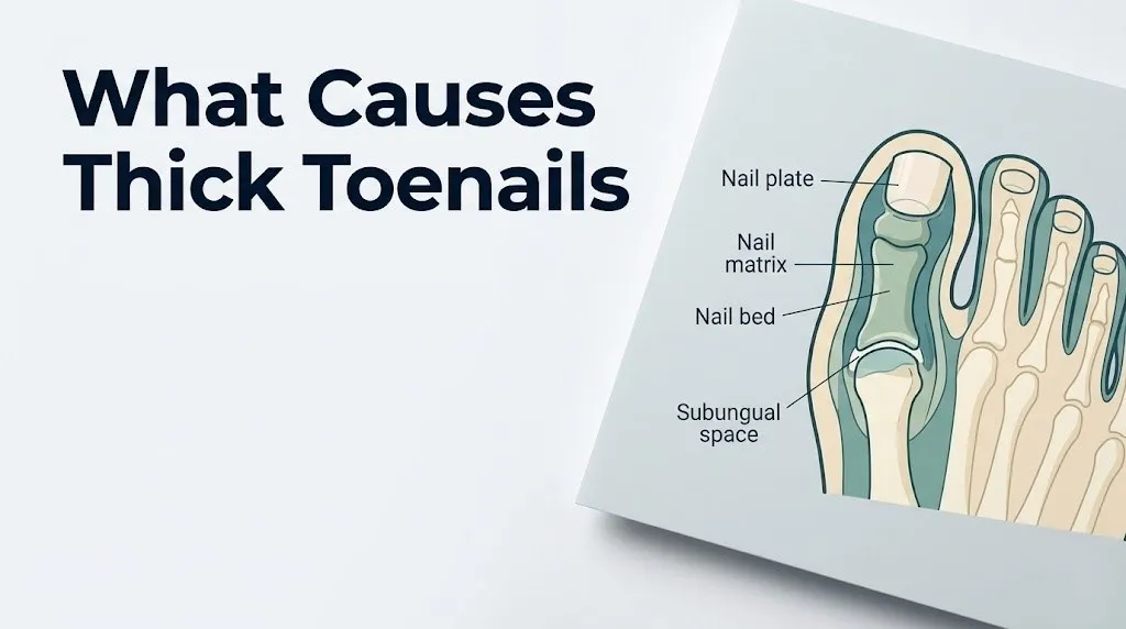
Understanding what causes thick toenails requires knowing the normal architecture of the nail unit. The nail plate — the hard, visible structure you trim — is produced entirely by the nail matrix, a crescent-shaped zone of germinal epithelium located beneath the base of the nail and extending slightly under the lunula (the pale half-moon at the nail base).
Nail plate keratin is synthesized by matrix keratinocytes, which divide, flatten, and progressively keratinize as they migrate distally from the matrix toward the nail tip. Think of the nail matrix as a keratin factory running on a conveyor belt: cells manufactured at one end move continuously forward, harden into flat keratin sheets, and are eventually cut away when you trim your nails.
The nail plate is normally 0.5 to 0.75 millimeters thick in healthy adults. When anything disrupts the matrix keratinocytes — infection, inflammation, trauma, vascular insufficiency, or systemic disease — the keratin output changes. Overactive or inflamed matrix cells produce too much keratin too fast, yielding a thickened plate. Alternatively, inflammatory cells and keratin debris accumulate in the subungual space between the nail plate and the nail bed, raising the plate upward and creating the appearance of thickness even when the plate itself is not pathologically enlarged.
According to the National Institute of Arthritis and Musculoskeletal and Skin Diseases (NIAMS), toenails grow approximately 1.5 to 2 millimeters per month, roughly three times slower than fingernails. This slow growth rate means that both damage and recovery manifest over many months, which is why persistent nail thickening rarely resolves quickly even after the underlying cause is treated.
| Nail Structure | Location | Role in Thickening |
|---|---|---|
| Nail matrix (germinal) | Under base of nail plate | Produces nail plate keratin cells |
| Nail plate | Visible hard nail | Becomes abnormally thick when matrix is overactive |
| Nail bed | Under nail plate | Contributes to subungual hyperkeratosis |
| Subungual space | Between nail plate and bed | Accumulates debris and fungal material |
| Hyponychium | Nail plate and skin junction | Entry point for dermatophyte invasion |
People who have had nail matrix injuries (crush injuries, repeated pressure from ill-fitting footwear) may have permanent alterations in nail plate production, meaning that some degree of thickening after significant matrix trauma can be irreversible even without ongoing infection or disease.
Fungal Nail Infection and Toenail Thickening
Fungal nail infection, clinically called onychomycosis or tinea unguium, is the single most common cause of thick toenails and accounts for approximately 50% of all nail disorders seen in clinical practice, according to a 2022 review published in the Journal of the American Academy of Dermatology. The great toenail is the most frequently affected nail because it has the largest subungual surface area and is most exposed to the warm, moist environment inside footwear.
Onychomycosis begins when dermatophyte organisms gain access to the nail unit through the hyponychium, the thin skin at the junction between the nail plate and the fingertip. Once inside the subungual space, the fungi colonize the nail bed and begin invading the nail plate itself.
Observable changes progress in a recognizable pattern:
- Early stage: yellowish or whitish discoloration starting at the nail tip or sides, mild thickening
- Intermediate stage: nail plate visibly thickened, crumbly texture, surface may appear rough or streaked
- Advanced stage: nail plate severely thickened, distorted, separating from the nail bed (onycholysis), often with a distinctive musty or foul odor from debris accumulation
The AAD notes that onychomycosis is more prevalent in males than females by approximately a 1.9 to 1 ratio, likely because male nail growth rate is faster (providing more rapidly available keratin substrate) and because male occupational footwear tends to create warmer, more occluded foot environments. Adults over age 60, athletes who use communal showers, and people who have had prior athlete’s foot (tinea pedis) face substantially higher rates of nail fungal infection.
Without treatment, onychomycosis does not resolve spontaneously. Topical antifungal agents alone have limited penetration into the nail plate and achieve cure rates below 10% for established infections without professional guidance, based on data from the British Journal of Dermatology.
Key Takeaway: Onychomycosis causes over half of all thick toenail cases in clinical practice, progresses without treatment, and requires either topical or oral antifungal therapy confirmed by a board-certified dermatologist or podiatrist — never assume toenail thickening is purely cosmetic until a fungal cause is ruled out.
How Dermatophytes Thicken the Nail Plate
The mechanism by which dermatophytes produce toenail thickening is more complex than simple overgrowth and is worth understanding precisely. Trichophyton rubrum accounts for approximately 70 to 80% of all onychomycosis cases in the United States, according to JAMA Dermatology, with Trichophyton mentagrophytes and non-dermatophyte molds such as Scopulariopsis brevicaulis responsible for most of the remaining cases.
Dermatophytes are obligate keratin-feeders. They secrete keratinase enzymes that digest the keratin proteins within the nail plate, allowing the fungal hyphae to penetrate progressively deeper into the nail structure. This enzymatic digestion creates structural channels and fragmentation within the nail plate itself.
Here is what happens at the tissue level:
- Dermatophyte hyphae penetrate the hyponychium and advance toward the nail matrix.
- Keratinase secretion dissolves keratin cross-links within the nail plate, creating a physically weakened and irregularly structured plate.
- The nail bed responds to fungal invasion with an inflammatory reaction, including recruitment of neutrophils and release of inflammatory cytokines.
- This inflammatory response drives hyperproliferation of nail bed keratinocytes, which produce excess subungual keratinous debris.
- The accumulating debris beneath the nail plate raises the plate upward (onycholysis) and adds apparent thickness.
- The compromised nail plate simultaneously loses its normal flat architecture, becoming irregular, crumbly, and thicker than normal.
The result is a nail that is thickened from both above (abnormal nail plate produced by an inflamed matrix) and below (subungual debris pushing the plate upward). This dual mechanism explains why advanced fungal nails are difficult to trim and why the texture is characteristically crumbly and fragmented rather than simply hard and smooth.
People with HIV infection, organ transplant recipients on immunosuppressive therapy, and individuals undergoing chemotherapy face significantly higher rates of Candida species nail involvement alongside or instead of dermatophyte infection, which requires different antifungal treatment strategies. A licensed podiatrist or board-certified dermatologist should supervise antifungal selection in any immunocompromised person with nail fungal infection.
Nail Psoriasis and Toenail Thickening
Nail psoriasis is a cause of thick toenails that is consistently underdiagnosed because many people with nail psoriasis have no obvious skin psoriasis, making the nail changes appear to come from nowhere. According to the National Institute of Arthritis and Musculoskeletal and Skin Diseases (NIAMS), psoriasis affects approximately 3% of U.S. adults, and up to 90% of people with psoriasis develop nail involvement at some point during their lifetime.
The mechanism is a T-cell mediated autoimmune inflammatory cascade. Interleukin-17 (IL-17) and interleukin-23 (IL-23) overexpression in the nail matrix and nail bed drives keratinocyte hyperproliferation at a rate that outpaces normal orderly nail plate formation. The result is a disordered, thickened nail plate with characteristic features that distinguish it from fungal thickening.
Classic features of psoriatic nail disease include:
- Nail pitting: small punctate depressions on the nail plate surface from abnormal nail matrix keratinization
- Onycholysis: separation of the nail plate from the nail bed, often with a yellowish-pink border called the “oil drop” sign
- Subungual hyperkeratosis: chalky white or yellowish buildup under the nail plate
- Leukonychia: white spots or lines within the nail plate
- Nail plate thickening: typically more irregular and less crumbly than fungal thickening
Psoriatic nail changes have significant clinical importance beyond cosmetics. A 2021 study published in the Journal of the American Academy of Dermatology found that nail psoriasis is a strong predictor of psoriatic arthritis development, affecting approximately 30% of people with psoriasis. Nail involvement correlates with active joint inflammation in the distal interphalangeal joints of the toes and fingers.
People with isolated nail psoriasis (no skin plaques visible) often wait years before diagnosis because their presentation is mistaken for onychomycosis. A key distinguishing feature: psoriatic nails show pitting (small surface dimples) while fungal nails typically do not. A board-certified dermatologist or rheumatologist should evaluate suspected nail psoriasis, particularly when joint pain accompanies nail changes.
Key Takeaway: Nail psoriasis involves IL-17 and IL-23-driven keratinocyte overproduction in the nail matrix and strongly predicts psoriatic arthritis development, which makes early evaluation by a dermatologist or rheumatologist clinically important rather than optional.
Toenail Trauma and Post-Injury Thickening
Traumatic toenail thickening is the second most common non-infectious cause of thick toenails and is frequently overlooked because the injury may have occurred months or even years before the thickening becomes noticeable. The nail matrix responds to physical injury the same way any tissue responds to trauma: with inflammation, disrupted cell division, and eventual scarring that permanently alters the structure of new nail plate produced.
Acute trauma such as a heavy object dropped on the toe, a door crush injury, or a single severe blow from a ball or fall typically causes a subungual hematoma (blood pooling beneath the nail plate) that creates visible darkening. As the damaged nail grows out over the following three to six months, the portion of nail produced by the traumatized matrix may be abnormally thick, ridged, or opaque.
Repetitive microtrauma is clinically more significant and more often missed:
- Running shoes that allow forward toe slide cause repeated nail tip impact with each stride
- Ill-fitting footwear where the nail’s lateral edges press against the shoe creates chronic lateral nail fold and matrix pressure
- Occupational standing on hard surfaces over years creates cumulative pressure loading on the nail unit
- Ballet dancers, soccer players, and long-distance runners develop post-traumatic nail thickening at rates substantially higher than the general population, based on data from the American Podiatric Medical Association (APMA)
The nail matrix, when repeatedly traumatized, eventually produces nail plate with irregular thickness and altered keratin layering. This is called traumatic onychopathy. Unlike fungal thickening, post-traumatic thickening does not respond to antifungal therapy because no pathogen is present. Management centers on footwear modification, offloading pressure, and in some cases surgical or chemical nail matrix ablation by a podiatrist when the nail becomes persistently painful or causes recurrent ingrown nail complications.
Older adults who cannot clearly recall an injury should not assume their nail thickening is non-traumatic. Peripheral neuropathy from diabetes reduces pain sensation, meaning significant nail trauma can occur without the person registering it clearly.
Onychogryphosis and Ram’s Horn Toenails
Onychogryphosis is one of the most visually dramatic forms of toenail thickening and represents a specific structural deformity rather than simply a thickened nail plate. In onychogryphosis, the nail plate grows in a curved, horn-like trajectory, sometimes twisting laterally and extending beyond the toe tip in a ram’s horn or claw configuration that can grow several centimeters if left completely untreated.
The nail matrix in onychogryphosis loses uniform production across its full width. One side of the matrix (often the medial edge) grows faster than the other, causing the nail plate to curve spirally rather than growing flat. This asymmetric growth combined with thickened, hardened nail plate production yields the characteristic appearance.
According to the Mayo Clinic, onychogryphosis predominantly affects the great toenail and most commonly occurs in older adults, particularly those who have difficulty performing regular nail care due to mobility limitations, visual impairment, or cognitive decline. It can also result from severe neglect of any nail condition, including untreated fungal infection, previous severe trauma, or prolonged pressure from tight footwear.
Contributing factors include:
- Peripheral vascular disease reducing blood supply to the nail matrix
- Long-term tight footwear causing lateral nail matrix pressure
- Neurological conditions impairing foot sensation (diabetes-related neuropathy, stroke)
- Prolonged immobility (extended hospitalization, nursing home residence)
- Previous severe nail trauma causing matrix scarring
Onychogryphosis itself is not an infection, but the distorted nail creates spaces where fungi can colonize secondarily. A licensed podiatrist should manage this condition. Treatment involves professional nail debridement and, in permanent cases, surgical nail matrix destruction (matricectomy) to prevent regrowth. Home trimming of an onychogryphotic nail is rarely possible with standard clippers and can cause nail plate splitting, nail fold injury, and secondary infection.
Key Takeaway: Onychogryphosis is a structural nail matrix deformity producing curved, thickened, ram’s horn-shaped toenails that require professional podiatric management, not home trimming, particularly in older adults with limited mobility or vascular disease.
Aging and Toenail Thickening in Seniors
Age-related toenail thickening is one of the most frequently encountered and least well-explained nail changes in adults over 60. The mechanism is not simply “nails get harder with age” — the physiology involves multiple parallel changes in the nail unit that alter both production rate and plate structure.
As the body ages, nail matrix keratinocyte division slows progressively. Research published in the British Journal of Dermatology found that toenail growth rate decreases by approximately 50% between early adulthood and the seventh decade of life. Slower keratinocyte transit time through the nail plate means each cell layer remains in place longer, accumulating additional cross-linking between keratin proteins and producing a denser, harder, more opaque nail plate.
Simultaneously, blood flow to the peripheral nail unit decreases as a result of arteriolar narrowing and reduced cardiac output associated with normal aging. This reduced vascular supply to the nail matrix impairs keratinocyte maturation, producing nail plate with irregular thickness and less smooth surface architecture.
Age-related changes that contribute to toenail thickening in seniors:
- Slowed nail growth rate producing denser keratin layering per unit length
- Reduced nail bed vascularity impairing normal keratinocyte maturation
- Cumulative lifetime microtrauma to nail matrix from decades of footwear use
- Increased susceptibility to onychomycosis due to age-related immune senescence
- Reduced flexibility of the nail plate increasing risk of splitting, which drives protective thickening responses
- Reduced ability to perform regular nail care, allowing nail plate to accumulate additional keratin layers
The distinction between purely age-related thickening and onychomycosis is clinically important because the two coexist frequently in seniors. A person over 70 with thickened toenails may have both aging-related nail changes and superimposed fungal infection. A potassium hydroxide (KOH) preparation or nail culture is necessary to confirm whether fungal treatment is warranted, even in older adults whose thickening seems age-consistent.
Peripheral Arterial Disease and Thick Toenails
Peripheral arterial disease (PAD) causes toenail thickening through a vascular mechanism that is distinct from both fungal and inflammatory causes. In PAD, atherosclerotic plaque narrows the peripheral arteries supplying blood to the lower legs and feet, reducing oxygen and nutrient delivery to the nail matrix and surrounding nail unit tissues.
The nail matrix is highly vascular and depends on continuous oxygen delivery to sustain the rapid cell division required for normal nail plate production. When arterial blood supply falls below the threshold needed for normal keratinocyte function, the nail matrix produces nail plate with impaired keratin organization. The resulting nail is typically thickened but also brittle, slow-growing, and often yellowish-brown or pale in color because the nail bed’s underlying vascular tone (which normally gives nails their pinkish appearance) is diminished.
According to the Centers for Disease Control and Prevention (CDC), PAD affects approximately 6.5 million Americans over age 40, with prevalence rising sharply after age 65. The condition is more common in people with atherosclerosis risk factors: cigarette smoking, type 2 diabetes mellitus, hypertension, and hyperlipidemia.
Associated findings that suggest PAD as a contributor to nail thickening:
- Cold, pale, or bluish toes alongside the nail changes
- Reduced or absent pedal pulses (dorsalis pedis or posterior tibial arteries)
- Calf pain or foot cramping with walking that resolves with rest (claudication)
- Non-healing cuts or sores on the feet or lower legs
- Hair loss from the lower legs and dorsum of the foot
Any person with thick toenails who also has risk factors for cardiovascular disease or diabetes should have vascular status evaluated. An ankle-brachial index (ABI) test, a non-invasive pressure measurement comparing ankle blood pressure to arm blood pressure, is the standard initial screening tool used by primary care physicians and vascular medicine specialists to detect PAD. An ABI below 0.9 indicates hemodynamically significant arterial obstruction.
Yellow Nail Syndrome and Nail Thickening
Yellow nail syndrome is a rare but clinically distinct cause of thick toenails that involves the lymphatic system rather than the nail matrix directly. Characterized by the triad of thickened and yellow-colored nails, lymphedema (swelling from lymphatic drainage impairment), and respiratory disease (chronic bronchiectasis, pleural effusion, or recurrent pneumonia), yellow nail syndrome is diagnosed in fewer than 1 in 1 million people annually based on case data from the British Journal of Dermatology.
The mechanism involves impaired lymphatic drainage in the nail bed and periungual tissues. Lymphatic vessel dysfunction reduces fluid clearance from the subungual space, producing chronic mild edema of the nail bed that disrupts normal keratinocyte function. The result is a nail plate that grows extremely slowly (sometimes less than 0.25 millimeters per month, compared to the normal 1.5 to 2 millimeters) and accumulates abnormal thickness over months to years.
The characteristic appearance of yellow nail syndrome nails differs from both fungal and psoriatic thickening:
- Uniform yellow or yellow-green discoloration across the entire nail plate
- Complete absence of the lunula in most cases
- Marked slowing of nail growth (the nail may appear not to grow for weeks)
- Nail plate thickened and excessively curved (increased transverse curvature)
- Loss of the nail cuticle
Yellow nail syndrome has documented associations with lymphedema, pleural effusions, chronic bronchiectasis, and titanium implants, though the exact causal mechanism for the titanium association remains under investigation as of 2026 clinical review. A person presenting with thick yellowish nails alongside unexplained limb swelling or chronic respiratory symptoms should prompt referral to a board-certified pulmonologist and a dermatologist experienced in nail disorders for systemic workup.
Key Takeaway: Yellow nail syndrome is rare but represents a systemic lymphatic dysfunction that produces uniformly yellow, extremely slow-growing, thickened nails alongside lymphedema and respiratory disease, requiring specialist evaluation rather than antifungal treatment.
Diabetes and Thick Toenails
Diabetes mellitus contributes to toenail thickening through three simultaneous physiological mechanisms, making people with poorly controlled diabetes among the highest-risk groups for nail pathology. The National Institutes of Health (NIH) reports that foot complications — including nail disorders — affect up to 50% of people with long-standing diabetes, representing one of the most common and costly manifestations of diabetic peripheral vascular and neurological disease.
First mechanism: immune dysfunction. Hyperglycemia (sustained elevated blood glucose) impairs neutrophil chemotaxis and reduces the production of reactive oxygen species used by white blood cells to kill pathogens. This creates an immunologically permissive environment for dermatophyte proliferation in the nail unit. People with diabetes are two to three times more likely to develop onychomycosis than age-matched non-diabetic adults, based on data from a multi-center study published in the International Journal of Dermatology.
Second mechanism: peripheral neuropathy. Sensory neuropathy from long-standing diabetes reduces pain sensation in the feet and toes. This means traumatic nail injuries that would cause significant pain in a healthy person go unnoticed in someone with diabetic neuropathy, allowing cumulative matrix damage that produces thickened, deformed nail plate without the person ever registering an injury.
Third mechanism: microvascular disease. Diabetic microangiopathy (disease of the small blood vessels) reduces capillary perfusion to the nail matrix, impairing keratinocyte function in the same way that PAD reduces large-vessel blood flow. The combination of microvascular and macrovascular disease in diabetes creates compounded nail matrix dysfunction.
For people with diabetes, thick toenails are not a minor cosmetic issue. Any nail changes in a diabetic patient, including thickening, discoloration, onycholysis, or periungual redness, should be evaluated promptly by a licensed podiatrist experienced in diabetic foot care. Untreated onychomycosis in people with diabetes can serve as an entry point for serious bacterial infections of the foot, including cellulitis and osteomyelitis (bone infection).
Medication-Induced Toenail Thickening
Several medications cause toenail thickening either directly (by altering nail matrix keratinocyte function) or indirectly (by causing onycholysis and nail bed changes that secondarily produce subungual debris and apparent thickening). This cause is consistently omitted from most publicly available health information on nail thickening, making it a source of significant diagnostic confusion for both patients and clinicians.
Medications documented to cause nail changes including thickening include:
- Hydroxyurea (used for sickle cell disease and certain cancers): directly inhibits nail matrix keratinocyte DNA synthesis, producing nail plate thinning in some patients and abnormal hyperkeratosis in others, with onycholysis and secondary subungual hyperkeratosis
- Oral retinoids (isotretinoin, acitretin, used for acne and psoriasis): alter keratinocyte differentiation throughout the skin and nails, sometimes producing nail plate abnormalities including brittleness and thickening
- Taxane chemotherapy agents (paclitaxel, docetaxel): cause onycholysis and subungual hemorrhage, and when the nail plate separates and re-adheres, the regrowth may be thickened and distorted
- Psoralens (used in PUVA phototherapy for psoriasis): can induce nail matrix photodamage with subsequent abnormal regrowth
- Beta-blockers: associated with nail changes including thickening in case reports, though the mechanism is not fully characterized
According to a 2020 review in Dermatologic Clinics, medication-induced nail changes are underreported and underrecognized in clinical practice. The key diagnostic clue is temporal correlation: nail changes that began within weeks to months of starting a new medication, particularly if multiple nails are affected simultaneously, should prompt review of the medication list with the prescribing provider.
Anyone taking a medication listed above who develops new or worsening toenail thickening should bring a complete medication list to their dermatology or podiatry appointment. Do not discontinue any prescription medication without consulting the prescribing physician, as stopping treatments for cancer, sickle cell disease, or psoriasis carries its own significant clinical risk.
Key Takeaway: Medication-induced toenail thickening from hydroxyurea, retinoids, and taxane chemotherapy agents is frequently missed because providers and patients don’t connect nail changes to drugs started months earlier, making a complete medication review an essential part of any toenail thickening workup.
Subungual Hyperkeratosis Explained
Subungual hyperkeratosis is the specific pathological term for the accumulation of excess keratin material in the subungual space, between the nail plate and the nail bed. It is not a diagnosis by itself but rather a tissue-level finding that can result from multiple underlying causes, including onychomycosis, nail psoriasis, reactive arthritis, and lichen planus of the nail.
The nail bed normally produces a thin layer of keratin (called the bed epithelium) that moves distally with the growing nail plate. When the nail bed is inflamed or irritated by any persistent stimulus — whether a fungal pathogen, psoriatic autoimmune activity, or chronic pressure — its keratinocytes overproduce keratin far faster than it can be cleared. This excess keratin accumulates as a chalky, white-to-yellowish, sometimes crumbly material visible when the nail plate is lifted or when it separates partially from the nail bed.
Subungual hyperkeratosis contributes to the sensation of toenail thickening in three ways:
- The accumulated material lifts the nail plate upward, increasing the total height of the nail unit
- The material hardens over time, making the nail feel solid and dense even when the nail plate itself is not significantly thicker than normal
- The debris provides additional substrate for secondary fungal colonization, perpetuating a cycle of inflammation and further hyperkeratosis
Subungual hyperkeratosis associated with psoriasis typically appears more silvery-white and less crumbly than the yellow-brown, friable debris associated with onychomycosis. Reactive arthritis (previously called Reiter syndrome) produces subungual hyperkeratosis that is often thicker and more keratotic than psoriatic involvement and may affect multiple nails simultaneously with associated joint and urogenital symptoms.
A nail biopsy with periodic acid-Schiff (PAS) staining can identify fungal hyphae within the subungual debris when microscopic examination is needed to distinguish infectious from inflammatory hyperkeratosis. A board-certified dermatologist or podiatrist performs this procedure in-office.
Risk Factors for Thick Toenails
Certain physiological, behavioral, and medical risk factors make toenail thickening substantially more likely. Knowing which risk factors apply to you helps prioritize whether evaluation is urgent, routine, or watchful.
| Risk Factor | Mechanism of Increased Risk | Relative Risk Elevation |
|---|---|---|
| Age over 60 | Slowed nail growth, immune senescence, cumulative microtrauma | 2 to 5 times general population |
| Male biological sex | Higher nail growth rate provides more keratin substrate for dermatophytes; occupational footwear differences | 1.9 times versus females |
| Diabetes mellitus | Hyperglycemia-driven immune impairment, peripheral neuropathy, microvascular disease | 2 to 3 times non-diabetic adults |
| Peripheral arterial disease | Reduced nail matrix blood supply | Substantially elevated |
| Psoriasis (skin or joint) | Nail matrix and bed involvement in 90% of lifetime cases | 90% lifetime risk |
| Immunosuppression | HIV, organ transplant, chemotherapy: reduced antifungal immunity | Substantially elevated |
| Athlete’s foot (tinea pedis) | Direct proximate source for hyponychial dermatophyte entry | 3 to 5 times those without tinea pedis |
| Communal shower use | High environmental dermatophyte exposure | Elevated |
| Hyperhidrosis (excessive sweating) | Warm moist foot environment accelerates fungal growth | Elevated |
| Tight or poorly fitting footwear | Chronic nail matrix pressure, microtrauma | Elevated |
Biological sex differences deserve specific note. Pre-menopausal women have slightly faster peripheral nail growth rates than age-matched males due to estrogen’s stimulating effect on keratinocyte proliferation. After menopause, this advantage disappears, and the prevalence of onychomycosis in post-menopausal women approaches that of males in the same age group. A 2019 study in JAMA Dermatology documented this convergence in nail fungal infection rates across the menopause transition.
Prolonged use of occlusive footwear, including steel-toed work boots and rubber-soled athletic shoes without moisture-wicking sock systems, creates a consistently warm and humid environment around the toes that supports dermatophyte survival and proliferation at the hyponychium.
When Thick Toenails Signal a Serious Condition
Most thick toenails are caused by onychomycosis, aging, or repetitive microtrauma, and while they require treatment, they are not emergencies. However, certain presentations of toenail thickening signal underlying conditions that require prompt medical evaluation beyond routine nail care.
Presentations that require evaluation within one to two weeks:
- Toenail thickening accompanied by any swelling, redness, or warmth in the surrounding toe or foot tissue (may indicate bacterial secondary infection)
- Multiple toenails thickening simultaneously in a person not previously diagnosed with psoriasis or a systemic condition
- New toenail thickening in a person with known diabetes, PAD, or any immunosuppressive condition
- Thickening accompanied by joint pain or swelling in the toes or fingers (may indicate psoriatic arthritis or reactive arthritis)
- Any nail change in a person being treated with chemotherapy (chemotherapy-induced nail toxicity requires oncology team notification)
Presentations that require evaluation by a primary care physician promptly:
- Thick toenails with unexplained leg swelling, especially bilateral (yellow nail syndrome or venous disease)
- Thick toenails with chronic cough, recurrent pneumonia, or unexplained shortness of breath (yellow nail syndrome triad)
- Thick toenails accompanied by cold, pale, or blue toes at rest (may indicate critical limb ischemia from severe PAD)
The American Podiatric Medical Association recommends that anyone with diabetes inspect their feet and toenails daily and report any nail changes, skin breaks, or foot sores to their podiatrist within 24 to 48 hours rather than monitoring at home. Diabetic foot complications, including those originating from nail disorders, account for the majority of non-traumatic lower limb amputations in the United States.
Key Takeaway: Thick toenails in a person with diabetes, peripheral arterial disease, or active immunosuppression should be evaluated by a licensed podiatrist within 48 to 72 hours of discovery rather than monitored at home, because the risk of secondary bacterial infection and serious foot complications is substantially elevated in these populations.
Emergency Symptoms: When to Call 911 or Go to the ER
Certain symptoms associated with severe foot infection or acute vascular events can occur alongside or in the setting of toenail pathology and require immediate emergency evaluation. Do not wait to see if these resolve on their own.
Call 911 or go to the nearest emergency room immediately if you experience:
- A toe or foot that suddenly becomes cold, pale, white, or blue with severe pain or complete loss of sensation (signs of acute arterial occlusion or critical limb ischemia)
- A spreading red streak running up the foot or leg from the toenail area, particularly with fever, chills, or confusion (signs of septic lymphangitis or systemic infection requiring immediate IV antibiotic treatment)
- Rapidly spreading redness, warmth, and swelling across the foot with fever above 101°F (38.3°C), especially in a person with diabetes or immunosuppression (signs of necrotizing fasciitis or severe cellulitis)
- Black or gangrenous discoloration of one or more toes with or without pain (sign of tissue death from critical ischemia, requiring emergency vascular surgical assessment)
- Any foot or toe symptoms in a person with diabetes accompanied by fever, elevated blood sugar, confusion, or inability to bear weight
These presentations can indicate life-threatening infectious or vascular emergencies, not cosmetic nail problems, and they require emergency medical assessment, not an urgent care appointment or a phone call to schedule a routine podiatry visit.
What Doctor Treats Thick Toenails and How Diagnosis Works
The right provider for thick toenails depends on what the clinical picture suggests as the most likely cause. A licensed podiatrist (doctor of podiatric medicine, DPM) is the most common first-line specialist for isolated toenail thickening without obvious systemic disease. A board-certified dermatologist is equally appropriate, particularly when nail psoriasis, lichen planus, or medication-related nail changes are suspected.
For toenail thickening in the context of known or suspected systemic disease, additional providers become relevant:
- Primary care physician: First appropriate contact for systemic workup (diabetes screening, vascular assessment, medication review, inflammatory marker testing)
- Rheumatologist: When nail changes accompany joint pain or swelling, to evaluate for psoriatic arthritis or reactive arthritis
- Vascular medicine specialist or vascular surgeon: When ankle-brachial index testing suggests peripheral arterial disease contributing to nail changes
- Board-certified pulmonologist: When yellow nail syndrome is suspected based on nail changes plus respiratory symptoms and limb swelling
How a nail thickening diagnosis typically unfolds:
- Clinical examination of all nails, not just the affected one, with assessment of color, texture, nail plate adherence, and presence of periungual inflammation
- Potassium hydroxide (KOH) preparation: A nail plate scraping is treated with KOH solution and examined under microscopy for fungal hyphae. This is the fastest initial test for onychomycosis.
- Nail culture on Sabouraud dextrose agar: Identifies the specific fungal species and guides antifungal selection. Results take two to four weeks.
- Periodic acid-Schiff (PAS) histological staining of nail clippings: More sensitive than KOH preparation for detecting fungal elements, particularly in cases where the KOH is negative but clinical suspicion remains high.
- Dermoscopy (onychoscopy): Non-invasive imaging of the nail plate and subungual structures using a dermatoscope, which can identify pattern features distinguishing fungal from psoriatic from traumatic causes.
- Nail biopsy: Reserved for diagnostically challenging cases where KOH, culture, and PAS staining are all inconclusive and lichen planus or other inflammatory nail disease is suspected.
- Ankle-brachial index testing: Ordered by a primary care physician or vascular specialist when PAD is suspected based on the history and examination.
- Blood tests: HbA1c (for diabetes screening), inflammatory markers (ESR, CRP for inflammatory causes), complete blood count if infection is suspected.
Before your appointment, bring a photograph taken in good lighting of the affected nail from multiple angles, a complete list of all current medications including over-the-counter supplements, and a timeline of when you first noticed the nail change and what it looked like at each stage.
Frequently Asked Questions About Thick Toenails
What is the most common cause of thick toenails?
Onychomycosis, a fungal nail infection caused most often by the dermatophyte Trichophyton rubrum, is the most common cause of thick toenails in adults.
The American Academy of Dermatology estimates onychomycosis accounts for approximately 50% of all nail disorders seen in clinical practice.
Aging-related nail plate changes and repetitive microtrauma are the next most common causes in adults over age 60.
Can thick toenails go away on their own?
Thick toenails caused by onychomycosis do not resolve without treatment, as dermatophytes do not self-eliminate from the nail unit once established.
Post-traumatic toenail thickening from a single acute injury may partially improve over 12 to 18 months as the damaged nail grows out, but the nail may retain some residual thickening if matrix scarring occurred.
Age-related and systemic-disease-related thickening will not reverse without addressing the underlying condition.
What causes thick toenails in seniors specifically?
Thick toenails in seniors result from a combination of slowed nail growth rate (reducing approximately 50% by age 70), reduced peripheral blood flow to the nail matrix, immune senescence that increases susceptibility to onychomycosis, and cumulative lifetime microtrauma to the nail unit.
The British Journal of Dermatology documented the progressive reduction in toenail growth rate with aging, which concentrates keratin layering and produces denser, harder nail plate.
Any senior with toenail thickening should have a KOH preparation performed to rule out superimposed fungal infection before attributing all changes to aging alone.
Are thick toenails a sign of diabetes?
Thick toenails in a person with diabetes most often indicate onychomycosis, which occurs two to three times more frequently in people with diabetes than in non-diabetic adults.
The underlying mechanisms include hyperglycemia-impaired immune function, peripheral neuropathy reducing awareness of nail trauma, and microvascular disease reducing nail matrix blood supply.
Any new nail change in a person with diabetes should be evaluated by a licensed podiatrist within 48 to 72 hours given the elevated risk of secondary bacterial infection and serious foot complications.
What does a thick toenail look like if it’s fungal?
A fungal toenail (onychomycosis) typically appears yellowish, brownish, or whitish, with a crumbly or powdery texture, and may have a distinctive unpleasant odor from subungual debris.
The thickening usually starts at the nail tip or along one side and advances toward the nail base over months, with the nail plate potentially separating from the nail bed (onycholysis) in advanced cases.
Nail pitting (small surface dimples) is absent in fungal infection but present in psoriatic nail disease, which can help distinguish the two causes before laboratory testing.
When should I see a doctor for thick toenails?
See a board-certified dermatologist or licensed podiatrist if toenail thickening has persisted for more than eight weeks, involves multiple nails, or is accompanied by discoloration, odor, or nail plate separation.
People with diabetes, peripheral arterial disease, psoriasis, or any form of immunosuppression should seek evaluation within one week of noticing any toenail changes because the risk of serious complications is substantially higher in these populations.
Go to an emergency room immediately if thick toenails are accompanied by spreading redness, a fever above 101°F, or a cold, pale, or blue toe with sudden severe pain or numbness.
Closing
Thick toenails are almost never just a cosmetic inconvenience. The cause, whether fungal, vascular, inflammatory, structural, or systemic, determines whether treatment is simple and topical, requires months of oral antifungal therapy, or signals a cardiovascular or autoimmune disease that needs its own workup.
The single most practical thing you can take from this article: if your toenail has been thickening for more than two months and you don’t know why, the answer is not more diligent filing. It’s a KOH preparation, a clinical nail examination, and a conversation with a podiatrist or dermatologist about what the specific pattern of changes suggests.
You now have enough anatomical, physiological, and clinical detail to walk into that appointment informed. You know which symptoms suggest fungal infection, which suggest psoriasis, which point toward vascular disease, and which require the emergency room. Use that knowledge to get the right care, from the right provider, at the right time.