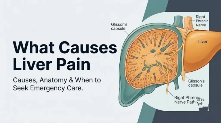
7 Root Causes of Mast Cell Activation Syndrome 2026
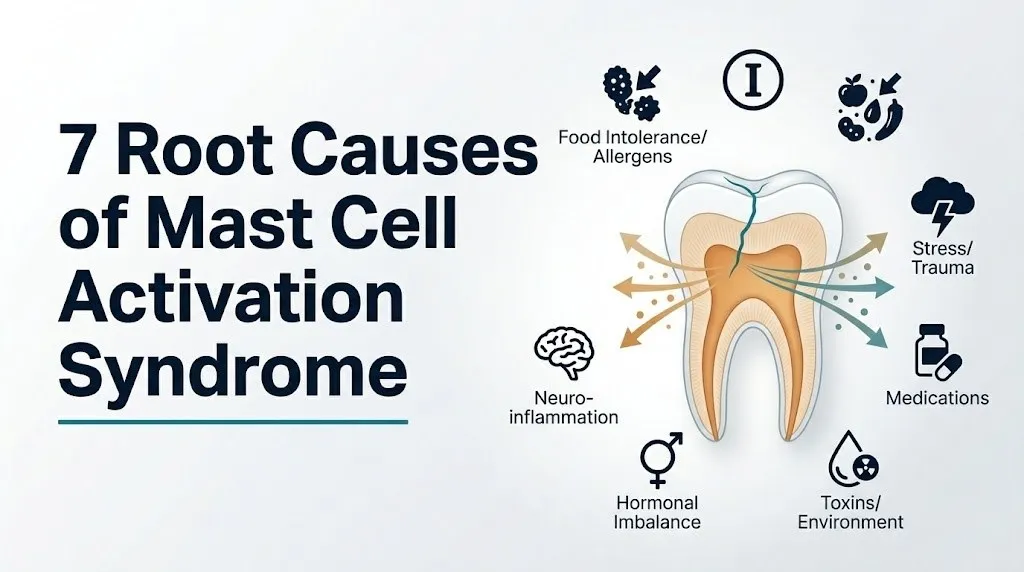
The 7 root causes of mast cell activation syndrome include clonal mast cell disease driven by KIT gene mutations, IgE-mediated allergic sensitization, autoimmune and chronic inflammatory conditions, autonomic nervous system dysregulation, connective tissue disorders, estrogen-driven hormonal activation, and chronic infection with gut microbiome disruption. Each root cause operates through a distinct molecular pathway that lowers the mast cell activation threshold or increases the total mast cell population, producing the same symptomatic picture through different underlying biology.
According to the National Institute of Allergy and Infectious Diseases (NIAID), mast cell disorders represent a heterogeneous group of conditions affecting multiple organ systems simultaneously, and identifying the specific root cause driving an individual’s MCAS is the foundation of targeted management. The Valent consensus diagnostic criteria, published in the International Journal of Molecular Sciences, establish that MCAS requires evidence of mast cell mediator release, a symptomatic response to those mediators, and exclusion of other explanatory diagnoses before root cause investigation begins.
This article covers each of the seven established root cause categories with the specific cellular and molecular mechanism that drives mast cell dysfunction in each case, how these causes differ in terms of diagnostic testing, who is most at risk for each, and exactly how to navigate the specialist system to identify which root cause applies to you.
7 Root Causes of Mast Cell Activation Syndrome: Overview
The seven root causes of mast cell activation syndrome fall into three major etiological categories: primary causes (driven by intrinsic mast cell abnormalities), secondary causes (driven by external conditions that chronically stimulate normal mast cells), and idiopathic causes (where neither primary nor secondary mechanisms are confirmed but mast cell activation is clearly occurring).
Understanding which category your MCAS falls into changes everything about the diagnostic workup and the clinical pathway that follows. A person with primary clonal MCAS needs a bone marrow biopsy and a hematologist. A person with secondary IgE-mediated MCAS needs a comprehensive allergy panel and an allergist. A person with autonomic nervous system-driven MCAS needs a tilt table test and an autonomic specialist.
The seven root causes in order of clinical frequency, based on clinical series reviewed in the Journal of Allergy and Clinical Immunology:
| Root Cause | Category | Primary Diagnostic Clue | Specialist Needed |
|---|---|---|---|
| 1. Clonal mast cell disease / KIT mutation | Primary | Elevated baseline tryptase, prior anaphylaxis | Hematologist |
| 2. IgE-mediated secondary MCAS | Secondary | Elevated total IgE, specific allergen sensitization | Allergist/immunologist |
| 3. Autoimmune and chronic inflammatory disease | Secondary | Elevated ANA, complement, inflammatory markers | Rheumatologist or immunologist |
| 4. Autonomic nervous system dysregulation / POTS | Secondary/Idiopathic | POTS on tilt table test, orthostatic symptoms | Autonomic specialist or cardiologist |
| 5. Connective tissue disorders / hEDS | Secondary/Idiopathic | Beighton score, skin and joint hyperextensibility | Geneticist or rheumatologist |
| 6. Hormonal triggers / estrogen activation | Secondary | Perimenstrual symptom worsening pattern | Gynecologist or endocrinologist |
| 7. Chronic infection / gut microbiome dysbiosis | Secondary/Idiopathic | Stool microbiome analysis, prior infection timeline | Gastroenterologist |
No root cause can be definitively identified without professional evaluation and specific testing. This framework is for educational orientation, not self-diagnosis.
What Is Mast Cell Activation Syndrome
Mast cell activation syndrome (MCAS) is a condition in which mast cells, the immune system’s tissue-resident surveillance cells, activate and release their chemical mediators excessively or in response to stimuli that would not trigger activation in a healthy person, producing symptoms across multiple organ systems simultaneously.
Mast cells are found throughout the body, particularly in the skin, gut mucosa, respiratory mucosa, and perivascular connective tissue. They function normally as first-responders to perceived threats: an allergen, a pathogen, or tissue injury causes them to release a suite of chemical signals that initiate inflammation, recruit other immune cells, and alter local blood vessel permeability. In MCAS, this release happens too easily, too often, or in response to harmless stimuli.
The ICD-10 code for MCAS is D89.42. MCAS is distinct from systemic mastocytosis, though both involve mast cell dysfunction. In systemic mastocytosis, there is a confirmed abnormal clonal proliferation of mast cells detectable on bone marrow biopsy. In MCAS, the mast cells themselves may be present in normal numbers but are functionally dysregulated. The distinction matters clinically because it determines the diagnostic workup and the treatment approach.
The World Health Organization (WHO) 2022 classification of mast cell diseases distinguishes cutaneous mastocytosis, systemic mastocytosis, and mast cell activation disorders as separate entities with overlapping features. MCAS falls within the mast cell activation disorder category.
Pediatric patients with MCAS frequently present with predominantly gastrointestinal symptoms (recurrent abdominal pain, food intolerance, diarrhea, and chronic nausea) rather than the skin and cardiovascular predominance more typical in adult presentations, making the diagnosis harder to recognize in children and adolescents.
How Mast Cells Cause Symptoms: The Mediator Mechanism
Mast cells cause symptoms by releasing specific chemical mediators from intracellular secretory granules and by synthesizing additional lipid mediators and cytokines after activation, with each mediator acting on a different organ system to produce a distinct symptom.
Think of a mast cell like a fully loaded fire extinguisher mounted on a hair trigger. In a healthy immune system, only a genuine fire trips that trigger. In MCAS, the trigger mechanism is faulty: it fires at smoke, at steam, at warmth, at stress. And when it fires, every chemical in the extinguisher releases at once into every nearby tissue.
The primary mediators and the symptoms they produce:
| Mediator | Release Mechanism | Primary Symptoms Produced |
|---|---|---|
| Histamine | Immediate granule release (seconds) | Flushing, urticaria, rhinorrhea, GI cramping, headache, brain fog via H3 receptors |
| Tryptase | Immediate granule release (seconds to minutes) | Tissue remodeling, fibrinogen cleavage; elevated in blood during acute episodes |
| Heparin | Immediate granule release | Anticoagulant effect, increased bleeding tendency |
| Prostaglandin D2 (PGD2) | De novo synthesis, COX pathway (minutes to hours) | Vasodilation, flushing, GI motility changes, bronchospasm |
| Leukotriene C4 (LTC4) | De novo synthesis, 5-LOX pathway (minutes to hours) | Bronchoconstriction, increased mucus secretion, vascular permeability |
| Platelet-activating factor (PAF) | De novo synthesis | Severe hypotension, anaphylaxis potentiation |
| TNF-alpha, IL-4, IL-13 | Cytokine secretion (hours) | Systemic inflammation, IgE class-switching amplification |
The reason MCAS produces symptoms in so many organ systems simultaneously is that mast cells reside in every vascularized tissue. When they degranulate, the mediators they release act locally and enter the circulation to act systemically.
Patients taking H1 antihistamines (such as cetirizine or loratadine) for unrelated allergies may have partially suppressed histamine symptoms that mask the full MCAS picture, making the condition harder to recognize and the clinical presentation atypical.
Root Cause 1: Clonal Mast Cell Disease and KIT Mutations
The first root cause of mast cell activation syndrome is clonal mast cell disease, in which a somatic mutation in the KIT proto-oncogene (most commonly the KIT D816V point mutation) causes constitutive, ligand-independent activation of the KIT tyrosine kinase receptor on mast cells, driving autonomous mast cell proliferation and a pathologically lowered activation threshold through constitutive SRC-family kinase and JAK2/STAT5 signaling.
The KIT receptor (also called CD117) is the cell surface receptor that normally binds stem cell factor (SCF) from stromal cells to signal mast cell survival, proliferation, and maturation. The D816V mutation in the KIT gene substitutes valine for aspartate at position 816 in the kinase domain, causing the receptor to signal continuously without needing SCF to bind. The result is a mast cell that proliferates without normal restraint and that fires at stimuli far below the normal activation threshold.
This root cause sits on a spectrum. At one end is full systemic mastocytosis (SM), where the bone marrow biopsy shows dense mast cell infiltration (greater than 25 mast cells per aggregate), aberrant CD25 or CD2 expression on mast cells, and elevated serum baseline tryptase typically exceeding 20 ng/mL. At the other end is clonal MCAS (also called monoclonal MCAS), where the KIT mutation is present at low allele frequency in mast cells but the bone marrow findings do not meet the full WHO criteria for systemic mastocytosis.
According to the WHO 2022 classification of mast cell diseases, the KIT D816V mutation is identified in over 90 percent of systemic mastocytosis cases and in a subset of MCAS patients who demonstrate other minor SM criteria without meeting full SM criteria.
Patients who have experienced unexplained anaphylaxis, particularly anaphylaxis triggered by Hymenoptera (bee or wasp) stings or occurring without an identifiable trigger, have a substantially higher probability of underlying clonal mast cell disease than the general MCAS population. A board-certified hematologist with experience in mast cell diseases should evaluate any MCAS patient with a baseline serum tryptase exceeding 20 ng/mL.
Key Takeaway: An elevated baseline serum tryptase above 20 ng/mL in an MCAS patient is a specific clinical red flag for clonal mast cell disease requiring bone marrow biopsy by a board-certified hematologist, not just an allergy workup.
Root Cause 2: IgE-Mediated Secondary MCAS
The second root cause of mast cell activation syndrome is IgE-mediated secondary MCAS, in which chronically elevated IgE (immunoglobulin E) from allergic sensitization, parasitic infection, or atopic disease loads the FcεRI (high-affinity IgE receptor) on mast cell surfaces so densely that even trace allergen exposure crosslinks multiple IgE-FcεRI complexes simultaneously, triggering the SYK/LAT/PLC-gamma pathway and producing calcium-dependent mast cell degranulation at exposure levels that would not activate a less sensitized mast cell.
The FcεRI receptor mechanism is the most thoroughly understood pathway in mast cell activation biology. IgE antibodies specific to a particular allergen (dust mites, foods, pollens, medications) bind to FcεRI on the mast cell surface during the initial sensitization phase. When the same allergen is encountered again, it crosslinks two or more of these primed IgE-FcεRI complexes, bringing SYK kinase (a cytoplasmic tyrosine kinase) into proximity with LAT (linker for activation of T cells). This activates PLC-gamma, which cleaves PIP2 into IP3 and DAG. IP3 releases calcium from the endoplasmic reticulum, and DAG activates protein kinase C. Together, calcium and PKC drive mast cell granule fusion with the plasma membrane and mediator secretion.
In secondary IgE-mediated MCAS, the sheer density of IgE-loaded FcεRI receptors on each mast cell means that the activation threshold drops substantially below what would trigger a less sensitized person’s mast cells. A barely detectable amount of an allergen that would produce no reaction in a non-sensitized person produces full mast cell degranulation in a heavily sensitized individual.
According to the Journal of Allergy and Clinical Immunology, the distinction between classical IgE-mediated allergic disease and secondary MCAS lies in the multisystem, multi-trigger nature of the MCAS presentation: a patient with true secondary MCAS reacts to dozens of triggers across several organ systems, not just the single allergen typically driving classical allergy.
Female patients with atopic disease (asthma, eczema, allergic rhinitis) have higher rates of secondary IgE-mediated MCAS than males, reflecting the generally higher IgE burdens and mast cell densities observed in women with atopic disease.
Root Cause 3: Autoimmune and Chronic Inflammatory Disease
The third root cause of mast cell activation syndrome is autoimmune and chronic inflammatory disease, in which IgG autoantibodies directed against the FcεRI receptor itself or against IgE activate mast cells through a non-allergen, non-IgE-dependent pathway, or in which chronic systemic inflammation from conditions like lupus erythematosus, rheumatoid arthritis, or inflammatory bowel disease maintains persistently elevated levels of complement activation products (C3a, C5a) that directly stimulate mast cells via their complement receptors.
The autoimmune root cause operates through a distinct but parallel pathway to IgE-mediated activation. In some patients, IgG antibodies target the alpha subunit of FcεRI or target IgE itself. These IgG antibodies crosslink FcεRI receptors (through their own binding) and trigger the same downstream SYK/LAT/PLC-gamma cascade as allergen-IgE crosslinking, without any allergen being present. This is why autoimmune MCAS can produce reactions seemingly from nowhere, without the patient being able to identify any trigger.
Complement-driven mast cell activation adds a second autoimmune mechanism. In systemic lupus erythematosus, antigen-antibody complex deposition activates the complement cascade, generating C3a and C5a. Both bind to specific receptors on mast cell surfaces (C3aR and C5aR), triggering degranulation via a G-protein coupled receptor pathway distinct from the FcεRI mechanism. This is why MCAS often worsens during lupus flares even when the patient is not exposed to any new allergen or food trigger.
Research published in the Annals of Allergy Asthma and Immunology has found an association between autoimmune thyroid disease (both Hashimoto’s thyroiditis and Graves’ disease) and MCAS, with the proposed mechanism involving thyroid peroxidase antibodies cross-reacting with mast cell surface proteins, though this association is currently supported by observational data rather than confirmed causation.
Patients with known autoimmune conditions who develop MCAS-like multisystem symptoms should be evaluated by a board-certified rheumatologist or immunologist who can assess disease activity and complement levels alongside mast cell mediator testing.
Key Takeaway: Autoimmune MCAS can produce reactions with no identifiable allergen trigger because the activation pathway bypasses IgE entirely; autoantibodies against FcεRI or complement activation products directly stimulate mast cells through a distinct molecular route.
Root Cause 4: Autonomic Nervous System Dysregulation and POTS
The fourth root cause of mast cell activation syndrome is autonomic nervous system dysregulation, particularly postural orthostatic tachycardia syndrome (POTS), in which dysregulated sympathetic nervous system activity drives mast cell activation through the release of corticotropin-releasing hormone (CRH) from sympathetic nerve terminals adjacent to mast cells in the skin, gut, and perivascular tissue, triggering mast cell degranulation via CRH receptor (CRHR1) signaling on the mast cell surface.
The nerve-mast cell communication unit is one of the most physiologically important but least-discussed aspects of mast cell biology in general health content. Mast cells are densely concentrated around sensory and autonomic nerve terminals throughout the body. During stress or autonomic dysregulation, sympathetic neurons release CRH locally from their nerve endings. CRH binds CRHR1 on adjacent mast cells, activating a cAMP-mediated signaling cascade that triggers granule exocytosis independently of any IgE or allergen. This is the mechanism by which psychological stress, orthostatic stress, and sympathetic surges in POTS directly provoke MCAS symptoms.
Substance P, a neuropeptide released from sensory C-fiber neurons, provides a parallel nerve-to-mast cell activation pathway. Substance P binds the NK1 receptor on mast cells and triggers degranulation via a calcium-dependent mechanism. In patients with POTS, where autonomic dysregulation produces heightened sensory nerve activation and elevated substance P levels, mast cells adjacent to those nerves are in a state of chronic partial activation.
Clinical observation documented in specialty MCAS and dysautonomia literature consistently shows that POTS and MCAS co-occur at rates substantially exceeding random chance. A 2019 report in the Annals of Allergy Asthma and Immunology noted the frequent clinical overlap of POTS, MCAS, and hEDS, suggesting shared pathophysiology rather than coincidental comorbidity.
Patients with POTS who also experience flushing, urticaria, GI cramping, and multisystem intolerance should have mast cell mediator testing (serum tryptase during acute episodes, 24-hour urine N-methylhistamine and prostaglandin D2 metabolites) ordered by a board-certified cardiologist or autonomic medicine specialist familiar with this overlap.
Root Cause 5: Connective Tissue Disorders and Hypermobile EDS
The fifth root cause of mast cell activation syndrome is connective tissue dysfunction, particularly hypermobile Ehlers-Danlos syndrome (hEDS), in which defective extracellular matrix composition produces abnormal mechanical stress on mast cells embedded in connective tissue, activating mechanosensitive ion channels (including Piezo1 and TRPV4) on mast cell membranes and triggering calcium-dependent degranulation in response to tissue movements that would not activate mast cells in structurally normal connective tissue.
Mast cells do not float freely: they are anchored within the extracellular matrix via integrin receptors that physically connect the mast cell cytoskeleton to the surrounding collagen and fibronectin scaffold. When this scaffold is normal and mechanically sound, the physical forces transmitted through the matrix during normal body movement remain below the mast cell activation threshold. When the extracellular matrix is structurally abnormal, as in hEDS where glycine substitution mutations in fibrillar collagen genes produce hypercompliant, mechanically unstable connective tissue, even normal daily movements transmit abnormal tensile and shear forces through the matrix to the anchored mast cells.
This mechanical activation pathway explains why hEDS patients with MCAS frequently notice that physical activity, barometric pressure changes, and even position changes provoke mast cell symptoms, while people without connective tissue abnormality would experience no reaction to the same stimuli.
The hEDS-MCAS co-occurrence is well-documented in specialty clinical literature. Research published in the Journal of Allergy and Clinical Immunology and referenced by The Mastocytosis Society describes the POTS-hEDS-MCAS triad as a clinically recognized entity that disproportionately affects women of reproductive age, with some clinical series reporting MCAS diagnosis rates of 50 percent or higher in patients presenting with hEDS.
Young women (ages 15 to 35) with joint hypermobility, chronic pain, dysautonomia, and multisystem sensitivity should be evaluated for MCAS as a concurrent diagnosis rather than attributing all symptoms to hEDS alone.
Root Cause 6: Hormonal Triggers and Estrogen Mast Cell Activation
The sixth root cause of mast cell activation syndrome is estrogen-driven hormonal mast cell activation, in which estrogen (17-beta-estradiol) binding to estrogen receptor alpha (ERα) expressed on the mast cell surface potentiates the calcium mobilization response to FcεRI crosslinking, increasing the amplitude of mast cell degranulation at any given allergen stimulus level and producing symptom worsening that correlates with estrogen peaks in the menstrual cycle.
Mast cells express ERα on their surfaces, a fact established by preclinical research and confirmed in human mast cell lines. When estrogen binds ERα, it activates a rapid, non-genomic signaling pathway that increases the sensitivity of the PLC-gamma/calcium mobilization system without requiring new gene transcription. The practical result is that the same allergen load or the same emotional stress that produces a mild mast cell response at low estrogen levels produces a substantially larger degranulation event when estrogen is high.
This estrogen potentiation explains two clinical patterns that are highly characteristic of MCAS in women but not explained in any competitor article. First, symptoms frequently worsen in the mid-follicular phase (days 7 to 12 of the cycle) when estrogen peaks before ovulation. Second, symptoms often become dramatically worse during the perimenopause, when estrogen levels fluctuate unpredictably, creating periods of very high estrogen exposure interspersed with drops, each transition potentially provoking a mast cell episode.
Progesterone, by contrast, has been shown in preclinical research to exert a mast cell-inhibitory effect by reducing IL-4-driven IgE class-switching and downregulating FcεRI surface expression. The luteal phase, when progesterone is highest relative to estrogen, often produces symptom relief in women with MCAS, followed by a premenstrual flare as progesterone drops and estrogen undergoes a secondary relative peak.
Women who notice a clear menstrual cycle correlation with their MCAS symptoms should document this pattern specifically (noting cycle day alongside symptom diary entries) and share it with a board-certified gynecologist or endocrinologist who can evaluate the hormonal contribution alongside an allergist/immunologist managing their MCAS.
Key Takeaway: Estrogen binds directly to mast cell surface receptors and amplifies their degranulation response, which is why MCAS symptoms track with estrogen fluctuations across the menstrual cycle and why perimenopausal women often experience dramatic MCAS onset or worsening.
Root Cause 7: Chronic Infection and Gut Microbiome Dysbiosis
The seventh root cause of mast cell activation syndrome is chronic infection and gut microbiome dysbiosis, in which chronic bacterial, viral, or parasitic infections or the loss of a healthy gut microbiome community produces persistent Toll-like receptor (TLR) stimulation of intestinal mast cells, chronic low-grade mast cell activation through TLR4 (lipopolysaccharide receptor) and TLR2 (peptidoglycan receptor) signaling, and a proinflammatory cytokine environment that progressively lowers the mast cell activation threshold throughout the gut and systemically.
The gut contains the highest density of mast cells in the human body, positioned immediately beneath the intestinal epithelial barrier where they serve as sentinels detecting luminal contents. In a healthy gut, the epithelial barrier prevents microbial products from reaching the subepithelial mast cell layer. When gut dysbiosis produces intestinal epithelial barrier dysfunction (sometimes called increased intestinal permeability), microbial products including lipopolysaccharide (LPS) from gram-negative bacteria and peptidoglycan from gram-positive bacteria translocate across the epithelium and directly contact subepithelial mast cells.
LPS binding to TLR4 on mast cells activates the MyD88 and TRIF intracellular signaling pathways, triggering de novo synthesis of TNF-alpha, IL-6, and IL-1beta without the calcium-dependent granule exocytosis of classical activation. This produces a slower but persistent mast cell inflammatory output that chronically primes the mast cell population for exaggerated responses to subsequent stimuli.
According to observational data cited in the International Journal of Molecular Sciences, patients with MCAS have been found to have altered intestinal microbiome compositions compared to healthy controls, with reductions in short-chain fatty acid-producing bacteria (which normally maintain intestinal epithelial barrier integrity) and increases in gram-negative bacterial populations that provide a higher LPS load.
Patients with MCAS who have a clear history of onset following a gastrointestinal infection (post-infectious MCAS), antibiotic courses that disrupted gut microbiome composition, or concurrent inflammatory bowel disease should discuss intestinal biopsy for mast cell density quantification with a board-certified gastroenterologist experienced in functional gastrointestinal disorders and mast cell disease.
MCAS Triggers vs Root Causes: Understanding the Difference
MCAS triggers are the immediate stimuli that provoke individual mast cell activation episodes, while root causes are the underlying biological mechanisms that make mast cells pathologically reactive in the first place, and confusing the two leads patients to focus exclusively on avoidance behavior rather than pursuing the diagnosis and treatment of the underlying condition.
A food trigger is not a root cause. Alcohol, high-histamine foods, exercise, heat, cold, vibration, and emotional stress are all documented MCAS triggers. They produce mast cell activation episodes. But none of them explains why the mast cells are reactive at a pathologically low threshold. A person without an underlying MCAS root cause eats the same foods, exercises in the same heat, and experiences the same emotional stress without having a systemic mast cell activation episode.
The distinction has direct clinical implications:
- Treating only triggers (eliminating foods, avoiding environments) may reduce symptom frequency but does not address the root cause biology. Clonal mast cell disease continues to expand. Autoimmune mast cell activation continues. Gut dysbiosis continues producing TLR stimulation.
- Identifying and treating the root cause can reduce the overall mast cell reactivity threshold, making triggers less provocative over time.
The practical priority for a patient new to MCAS is not to compile the longest possible trigger avoidance list. The practical priority is to establish which root cause category applies, because that determines who needs to be on the clinical team and what treatment approaches are evidence-based for that specific root cause.
A comprehensive trigger diary kept over 4 to 6 weeks, documenting symptoms, timing, associated activities, foods, environmental exposures, cycle phase (for women), and stress levels, provides the data a physician needs to begin differentiating root cause categories before formal testing.
MCAS in Females: Why Women Are Disproportionately Affected
Women are disproportionately diagnosed with mast cell activation syndrome for three converging reasons: estrogen receptor alpha expression on mast cells potentiates degranulation, female sex hormones influence both mast cell number and threshold, and the conditions that most commonly drive secondary MCAS (hEDS, POTS, autoimmune disease) are themselves more prevalent in women.
Clinical series consistently report female predominance in MCAS diagnoses. A review of MCAS patient populations published in the Annals of Allergy Asthma and Immunology found that women represent approximately 70 to 80 percent of diagnosed MCAS cases in most specialty clinical series, a disproportionality that cannot be explained by diagnostic bias alone.
Beyond the estrogen receptor mechanism detailed in the hormonal root cause section, estrogen also increases the total number of mast cells in tissue. Animal model research has found that estrogen promotes mast cell proliferation and survival through ERα-mediated upregulation of KIT receptor expression, increasing the mast cell population density in estrogen-rich tissues. This means women of reproductive age may have both more mast cells and more reactive mast cells than males with the same root cause, amplifying the clinical expression of MCAS.
The autoimmune disease burden also falls heavily on women. Hashimoto’s thyroiditis, lupus, Sjogren’s syndrome, and rheumatoid arthritis, all of which can drive autoimmune secondary MCAS, affect women at 7 to 9 times the rate of men.
Perimenopausal women represent a population in whom MCAS frequently either first presents or dramatically worsens. The unpredictable estrogen fluctuations of perimenopause, before the sustained lower estrogen of postmenopause, create a period of maximal mast cell instability. Women in their 40s experiencing new-onset multisystem intolerance, flushing, GI symptoms, and urticaria during perimenopause should have mast cell mediator testing evaluated alongside hormonal assessment by a gynecologist or endocrinologist.
MCAS Diagnostic Criteria and How Root Cause Affects Testing
The Valent consensus diagnostic criteria for mast cell activation syndrome require three elements: documented mast cell mediator release during symptomatic episodes (via serum tryptase, urine N-methylhistamine, or urine prostaglandin D2 metabolites), symptomatic involvement of multiple organ systems consistent with mediator release, and a clinical response to mast cell-directed therapy, and the specific tests ordered to establish root cause differ substantially depending on which of the seven categories is suspected.
The Hahn tryptase formula is the most widely used objective criterion for documenting acute mast cell activation: serum tryptase measured within 4 hours of a symptomatic episode must exceed the patient’s own baseline tryptase by at least 20 percent plus 2 ng/mL. This formula accounts for individual variation in baseline tryptase and avoids false positives from the hereditary alpha-tryptasemia (HAT) variant, in which baseline tryptase is constitutively elevated without active MCAS.
Root cause-specific testing after baseline criteria are confirmed:
- For suspected clonal (primary) MCAS: serum baseline tryptase, c-KIT D816V mutation testing in peripheral blood (highly sensitive assay), and referral to a board-certified hematologist for bone marrow biopsy with CD117, CD25, and CD2 immunostaining.
- For suspected IgE-mediated (secondary) MCAS: total serum IgE, specific IgE panel to environmental and food allergens, intradermal skin testing under physician supervision.
- For suspected autoimmune MCAS: antinuclear antibody (ANA), anti-FcεRI antibody testing where available, complement C3 and C4 levels, complete blood count with differential.
- For suspected autonomic/POTS-driven MCAS: tilt table test under cardiologist supervision, 24-hour urine catecholamines, heart rate variability testing.
- For suspected hEDS-driven MCAS: Beighton hypermobility scoring by a trained clinician, echocardiogram for aortic root dilation, referral to a board-certified clinical geneticist.
- For suspected hormonal MCAS: symptom diary correlated to menstrual cycle, serum estrogen and progesterone across cycle phases, thyroid function testing.
- For suspected gut dysbiosis MCAS: gut microbiome analysis, intestinal biopsy for mast cell density and tryptase staining by a board-certified gastroenterologist.
Patients being tested for MCAS should ask their physician whether they should hold H1 and H2 antihistamines before 24-hour urine collections, as antihistamine use can suppress urine histamine metabolite levels and produce false-negative results.
Key Takeaway: The Hahn tryptase formula (acute tryptase greater than baseline times 1.2 plus 2 ng/mL) is the most specific objective test for documenting active mast cell degranulation, and it should be measured within 4 hours of a symptomatic episode to capture the tryptase elevation before it clears.
Acute vs Chronic MCAS Activation Patterns
MCAS produces two clinically distinct activation patterns: acute episodic flares in which sudden mast cell degranulation produces dramatic multisystem symptoms over minutes to hours, and chronic baseline activation in which persistent low-level mediator release produces a steady-state pattern of fatigue, brain fog, pain, and gastrointestinal dysfunction that fluctuates but rarely fully resolves.
Acute MCAS episodes have a characteristic temporal signature. They begin within seconds to minutes of a triggering stimulus (a food, a scent, exercise, stress, or temperature change) as pre-formed granule mediators (histamine, tryptase, heparin) release immediately. Flushing, urticaria, abdominal cramping, and cardiovascular changes (tachycardia, hypotension, or paradoxical hypertension) peak within 15 to 60 minutes. De novo-synthesized mediators (prostaglandin D2, leukotriene C4) reach peak levels within 30 to 90 minutes, producing delayed bronchoconstriction, further GI effects, and prolonged flushing. The episode typically resolves over 1 to 4 hours but may leave a post-episode period of fatigue lasting 12 to 48 hours from the metabolic cost of the degranulation event and subsequent mast cell recovery.
Chronic baseline MCAS is more difficult to recognize because it lacks the dramatic episodic character. Patients describe persistent fatigue, cognitive difficulties (described as “brain fog”), chronic pain, daily gastrointestinal symptoms, and persistent dermatographic urticaria, all at a level that fluctuates but does not return to a normal asymptomatic baseline between episodes.
The root cause substantially influences which pattern predominates. Clonal MCAS with KIT mutation often produces more dramatic acute episodes, including anaphylaxis, because the autonomous mast cell hyperproliferation creates a large, primed mast cell population. Gut dysbiosis-driven and autonomic-driven MCAS more often produces the chronic low-grade pattern with episodic flares.
Patients with clonal MCAS who have experienced anaphylaxis should carry an epinephrine autoinjector (prescribed by their managing physician) at all times and know how to use it, based on clinical guidelines reviewed by The Mastocytosis Society.
When MCAS Symptoms Become a Medical Emergency
Certain mast cell activation episodes escalate to anaphylaxis, a life-threatening systemic hypersensitivity reaction requiring immediate emergency intervention, and patients with MCAS should know the specific threshold at which symptoms require calling 911 rather than self-managing at home.
Emergency Symptoms: When to Call 911 or Go to the ER
Certain MCAS episodes progress to anaphylaxis and represent a life-threatening medical emergency. Do not wait to see if these symptoms resolve on their own.
Call 911 or go to the nearest emergency room immediately if you experience:
- Throat tightening, difficulty swallowing, or a sensation that your airway is closing: indicates laryngeal or pharyngeal edema from mast cell mediator release, which can cause complete airway obstruction within minutes
- Sudden severe difficulty breathing or wheezing that does not improve with your usual medications: indicates bronchospasm from leukotriene-mediated airway constriction, which can progress to respiratory failure
- Sudden severe drop in blood pressure, lightheadedness, or loss of consciousness: indicates histamine and PAF-mediated vasodilation producing anaphylactic shock with cardiovascular collapse
- Rapid progression of hives or swelling combined with any of the above symptoms: multi-system anaphylaxis requiring epinephrine, not antihistamines alone
- Loss of consciousness or near-syncope during or after a suspected MCAS episode: indicates severe cardiovascular compromise requiring emergency evaluation
These presentations indicate anaphylaxis, which can be fatal without immediate intramuscular epinephrine and emergency medical assessment.
Patients with clonal MCAS have a higher risk of anaphylaxis than the general MCAS population. Any MCAS patient who has experienced an episode meeting anaphylaxis criteria should be evaluated by a board-certified allergist/immunologist for epinephrine autoinjector prescription and anaphylaxis action plan development.
How to Identify Your MCAS Root Cause and Find the Right Specialist
Identifying your specific MCAS root cause requires a systematic clinical evaluation that begins with a primary care physician establishing the baseline MCAS diagnosis and then directing you to the appropriate specialist based on your clinical history, trigger pattern, and initial laboratory results.
The diagnostic pathway from suspected MCAS to confirmed root cause:
- Primary care physician evaluation: document multisystem symptom history, request serum baseline tryptase (collected when asymptomatic), and request 24-hour urine N-methylhistamine and 11-beta-prostaglandin F2-alpha collected during or shortly after a symptomatic episode. Bring a written symptom diary covering at least 4 weeks, noting symptoms, timing, and associated circumstances.
- Establish baseline MCAS diagnosis: if baseline tryptase is elevated and/or urine mediators are elevated above reference range during symptoms, the Valent criteria begin to be satisfied. A response to a trial of H1 antihistamine (prescribed by your physician) provides the third criterion.
- Root cause investigation by history: review whether you have a prior anaphylaxis history (clonal MCAS path), an atopic disease history (secondary IgE path), an autoimmune diagnosis (autoimmune path), POTS or orthostatic symptoms (autonomic path), joint hypermobility (hEDS path), menstrual cycle correlation (hormonal path), or post-infectious onset (gut dysbiosis path).
- Specialist referral by root cause: the appropriate specialist for each root cause is identified in the overview table earlier in this article. Your primary care physician can generate the referral, or you can contact a board-certified allergist/immunologist as a coordinating specialist who can direct further root cause investigation.
- Root cause confirmation testing: performed by the appropriate specialist as detailed in the diagnostic criteria section above.
- Integrated management plan: once the root cause is identified, management addresses both the root cause (when treatable) and the mast cell hyperreactivity itself.
The Mastocytosis Society (TMS) maintains a list of physicians experienced in mast cell disorders across the United States that can help patients locate appropriate specialist care in their geographic area.
Frequently Asked Questions About MCAS Root Causes
What is the most common root cause of mast cell activation syndrome?
Idiopathic MCAS (where no primary or clearly identifiable secondary cause is confirmed) accounts for the largest proportion of MCAS diagnoses in most clinical series, according to the Valent consensus classification.
Secondary MCAS driven by IgE-mediated allergic sensitization is the most common identifiable cause, followed by autoimmune conditions and the POTS-hEDS overlap.
Clonal mast cell disease (primary MCAS with KIT mutation) is less common but clinically important to identify because it requires bone marrow evaluation and carries a different long-term prognosis.
Can MCAS be caused by a genetic mutation?
Yes, the KIT D816V somatic mutation causes clonal mast cell disease, the primary genetic root cause of MCAS, by producing constitutive KIT tyrosine kinase activation that drives autonomous mast cell proliferation.
Hereditary alpha-tryptasemia (HAT), caused by increased copy number of the alpha-tryptase gene TPSAB1, is a separate genetic variant associated with elevated baseline tryptase and increased MCAS symptom severity, though it does not directly cause MCAS.
Both conditions are identified through blood testing (c-KIT D816V assay, tryptase gene copy number analysis) ordered by a board-certified hematologist or allergist/immunologist.
Why do MCAS symptoms often get worse around my period?
MCAS symptoms worsen perimenstrually because estrogen binds estrogen receptor alpha (ERα) on mast cell surfaces and potentiates calcium mobilization during FcεRI activation, amplifying the degranulation response at higher estrogen levels.
The perimenstrual symptom flare typically corresponds to the late follicular phase estrogen peak (days 7 to 12) and then again to the premenstrual progesterone drop when estrogen becomes relatively dominant.
Documenting cycle day alongside your symptom diary and sharing this pattern with a board-certified gynecologist or endocrinologist allows hormonal contribution to be formally evaluated.
What is the connection between MCAS and POTS or Ehlers-Danlos syndrome?
POTS, hEDS, and MCAS co-occur at rates substantially exceeding coincidence, forming a clinically recognized triad in which shared pathophysiology rather than independent diseases likely drives the overlap.
POTS drives mast cell activation through sympathetic nervous system CRH release at nerve-mast cell communication units, while hEDS drives mast cell activation through abnormal mechanical stress on mast cells embedded in defective extracellular matrix.
Any patient with confirmed POTS or hEDS who develops multisystem intolerance, flushing, urticaria, or GI symptoms should be evaluated for concurrent MCAS by a board-certified allergist/immunologist or by an autonomic specialist familiar with this triad.
How do doctors figure out which root cause is driving MCAS?
Identifying the MCAS root cause requires combining clinical history (onset pattern, trigger types, prior diagnosis history, menstrual correlation) with targeted laboratory testing (serum tryptase, urine mediators, IgE panel, ANA, c-KIT mutation assay) and specialist evaluation specific to the suspected root cause category.
No single test identifies all root causes; the diagnostic pathway is directed by the clinical picture rather than a fixed panel.
A board-certified allergist/immunologist is typically the most appropriate coordinating specialist for initiating the root cause investigation, with referral to hematology, rheumatology, gastroenterology, or autonomic medicine as the history directs.
Can MCAS go away if the root cause is treated?
Treating the root cause can substantially reduce MCAS severity and trigger sensitivity in secondary MCAS categories, particularly when autoimmune disease is controlled, gut dysbiosis is addressed, or autonomic dysregulation is improved.
Clonal MCAS from KIT mutation does not resolve with symptomatic treatment alone; targeted therapies affecting KIT signaling are managed by a board-certified hematologist for appropriate candidates.
Complete resolution of MCAS is not guaranteed even with root cause treatment, because chronic mast cell activation can produce epigenetic changes that maintain a lowered activation threshold even after the original driver is addressed.
Closing
The seven root causes of mast cell activation syndrome each operate through a distinct molecular mechanism, and that distinction is not academic. It determines which specialist you need, which tests are ordered, which treatments are evidence-based for your situation, and what realistic improvement looks like with targeted management.
The most actionable step from this article is to identify which root cause category most closely matches your clinical history, bring that analysis to a primary care physician or board-certified allergist/immunologist, and request testing appropriate to that category rather than pursuing only trigger avoidance.
MCAS is a manageable condition for most patients when the root cause is identified and addressed systematically. You are in a better position to advocate for that process now than you were before you understood what is actually happening at the cellular level.