Blood Blister in Mouth Causes: What’s Behind Each One
The most common blood blister in mouth causes are minor trauma events like accidentally biting the inner cheek, but the same appearance can also signal a platelet disorder, a vitamin deficiency, an autoimmune disease, or, in rare cases, early oral cancer. Knowing which cause fits your situation is what determines whether you can leave the blister alone or need a clinical evaluation this week.
Most blood blisters inside the mouth resolve without treatment when the cause is mechanical. According to the National Institute of Dental and Craniofacial Research (NIDCR), oral mucosal tissue has a high regenerative capacity and typically heals faster than skin. That said, a blood blister that appeared suddenly without any recalled injury, is growing, has recurred multiple times, or develops alongside other mouth sores is a clinically different presentation and should not be managed the same way as a bite injury.
This article covers every clinically recognized cause of mouth blood blisters in order of likelihood, the specific physiological mechanism behind each cause, which populations face elevated risk, and exactly which symptom patterns warrant evaluation by a primary care physician, dentist, or specialist.
What Is a Blood Blister in the Mouth
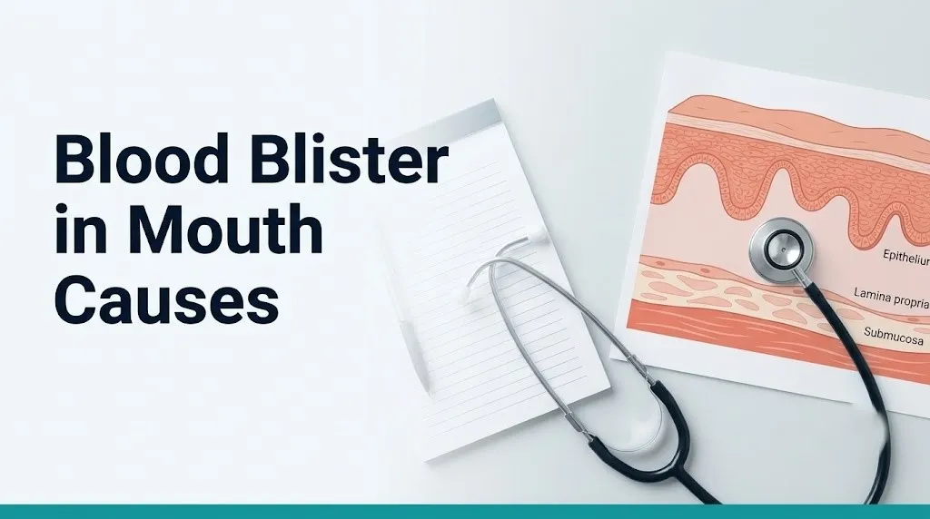
A blood blister in the mouth is a raised, fluid-filled lesion containing blood rather than clear serum, forming when blood pools in the space between the oral mucosa’s stratified squamous epithelial layer and the underlying lamina propria (the connective tissue layer directly beneath the epithelium).
The oral cavity is lined by specialized mucosal tissue. On the inner cheeks (buccal mucosa), inner lips (labial mucosa), roof of the mouth (palatal mucosa), and the floor of the mouth and underside of the tongue (sublingual mucosa), this lining consists of stratified squamous epithelium supported by a vascular connective tissue bed. Tiny capillaries and venules run through the lamina propria close to the epithelial surface.
When these vessels rupture and blood cannot immediately be contained by normal hemostasis, it accumulates beneath the overlying epithelium. The pressure from the pooling blood elevates the epithelial surface, creating the characteristic raised, dark red or purple blister visible to the naked eye. Think of it like pressing your thumb under a piece of thin plastic wrap stretched over a small pool of red liquid: the plastic lifts, and the liquid sits trapped beneath it.
The color ranges from bright red in fresh blisters to dark purple or nearly black in older ones where the blood has undergone deoxygenation and partial breakdown. Size ranges from a few millimeters to over a centimeter in diameter depending on how much blood has accumulated.
Quick Tip:
- Fresh blood blisters appear bright red and feel tense when pressed gently with the tongue.
- Older blisters turn dark purple or brownish as the hemoglobin within the collected blood breaks down over days.
- A blister that feels firm and does not move freely when pressed may warrant evaluation to rule out a solid lesion rather than a fluid-filled one.
Blood Blister in Mouth Causes: A Ranked Overview
The causes of blood blisters in the mouth range from the completely benign and self-resolving to the medically serious, and ranking them by likelihood helps frame the clinical picture before any individual cause is examined in detail.
| Cause Category | Likelihood in the General Population | Resolves Without Treatment |
|---|---|---|
| Mechanical trauma (cheek bite, sharp food, dental appliance) | Most common | Yes, in 7 to 14 days |
| Thermal or chemical injury (hot food, spicy foods) | Common | Yes, in 7 to 10 days |
| Nutritional deficiency (B12, iron, folate) | Moderate | Yes, once deficiency corrected |
| Anticoagulant or antiplatelet medication | Moderate in medication users | Varies; underlying treatment needed |
| Thrombocytopenia (low platelet count) | Less common in general population | No; requires medical treatment |
| Angina bullosa haemorrhagica | Less common; middle-aged to older adults | Generally yes; recurs |
| Allergic reaction or contact stomatitis | Less common | Yes, once trigger removed |
| Autoimmune blistering disease | Rare | No; requires specialist treatment |
| Oral cancer | Rare; blood blisters are not a classic presentation | No; requires oncological treatment |
According to the American Academy of Oral Medicine (AAOM), the vast majority of oral blood blisters encountered in clinical practice are traumatic in origin and require no treatment beyond monitoring. The clinical concern arises when the blister pattern does not fit the trauma profile: spontaneous onset, recurrence without a clear injury event, or accompaniment by other mucosal changes.
Children who develop oral blood blisters without any clear trauma event should be evaluated by a pediatrician to rule out immune thrombocytopenic purpura (ITP), a platelet disorder seen more frequently in pediatric populations following a viral infection.
Blood Blister in Mouth From Biting or Trauma
A blood blister from biting the inside of the cheek forms when the teeth compress and shear the buccal mucosa against itself or against the dental surfaces, rupturing submucosal capillaries and venules and producing a localized hematoma beneath the epithelial surface.
The buccal mucosa is about 0.5 to 1.5 millimeters thick, depending on location and individual anatomy. During the compression of a cheek bite, the thin tissue is caught between upper and lower molars or premolars. The mechanical shear force severs the small blood vessels (capillaries and post-capillary venules) running through the lamina propria. Blood immediately begins extravasating into the subepithelial space. Because the epithelium is still intact, the blood cannot escape outward. It collects beneath the surface and the blister forms within minutes to hours.
Dental appliances (orthodontic braces, ill-fitting dentures, night guards) cause the same mechanism through repeated low-level friction rather than a single compression event. A denture flange that rubs the same spot on the labial mucosa repeatedly will produce a chronically irritated area that blisters more easily than surrounding tissue.
People who clench or grind their teeth (bruxism) are at elevated risk for cheek-bite blood blisters because forceful tooth contact increases the probability of trapping mucosal tissue. Those with poorly fitting dental prosthetics should discuss adjustment with their dentist rather than treating recurring blisters at home, because the underlying mechanical cause will continue to produce new lesions.
To accurately describe a trauma-related blood blister to your dentist or physician:
- Note the exact location inside the mouth (inner cheek, inner lip, tongue border, roof of mouth).
- Recall whether a specific bite event, dental appointment, or prosthetic adjustment preceded the blister by hours to one day.
- Assess the size (smaller than a pea, pea-sized, or larger).
- Note whether the blister is tender, painless, or actively painful.
- Record how long it has been present and whether it has grown or stayed the same size since appearing.
What Causes Blood Blisters in the Mouth From Food or Heat
Blood blisters in the mouth caused by food or heat form when palatal mucosa or labial mucosa is exposed to temperatures or chemical irritants sufficient to rupture submucosal vessels or cause acute epithelial separation without breaking the outer surface.
Extremely hot foods and beverages, particularly dense foods like pizza, melted cheese, or hot soup that retain heat longer than liquids, contact the mucosal surface long enough to cause thermal injury at the lamina propria level. The heat damages the endothelium of submucosal capillaries, increasing their permeability and causing blood to leak out into the surrounding tissue. The result is a subepithelial hematoma that appears clinically identical to a traumatic bite blister.
Chemically irritating foods, including very spicy foods containing high concentrations of capsaicin, very acidic foods (citrus juices, vinegar-based foods), and some food preservatives, can trigger localized inflammatory responses in the oral mucosa through mast cell degranulation. The released histamine and prostaglandins increase local vascular permeability, allowing blood plasma and red blood cells to leak into the subepithelial space. This mechanism is distinct from direct thermal damage, and the blister may appear slightly later (several hours after eating rather than immediately).
According to MedlinePlus, thermal and chemical mucosal injuries in the oral cavity are among the most frequently self-reported oral injuries in adults. The palate (roof of the mouth) is the most commonly affected site for heat-related blisters because it makes sustained contact with hot food during chewing and swallowing.
Older adults face a higher risk from both food-related mechanisms. Reduced mucosal thickness with age means there is less tissue buffering between the surface and the submucosal vessels. Additionally, reduced heat perception in older individuals (due to declining sensory nerve density) means they may not register that food is dangerously hot until the damage is already done.
Key Takeaway: Most blood blisters from cheek biting, food trauma, or heat injury resolve within 7 to 14 days without treatment; the diagnostic concern shifts when no trauma event can be identified or when blisters keep recurring in the same spot.
Vitamin Deficiency and Blood Blisters in the Mouth
Deficiencies in vitamin B12 (cobalamin), iron, and folate cause blood blisters in the mouth by producing mucosal atrophy: a thinning and structural weakening of the oral epithelium that makes it far more vulnerable to rupture from ordinary mechanical forces that would not damage healthy tissue.
Here is the mechanism. Vitamin B12 and folate are both required for DNA synthesis in rapidly dividing cells. The oral epithelium is one of the body’s most actively renewing tissues, replacing itself approximately every 5 to 10 days. When B12 or folate is deficient, the rapidly dividing basal keratinocytes at the base of the epithelium cannot replicate normally. This produces a thinner, structurally weaker epithelial layer with fewer cell layers between the surface and the underlying blood vessels.
Iron deficiency impairs hemoglobin production in red blood cells, but it also reduces cytochrome c oxidase activity in mucosal epithelial cells, impairing their metabolic function and reducing tissue integrity. The result, again, is a thinner, more fragile mucosal surface.
A practical way to understand this: healthy oral mucosa is like a well-layered foam mat. Nutritional deficiency thins that mat to a sheet of paper. The same force that leaves a healthy mat undamaged tears right through the paper.
According to the National Institutes of Health Office of Dietary Supplements, adults over 50 are at substantially higher risk for vitamin B12 deficiency because the gastric atrophy that commonly develops with age reduces the production of intrinsic factor, the protein required for B12 absorption in the terminal ileum. Vegans and strict vegetarians face comparable risk through dietary absence of B12, which is found almost exclusively in animal-sourced foods.
People who have had gastric bypass surgery are at elevated risk for multiple nutritional deficiencies simultaneously (B12, iron, and folate), potentially making oral mucosal fragility more severe and more difficult to manage.
Blood Thinners and Low Platelet Count Causing Blood Blisters in the Mouth
Anticoagulant medications and thrombocytopenia (low platelet count) cause blood blisters in the mouth by impairing the normal hemostatic response that would otherwise stop bleeding immediately when oral mucosal capillaries are injured by ordinary, everyday forces.
Normal hemostasis in the oral mucosa works through a two-stage process. First, platelets adhere to the site of vessel injury and aggregate into a platelet plug, slowing blood flow at the rupture site. Second, the coagulation cascade produces a fibrin mesh that stabilizes the plug into a clot. Anticoagulant medications (warfarin, apixaban, rivaroxaban, dabigatran) interrupt the coagulation cascade. Antiplatelet medications (aspirin, clopidogrel) reduce platelet aggregation. Either interference means that a trivial injury to a tiny oral capillary (the kind that would be immediately sealed in someone with normal hemostasis) can instead allow enough blood to accumulate beneath the epithelium to produce a visible blister.
Thrombocytopenia impairs the first stage: platelet plug formation. When the platelet count falls below approximately 50,000 platelets per microliter of blood, the risk of mucosal bleeding increases substantially, according to the Cleveland Clinic. When counts fall below 20,000 per microliter, spontaneous mucosal hemorrhage (without any triggering trauma at all) can occur.
Medications to disclose to your provider when reporting oral blood blisters:
- Warfarin (Coumadin) with your current INR value if you have one
- Apixaban (Eliquis) or rivaroxaban (Xarelto): timing of doses relative to blister appearance
- Aspirin or clopidogrel (Plavix): dose and frequency
- Corticosteroids (oral or inhaled): can impair mucosal immune defense
- Chemotherapy agents: many reduce platelet production directly
- Any herbal supplements known to affect bleeding (fish oil, ginkgo biloba, vitamin E in high doses)
People on warfarin with a supratherapeutic INR (above the target therapeutic range) are at particularly high risk for oral blood blisters appearing suddenly after minimal or unrecalled mouth contact. This scenario warrants same-day contact with the prescribing physician.
Stress and Immune System Triggers for Blood Blisters in the Mouth
Psychological stress does not directly rupture blood vessels in the oral mucosa, but it triggers a neuroimmune cascade that increases mucosal vulnerability through two distinct physiological pathways: cortisol-mediated immune suppression and sympathetic nervous system-driven vascular dysregulation.
The hypothalamic-pituitary-adrenal (HPA) axis responds to psychological stress by increasing cortisol output from the adrenal cortex. Elevated cortisol has a well-documented suppressive effect on mucosal immune defenses, including reducing secretory IgA in saliva, impairing T-lymphocyte function, and reducing the mucosal barrier’s ability to resist opportunistic infection and mechanical injury. Research published in Psychosomatic Medicine has found associations between psychological stress and increased frequency of oral mucosal lesions, including aphthous ulcers and oral herpetic reactivation, both of which can occasionally present with blood-tinged blisters.
Additionally, sympathetic nervous system activation during stress alters peripheral vascular tone, transiently increasing capillary fragility in mucous membranes. This is the same mechanism behind stress-related nosebleeds: not a single vessel rupture, but a pattern of temporarily increased capillary vulnerability during high autonomic arousal.
Stress is unlikely to be the sole cause of a blood blister in a healthy adult with intact hemostasis and no nutritional deficiency. It is more accurately understood as a contributing factor that lowers the threshold at which minor mechanical events produce visible mucosal bleeding. A person under high stress who bites their cheek once may develop a blister, where the same bite during a period of low stress would not have caused one.
Adults with autoimmune conditions affecting the oral mucosa (discussed in detail in a subsequent section) should be aware that stress is a recognized flare trigger for conditions like recurrent aphthous stomatitis and can also trigger herpes simplex virus oral reactivation.
Key Takeaway: Anticoagulant medications and thrombocytopenia can make even ordinary mouth contact enough to produce a visible blood blister; any person on blood thinners who develops recurrent or spontaneous oral blood blisters should contact their prescribing physician for hemostasis assessment.
Angina Bullosa Haemorrhagica: Sudden Blood Blisters in the Mouth
Angina bullosa haemorrhagica (ABH) is a distinct clinical condition characterized by the sudden, spontaneous appearance of a blood-filled blister on the oral mucosa, most commonly on the soft palate, without any identified trauma, bleeding disorder, or autoimmune disease.
ABH is not widely recognized outside of specialist practice, and it is genuinely missing from most consumer health content on oral blood blisters. Research published in the Journal of Oral Pathology and Medicine describes ABH as a benign but alarming condition: the blister appears rapidly (often within minutes), can reach 1 to 2 centimeters in diameter, and typically ruptures spontaneously within minutes to hours, leaving a superficial raw erosion that heals within 7 to 10 days.
The precise mechanism of ABH remains incompletely understood. Current evidence supports a local vascular fragility hypothesis: submucosal venules in the soft palate region rupture under ordinary pressures (swallowing, suction from drinking, or even minor contact) in individuals with some form of localized vessel wall weakness. No systemic coagulation defect or platelet disorder is found on laboratory testing.
ABH is most commonly reported in middle-aged and older adults, with many cases occurring in the fifth and sixth decades of life. Some case reports in the Journal of Oral Pathology and Medicine describe a temporal association with inhaled corticosteroid use (for asthma management), suggesting that long-term corticosteroid exposure to the palatal mucosa may contribute to local tissue fragility.
The clinical significance of ABH lies primarily in its differential diagnosis. A sudden, painless, large blood blister on the soft palate must be distinguished from the initial presentation of pemphigus vulgaris, mucous membrane pemphigoid, and, much more rarely, early oral malignancy. A board-certified oral medicine specialist or board-certified dermatologist should evaluate any first episode of a spontaneous oral blood blister to confirm the ABH diagnosis and rule out these more serious conditions.
Autoimmune Conditions That Cause Blood Blisters in the Mouth
Two primary autoimmune blistering diseases cause blood blisters in the mouth: pemphigus vulgaris and mucous membrane pemphigoid (MMP), both of which produce oral blisters through fundamentally different immune attack mechanisms.
Pemphigus vulgaris is caused by IgG autoantibodies targeting desmoglein-3, a protein that forms the adhesive bonds (desmosomes) between adjacent keratinocytes in the oral epithelium. When IgG antibodies bind desmoglein-3, they disrupt these bonds through a process called acantholysis: the keratinocytes lose adhesion to each other and separate. The separation occurs within the epithelial layer itself (intraepithelial split), and the resulting blister is thin-walled, fragile, and often contains blood-tinged fluid. Oral lesions are the presenting feature in over 50 to 70 percent of pemphigus vulgaris cases, according to research published in the British Journal of Dermatology, often appearing months before skin involvement.
Mucous membrane pemphigoid (MMP) involves IgG and IgA autoantibodies targeting proteins at the basement membrane zone between the epithelium and lamina propria, including bullous pemphigoid antigen 180 (BP180) and laminin-332. This produces a subepithelial split (below the epithelium rather than within it), creating thicker, more tense blisters. In the mouth, MMP produces erosive gingivitis and blisters that frequently have blood-tinged fluid. MMP can also affect the eyes, nasal mucosa, esophagus, and genitals.
Neither condition resolves without specialist treatment. Both require evaluation by a board-certified dermatologist or board-certified oral medicine specialist, and diagnosis requires a tissue biopsy with direct immunofluorescence to identify the specific autoantibody pattern.
People over 50 years of age are at substantially elevated risk for both conditions, with MMP having a strong female predominance in clinical series reported in the Journal of Oral Pathology and Medicine.
Key Takeaway: Pemphigus vulgaris and mucous membrane pemphigoid both require biopsy with direct immunofluorescence for diagnosis; if your blood blisters are recurring, spreading, or accompanied by gum erosion or blisters elsewhere on the body, evaluation by a board-certified dermatologist is the appropriate next step.
Can Blood Blisters in the Mouth Be a Sign of Cancer
A blood blister in the mouth is rarely a presentation of oral cancer, but certain features of an oral lesion can overlap with early oral squamous cell carcinoma, and those features warrant prompt evaluation.
True blood-filled blisters are not a classic presentation of oral squamous cell carcinoma (ICD-10: C06.9). Oral cancer typically presents as a persistent ulcer with indurated (hardened) edges, a red patch (erythroplakia), a white patch (leukoplakia), or a mixed red-and-white lesion. However, some vascular oral cancers, including rare variants with extensive surface vascularity, or tumors that have eroded into submucosal blood vessels, can produce bloody or blood-tinged lesions that a patient might initially mistake for a blood blister.
The features that most reliably distinguish a benign blood blister from a lesion that may require biopsy to exclude malignancy include:
- Persistence beyond 2 to 3 weeks without showing signs of healing
- Hard or indurated tissue felt beneath or around the lesion when pressed gently
- Irregular or poorly defined edges rather than the smooth, distinct border of a typical blister
- No recalled trauma event preceding the lesion
- A lesion that grows rather than staying the same size or slowly shrinking
- Associated numbness, referred pain to the jaw or ear, or difficulty opening the mouth fully
- Tobacco or alcohol use, which are the primary risk factors for oral squamous cell carcinoma, according to the National Institute of Dental and Craniofacial Research (NIDCR)
A benign traumatic blood blister shrinks and darkens over days as the collected blood breaks down. It does not grow, does not have hard margins, and resolves completely. Any oral lesion that does not follow this pattern should be evaluated by a primary care physician, general dentist, or board-certified oral and maxillofacial surgeon, and biopsy is the only definitive way to rule out malignancy.
Blood Blister in Mouth During Pregnancy and Hormonal Changes
Blood blisters in the mouth during pregnancy occur because estrogen and progesterone both increase the permeability and fragility of the gingival and mucosal vasculature, substantially reducing the threshold at which ordinary oral forces produce submucosal bleeding.
Estrogen upregulates vascular endothelial growth factor (VEGF) expression in oral mucosal tissue. VEGF is the primary signaling protein that drives new blood vessel formation and increases vascular permeability in existing vessels. During pregnancy, VEGF-driven vascular changes in the gingiva and oral mucosa produce a well-documented condition called pregnancy gingivitis, affecting an estimated 60 to 75 percent of pregnant people, according to the American College of Obstetricians and Gynecologists (ACOG). The same vascular permeability that makes gums bleed more easily also makes oral mucosal capillaries more prone to rupture under ordinary biting forces.
Progesterone compounds this effect by increasing the sensitivity of oral mucosal mast cells, leading to increased histamine release in response to minor irritants. This further increases local vascular permeability.
The practical implication: a pregnant person who has never previously had blood blisters in their mouth may develop them in the second or third trimester from eating ordinary foods, brushing their teeth, or biting their cheek with much less force than would have previously caused any injury. This is expected given the hormonal physiology involved, and the blisters are typically benign.
However, a blood blister in the mouth during pregnancy that does not resolve within two weeks, that grows rapidly, or that is accompanied by gum lesions bleeding heavily without any apparent cause warrants evaluation by an obstetrician-gynecologist or a dentist with experience in pregnancy-related oral conditions. Certain hormonal changes can also unmask previously subclinical autoimmune conditions, making specialist evaluation appropriate for unusual presentations.
Recurring Blood Blisters in the Mouth: What Causes Them to Keep Coming Back
Recurring blood blisters in the mouth that come back repeatedly in the same or different locations are a distinct clinical pattern from a single isolated blister, and the most likely underlying causes are substantially different.
A single blood blister from a bite injury does not recur in a reliable pattern. When blood blisters recur multiple times over weeks or months, the cause is almost always systemic rather than incidental trauma. The main causes of recurrent oral blood blisters include:
- Angina bullosa haemorrhagica (ABH): by definition a recurrent condition; each episode is spontaneous and self-resolving, but episodes repeat over months to years
- Thrombocytopenia or coagulopathy: ongoing hemostatic impairment means any minor contact repeatedly produces blistering, rather than a single isolated event
- Nutritional deficiency: ongoing B12, iron, or folate deficiency maintains the mucosal fragility that makes recurrent blistering possible until the deficiency is corrected
- Recurrent oral herpes simplex virus (HSV-1) infection: reactivation of herpes simplex virus type 1 in the trigeminal ganglion produces recurrent clusters of small blood-tinged vesicles, typically near the lips or on the hard palate; these are distinct from solitary blood blisters but can be mistaken for them
- Active autoimmune blistering disease (pemphigus vulgaris, mucous membrane pemphigoid): produces continuous or episodic oral lesions that recur as long as the autoimmune process is active and untreated
- Continued use of an ill-fitting dental appliance without adjustment
According to research published in Oral Surgery Oral Medicine Oral Pathology Oral Radiology, recurrent oral mucosal lesions of any type that cannot be attributed to a persistent mechanical cause warrant laboratory evaluation, including a complete blood count (CBC), serum B12, serum iron studies, and folate level at minimum.
People who have been diagnosed with an autoimmune condition elsewhere in the body (rheumatoid arthritis, systemic lupus erythematosus, celiac disease) are at elevated risk for autoimmune oral blistering conditions as well, and their recurrent oral blisters should be evaluated in that context.
Key Takeaway: Recurrent blood blisters without a clear mechanical cause are not a minor inconvenience: they are a clinical signal for an underlying systemic cause requiring laboratory evaluation and often specialist assessment.
How Doctors Diagnose the Cause of a Blood Blister in the Mouth
Diagnosing the cause of a blood blister in the mouth follows a structured clinical sequence, beginning with history and visual examination and escalating to laboratory testing and tissue biopsy depending on the clinical picture.
The diagnostic process typically unfolds as follows:
- Clinical history: The provider asks about recalled trauma events, onset timing, current medications (especially anticoagulants and antiplatelets), dietary habits, prior similar episodes, and any history of autoimmune or blood disorders.
- Visual oral examination: The provider examines the blister’s location, size, border characteristics, surrounding tissue appearance, and whether other lesions are present elsewhere in the mouth, on the lips, or on the skin. A single tense, smooth-bordered blood blister after a recalled bite injury has a clear diagnosis. Multiple irregular lesions or lesions with indurated borders require further workup.
- Complete blood count (CBC): Evaluates platelet count (to screen for thrombocytopenia), white blood cell count (to screen for infection or immune dysregulation), and red blood cell indices (to screen for iron deficiency anemia or B12/folate deficiency macrocytosis).
- Coagulation panel (PT/INR, aPTT): Ordered when anticoagulant use or a coagulation disorder is suspected.
- Serum B12, folate, and iron studies (serum ferritin, serum iron, total iron-binding capacity): Ordered when nutritional deficiency is suspected based on history or CBC findings.
- Biopsy with direct immunofluorescence (DIF): The definitive test for autoimmune blistering diseases. A small tissue sample from the edge of the blister or from perilesional tissue is sent for standard histopathology and DIF staining. This is performed by a board-certified oral medicine specialist, board-certified oral and maxillofacial surgeon, or board-certified dermatologist.
A board-certified oral medicine specialist is the most appropriate referral for persistent, recurrent, or diagnostically unclear oral mucosal lesions, particularly when autoimmune disease is suspected.
When to See a Doctor for a Blood Blister in Your Mouth
Certain presentations of a blood blister in the mouth require prompt evaluation rather than home monitoring, regardless of whether the cause seems obvious.
See a primary care physician or general dentist within 2 to 3 days if:
- The blister has not shown any sign of shrinking or healing after 2 weeks
- This is a second or subsequent episode with no clear repeated trauma cause
- The blister appeared without any recalled injury, hot food, or bite event
- You are currently taking warfarin, apixaban, rivaroxaban, clopidogrel, aspirin, or any other anticoagulant or antiplatelet medication
- You have a known history of thrombocytopenia, a bleeding disorder, or a blood cancer
- The blister is accompanied by unusual fatigue, easy bruising elsewhere, or small red dots (petechiae) on the skin
See a primary care physician or dentist the same day if:
- The blister is rapidly enlarging within hours of appearing
- Multiple blisters have appeared simultaneously in the mouth
- Blisters are appearing on the gums, lips, or skin as well as inside the mouth
Go to the nearest emergency room immediately if:
- A blood blister in your mouth is large enough, or located in a position (base of tongue, soft palate), that it is affecting your ability to breathe or swallow
- You have known thrombocytopenia and are developing multiple simultaneous oral blood blisters (this may indicate a platelet crisis)
See a board-certified oral medicine specialist or board-certified dermatologist within 1 to 2 weeks if a primary care provider or dentist suspects an autoimmune blistering condition based on the clinical picture, so that biopsy and direct immunofluorescence can be performed to confirm the diagnosis.
How Long Do Blood Blisters in the Mouth Last and How to Manage Them
A traumatic blood blister in the mouth typically resolves within 7 to 14 days in a healthy adult without any specific treatment, following a predictable natural history driven by normal tissue repair.
In the first 24 to 72 hours, the blister remains tense as collected blood continues to organize. Between days 3 and 7, the blister typically ruptures on its own (often during eating or drinking), leaving a raw, shallow erosion on the mucosal surface. This erosion heals from the edges inward as the stratified squamous epithelium regenerates. By 10 to 14 days, the site is typically fully re-epithelialized.
Home management during the healing period:
- Avoid pressing, poking, or deliberately rupturing the blister. An intact blister provides a biological barrier against bacterial contamination of the raw tissue beneath.
- Eat soft, room-temperature foods to avoid applying mechanical pressure to the blister site.
- Rinse gently with warm salt water (one-quarter teaspoon of salt in 8 ounces of water) once or twice daily to reduce bacterial load in the oral environment around the healing site.
- Avoid very hot, very spicy, or very acidic foods until the site has fully healed, as these will irritate the raw erosion left after the blister ruptures.
- Do not use alcohol-containing mouthwashes directly on or near the blister, as alcohol is desiccating and irritating to healing mucosal tissue.
- Check the healing site daily: it should show steady, progressive improvement. If it does not, or if it worsens, contact a primary care physician or dentist promptly.
Blood blisters caused by nutritional deficiency, anticoagulant medication, thrombocytopenia, or autoimmune disease will not resolve permanently with home management alone. Addressing the underlying systemic cause is required for lasting resolution.
People with diabetes mellitus should be aware that oral mucosal healing is slower than in non-diabetic individuals. This is because advanced glycation end products (AGEs) impair collagen remodeling and reduce mucosal regeneration speed. A diabetic person whose blood blister has not healed within three weeks should contact their primary care physician.
Emergency Symptoms: When to Call 911 or Go to the ER
Certain symptoms associated with a blood blister in the mouth require immediate emergency evaluation. Do not wait to see if these resolve on their own.
Call 911 or go to the nearest emergency room immediately if you experience:
- Difficulty breathing, a sensation of throat tightening, or audible stridor from a blood blister that is located at the back of the mouth, base of the tongue, or soft palate and is large enough to threaten the airway (a rapidly expanding blister in this location can obstruct breathing and constitutes an airway emergency)
- Difficulty swallowing your own saliva due to a blister blocking the oropharynx
- Multiple simultaneous blood blisters throughout the mouth combined with excessive bruising elsewhere on the body and extreme fatigue, which can indicate a severe platelet crisis or acute leukemia with thrombocytopenia as a presenting feature
- Sudden onset of a large blood blister accompanied by swelling of the lips, tongue, or throat that spreads rapidly, which may indicate angioedema (a distinct vascular swelling emergency) rather than a mucosal blister, requiring epinephrine administration
These presentations can indicate airway obstruction, severe hematological emergency, or anaphylaxis-related angioedema, and they require emergency medical assessment, not an urgent care appointment or a phone call to a nurse line.
Frequently Asked Questions About Blood Blister in Mouth Causes
What does a blood blister in the mouth look like?
A blood blister in the mouth appears as a raised, dome-shaped lesion filled with dark red or purple fluid, located on the inner cheek, inner lip, roof of the mouth, or the tongue’s border.
Fresh blisters are bright red and feel tense; older ones turn dark purple to brownish as the contained blood breaks down over days.
Size ranges from a few millimeters to over a centimeter depending on how much blood has collected beneath the oral mucosa’s epithelial surface.
Can a blood blister in the mouth appear without any injury?
Yes, a blood blister in the mouth can appear without any recalled injury, and this spontaneous pattern is the defining feature of angina bullosa haemorrhagica, a benign but alarming condition most common in middle-aged and older adults.
Spontaneous oral blood blisters can also indicate thrombocytopenia, an autoimmune blistering disease, or, when accompanied by other symptoms, a hematological disorder.
Any blood blister appearing without a clear trauma cause should be evaluated by a primary care physician or dentist, particularly if it is large, recurrent, or accompanied by other mucosal changes.
Should I pop a blood blister in my mouth?
A blood blister in the mouth should not be deliberately ruptured, because the intact epithelium over the blister acts as a natural barrier against bacterial contamination of the raw tissue beneath.
Most oral blood blisters rupture spontaneously within days during normal eating or drinking, after which the remaining erosion heals on its own.
If a blister is causing pain that prevents eating, contact a primary care physician or dentist rather than rupturing it at home, because a clinical setting provides better sterile conditions and proper wound care guidance.
How long does it take for a blood blister in the mouth to go away?
A traumatic blood blister in the mouth typically resolves completely within 7 to 14 days in a healthy adult without any specific treatment.
The blister usually ruptures spontaneously within the first 3 to 7 days, leaving a shallow erosion that re-epithelializes over the following week.
A blood blister that has not healed within 2 to 3 weeks warrants evaluation by a primary care physician or dentist to assess whether an underlying systemic cause is preventing normal tissue repair.
What vitamin deficiency causes blood blisters in the mouth?
Vitamin B12 (cobalamin) deficiency, iron deficiency, and folate deficiency can all cause blood blisters in the mouth by producing mucosal atrophy: a thinning of the oral epithelium that makes it vulnerable to rupture under ordinary forces.
B12 and folate are required for normal DNA synthesis in the rapidly dividing cells of the oral epithelium; deficiency impairs cell renewal, producing a thinner, more fragile mucosal surface.
A complete blood count, serum B12, serum ferritin, and folate levels measured by a primary care physician can confirm which deficiency is present and guide supplementation with medical oversight.
When is a blood blister in the mouth a medical emergency?
A blood blister in the mouth becomes an emergency when it is large enough, or positioned far enough back in the mouth, to compromise the airway: if you notice difficulty breathing, stridor, or inability to swallow your own saliva, call 911 immediately.
Multiple simultaneous blood blisters appearing throughout the mouth alongside bruising, petechiae on the skin, and extreme fatigue may indicate a severe platelet crisis, which also requires emergency room evaluation.
A single small blood blister with a clear bite injury history in a healthy adult is not an emergency, but any presentation that does not fit a clear benign pattern warrants same-day or next-day professional evaluation rather than home monitoring alone.
Closing
Most blood blisters inside the mouth are minor. A bite injury, a sip of too-hot coffee, a sharp chip of hard food: these cause the same visible dark blister as conditions that are genuinely serious, which is exactly why the history and pattern matter more than the appearance alone.
The practical rules to take from this: a single blister with a clear trauma cause in a healthy adult not on blood thinners can be monitored at home for up to two weeks. A blister that appeared without injury, keeps coming back, grows rather than shrinks, or occurs in someone on anticoagulant medication or with a known blood disorder needs a clinical evaluation, not more waiting.
Ask your primary care physician or dentist specifically about the Spec fPL test if blisters are recurrent and about a CBC with platelets and B12 level if no cause is apparent. Those two steps will answer most of the clinical questions a blood blister raises.