Dog Torn ACL Symptoms: Key Signs and Warning Guide 2026
Dog torn ACL symptoms include sudden or progressive limping on a rear leg, visible swelling around the knee joint, reluctance to bear weight, muscle wasting in the affected thigh, and changes in the way your dog sits or rises from the floor. Most dogs do not cry out dramatically when this injury occurs, which is why many owners miss it until the lameness becomes persistent or severe.
The injury is correctly called a cranial cruciate ligament (CCL) rupture in veterinary medicine, not an ACL tear (which is the human term). According to the American College of Veterinary Surgeons, cranial cruciate ligament disease is the most frequently performed orthopedic surgery in dogs and costs pet owners more than two billion dollars annually in the United States alone. The condition is primarily degenerative in dogs, not traumatic, which is one of the most important distinctions this article will explain.
This article covers every observable symptom of a dog CCL tear, explains why each symptom happens inside the canine knee joint, distinguishes partial from complete tears, gives you a clear severity triage guide, explains which dogs are most at risk, and tells you exactly when the situation requires immediate veterinary evaluation.
Dog Torn ACL Symptoms: What Owners Need to Know First
Dog torn ACL symptoms are the observable behavioral and physical changes that occur when the cranial cruciate ligament (CCL) inside the stifle joint (the canine knee) is partially or completely ruptured.
The most important thing to understand from the start is that most dogs do not have one dramatic injury moment that clearly signals a CCL tear. Unlike a human athlete who tears their ACL in an obvious sports collision, the majority of dogs experience progressive ligament degeneration over months before the final rupture. Owners frequently describe their dog as “just starting to limp” or “seeming off on the back leg” without any identifiable incident.
The core observable symptoms, in order of clinical significance:
- Sudden rear leg lameness ranging from mild to complete non-weight bearing
- Carrying one rear leg up when standing or walking (intermittent or continuous)
- Visible swelling of the stifle (knee) joint, particularly on the medial (inner) side
- Reluctance to rise from lying down, or difficulty getting up and down stairs
- Muscle wasting in the thigh of the affected rear leg
- Sitting with one rear leg extended to the side rather than tucked normally
- Clicking or crunching sensation in the knee during movement
- Stiffness that is worst after rest and improves somewhat with gentle movement
Large breed dogs are disproportionately affected by CCL disease. Research published in the Journal of the American Veterinary Medical Association found that Labrador Retrievers, Rottweilers, and Newfoundlands are among the breeds with the highest lifetime CCL rupture risk. However, small breed dogs including West Highland White Terriers and Bichon Frises also face elevated risk compared to mixed-breed dogs of similar size.
No online symptom description replaces a physical veterinary examination. Only a licensed veterinarian performing the cranial drawer test and tibial compression test can confirm CCL instability and guide the treatment plan.
What Is a Dog ACL Tear and How Does It Happen
A dog ACL tear is the lay term for rupture of the cranial cruciate ligament (CCL), the primary stabilizing ligament inside the stifle joint (the canine equivalent of the human knee), which connects the lateral condyle of the femur to the cranial intercondyloid area of the tibia.
In healthy dogs, the CCL prevents the tibia from sliding forward (cranially) relative to the femur during weight bearing. It also limits internal rotation of the tibia and prevents hyperextension of the stifle joint. When the CCL is partially or completely torn, the tibia can slide abnormally forward during weight bearing, producing joint instability, pain, and progressive cartilage and meniscal damage.
The reason most CCL tears occur without a single obvious traumatic event is the tibial plateau angle (TPA). A dog’s tibial plateau is angled steeply at approximately 24 to 27 degrees (compared to 9 to 12 degrees in humans). This steep angle creates a persistent cranial tibial thrust force during every weight-bearing step: the tibia continuously tries to slide forward relative to the femur, and the CCL must constantly resist this force. Over time, this repetitive loading causes microscopic ligament fiber tears, progressive collagen degradation driven by matrix metalloproteinases (MMP-1, MMP-3, MMP-13), and concurrent lymphocytic-plasmacytic synovitis (immune-mediated joint inflammation) that further weakens the ligament.
The result is that a seemingly minor activity, a jump off the sofa, a turn while playing, or simply running in the yard, can trigger complete rupture of a CCL that was already substantially weakened. The “incident” the owner witnesses is rarely the real cause. The real cause was months of progressive degeneration.
According to the American College of Veterinary Surgeons, cranial cruciate ligament disease accounts for the majority of stifle-related problems diagnosed in veterinary practice, with the degenerative model confirmed by pathological studies demonstrating CCL degeneration in dogs without clinical lameness, indicating that subclinical disease precedes symptomatic rupture by a measurable period.
Dog Limping on Back Leg: The Primary Symptom of a Torn ACL
Limping on a rear leg is the most consistent and clinically prominent symptom of a dog ACL tear, and it results directly from the instability, inflammation, and pain produced by CCL failure inside the stifle joint.
When the CCL is partially or completely torn, the tibia slides cranially during weight bearing with each step. The synovial membrane lining the joint capsule, richly supplied with nociceptive nerve fibers transmitting pain signals via substance P, registers this abnormal movement as painful mechanical stress. The result is an immediate gait modification: the dog reduces how much weight it places on the affected rear leg, producing the visible limp.
The character of the limping varies depending on whether the tear is partial or complete. A partial CCL tear produces a mild to moderate lameness that may appear intermittent, worsening after exercise and improving with rest. A complete CCL tear typically produces a more pronounced, consistent lameness. In acute complete ruptures, many dogs hold the affected rear leg entirely off the ground.
The lameness from a CCL tear has a specific gait pattern that an experienced veterinarian will recognize. The dog tends to swing the affected leg out slightly to the side during the swing phase of gait (abduction), rather than bringing it directly forward. This external rotation reduces the internal rotational torque on the unstable stifle during weight bearing.
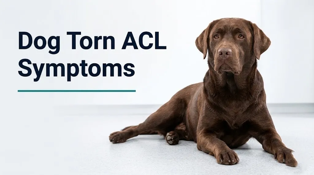
According to Veterinary Clinics of North America: Small Animal Practice, rear leg lameness that persists beyond 5 to 7 days warrants veterinary examination with orthopedic assessment, because self-resolving causes of rear leg lameness (minor muscle strains, bruising) typically improve within this window while CCL tears do not.
Older dogs over age 8 with chronic rear leg lameness may have concurrent degenerative joint disease (osteoarthritis) secondary to a chronic partially torn CCL that has been present for months without diagnosis. The lameness in these cases may be chronically present at a low level with periodic worsening rather than presenting acutely.
Key Takeaway: Rear leg limping that does not improve within 5 to 7 days requires veterinary evaluation, because CCL tears do not self-resolve and untreated joint instability produces progressive and irreversible meniscal and cartilage damage.
Dog Knee Swelling and Joint Effusion Symptoms
Swelling of the stifle joint following CCL rupture reflects two distinct processes: acute joint effusion (fluid accumulation within the joint capsule) and, over time, periarticular fibrosis (fibrous tissue thickening around the joint) that produces the characteristic “medial buttress” palpable along the inner stifle.
When the CCL tears, blood vessels within and around the ligament rupture, producing hemarthrosis (bleeding into the joint space). This hemorrhage, combined with synovial membrane inflammation, causes rapid fluid accumulation within the joint capsule that is often visible within hours of a complete rupture as puffiness around the knee, most prominent just medial (inside) and distal to the patella.
Over days to weeks, the acute hemarthrosis transitions to a serous or serosanguineous joint effusion as the chronic inflammatory response produces elevated synovial fluid from the inflamed synovial membrane. Prostaglandin E2 and TNF-alpha from activated macrophages in the synovial membrane drive continued fluid production and maintain joint warmth and swelling.
A distinct and diagnostically useful finding in chronic CCL instability is the medial buttress: a firm, thickened tissue response along the medial (inner) aspect of the stifle that develops as fibrous tissue accumulates in response to the chronic cranial tibial translation. Veterinarians palpate for medial buttress as a reliable indicator of CCL disease, especially in dogs with chronic or partial tears where the cranial drawer sign may be subtle.
To observe whether your dog’s stifle appears swollen compared to the other side:
- Have your dog stand naturally on all four legs on a firm, level surface
- Crouch or kneel to stifle level and compare both knees simultaneously from the front and side
- Look for visible fullness or puffiness around one knee compared to the other, particularly on the inner (medial) aspect of the joint
- Gently place a flat hand on each stifle and compare for warmth; an inflamed joint feels warmer than its counterpart
- Do not press hard or manipulate the joint, which may cause pain and will not add useful information without veterinary training
Small breed dogs may show less visible swelling due to their smaller joint volumes, even with significant joint effusion. A licensed veterinarian can palpate effusion reliably even when it is not visible to the owner.
Partial ACL Tear Symptoms in Dogs vs Complete Tear
A partial CCL tear in dogs produces milder, often intermittent symptoms compared to a complete tear, and the distinction matters because both require veterinary attention but with different urgency and treatment implications.
| Feature | Partial CCL Tear | Complete CCL Tear |
|---|---|---|
| Lameness severity | Mild to moderate; intermittent | Moderate to severe; often consistent or non-weight bearing |
| Weight bearing | Usually maintains weight bearing | May be non-weight bearing acutely |
| Lameness pattern | Worse after exercise, better with rest | Consistent regardless of rest |
| Stifle swelling | Mild, may not be visible to owner | Often visible, sometimes dramatic |
| Cranial drawer sign | Subtle or inconsistent | Clear, reproducible forward tibial translation |
| Medial meniscal risk | Lower initially; increases over time without treatment | Higher at time of rupture; approximately 30 to 40% concurrent meniscal tear |
| Progression risk | High: most partial tears progress to complete rupture without treatment | N/A: already complete |
| Natural history | Progressive worsening over weeks to months | Persistent instability, progressive joint damage |
Partial CCL tears are particularly difficult for owners to detect because the lameness is mild and intermittent. A dog with a partial tear may appear nearly normal at rest, limp subtly after a run, and then seem to recover. This misleads owners into thinking the problem has resolved when in fact the ligament is continuing to degenerate.
According to research published in Veterinary Surgery, the majority of partial CCL tears progress to complete rupture within months if the underlying degenerative disease process and tibial plateau biomechanics are not addressed. This is why a dog showing mild but recurrent rear leg lameness should be evaluated by a licensed veterinarian even when the dog is currently moving reasonably well.
Young, active dogs under age 3 may show a more acute partial tear presentation from high-impact activities, but this is less characteristic than the degenerative partial tear pattern seen in middle-aged dogs in affected breeds.
Dog Back Leg Weakness and Muscle Wasting From ACL Injury
Muscle wasting (atrophy) of the rear limb is a consistent finding in dogs with chronic CCL disease and reflects the body’s response to reduced weight bearing on the affected leg over days to weeks.
When a dog reduces weight bearing on a rear leg due to CCL-related pain and instability, the quadriceps muscle group (vastus lateralis, vastus medialis, vastus intermedius, and rectus femoris) on the affected side begins to lose muscle mass from disuse. The semimembranosus, semitendinosus, and biceps femoris muscles of the hamstring group also atrophy. The visible result is a noticeably thinner, less muscular upper thigh on the affected side compared to the healthy side, often apparent to the owner within 2 to 4 weeks of onset.
Think of it like this: the body knows the joint is painful and unstable, so it reduces the demand on those muscles. Without the regular weight-bearing contraction that normally maintains muscle bulk, the muscle fibers begin to shrink from disuse atrophy, following the principle that muscle requires loading to maintain its mass.
The weakness from muscle atrophy compounds the instability from the CCL tear itself. A dog with both CCL instability and quadriceps atrophy has double the functional impairment: the primary joint stabilizer is gone and the secondary dynamic stabilizers (muscles) are also compromised. This is why prompt veterinary evaluation and treatment matter for long-term functional outcomes.
According to the American College of Veterinary Surgeons, return of normal muscle mass is one of the key outcome measures after CCL repair surgery, and pre-surgical muscle atrophy is associated with longer rehabilitation timelines after TPLO or TTA procedures.
Older dogs over 9 years with concurrent hypothyroidism face a higher rate of muscle atrophy progression because hypothyroidism reduces muscle protein synthesis and may also have an independent association with CCL disease in some clinical observations.
Key Takeaway: Visible thigh muscle thinning on one rear leg compared to the other, developing over 2 to 4 weeks of rear leg lameness, is a consistent marker of chronic CCL disease and should prompt a veterinary appointment within 1 to 3 days rather than continued watchful waiting.
Dog Sitting Posture Changes and Stiffness From ACL Injury
Abnormal sitting posture is one of the most specific and underappreciated early signs of CCL disease in dogs, and most owners who recognize it are dogs who sit with one rear leg extended to the side rather than folded normally beneath the body.
Normal dogs sit with both rear legs symmetrically folded, the stifles flexed, and the hocks tucked in close to the body. A dog with CCL disease finds that flexing the affected stifle to a normal seated position produces pain from the unstable joint and the inflamed synovial membrane. The dog compensates by extending the affected rear leg out to the side in what veterinarians call the “lazy sit” or “puppy sit”: the affected leg splays outward so the stifle does not need to flex fully.
This posture change can appear before significant lameness develops, making it a valuable early clinical clue. Owners who notice their dog has started sitting “differently” on one side, even without obvious limping, should consider this a reason for veterinary evaluation rather than dismissing it as a quirk.
Stiffness after rest is another consistent pattern. Dogs with CCL disease are often most stiff and lame in the first few minutes after rising from sleep or after a period of inactivity. As the dog moves around and warms up the joint, the lameness may appear to improve slightly. This warm-up phenomenon reflects the reduced synovial fluid viscosity with movement and partial relief of periarticular muscle spasm. It does not mean the injury has resolved.
According to the Merck Veterinary Manual, stiffness that is most pronounced after rest and improves with mild movement is a hallmark pattern of inflammatory joint disease in dogs and should be reported to a licensed veterinarian along with any lameness history, dietary history, and recent activity changes.
Overweight dogs tend to show more pronounced stiffness after rest because increased joint loading amplifies the pain from synovial inflammation and cartilage damage in an already unstable stifle.
Dog ACL Symptoms by Severity: Triage Guide
| Symptom Pattern | Severity Level | Recommended Action |
|---|---|---|
| Occasional mild lameness after vigorous exercise, resolves with rest, no swelling | Monitor | Schedule veterinary appointment within 1 week; restrict running and jumping in the interim |
| Mild consistent lameness on a rear leg, present even after rest, minimal or no visible swelling | Prompt appointment | Schedule appointment within 2 to 3 days; keep dog on leash only |
| Moderate lameness with visible knee swelling, dog weight-bearing but favoring the leg clearly | Urgent appointment | Veterinary evaluation within 24 to 48 hours; restrict all off-leash activity |
| Intermittent leg carrying (dog holds leg up then puts it down), swelling present | Urgent appointment | Veterinary evaluation within 24 hours; leash-only, no stairs |
| Consistent non-weight bearing: dog holds rear leg up continuously | Same-day evaluation | Veterinary appointment the same day; contact veterinary emergency facility if regular vet unavailable |
| Inability to use both rear legs simultaneously, dog cannot rise | Emergency | Go to a veterinary emergency facility immediately |
| Sudden acute complete non-weight bearing after obvious audible pop or fall | Same-day evaluation | Same-day veterinary evaluation; suspected acute complete CCL rupture |
This triage guide reflects general clinical guidance based on CCL disease presentation patterns. A licensed veterinarian is the only person qualified to confirm CCL rupture and determine the appropriate treatment approach. No triage guide replaces a physical examination.
Dogs taking non-steroidal anti-inflammatory medications (NSAIDs) prescribed by a veterinarian for other conditions may show reduced lameness severity despite CCL rupture because the anti-inflammatory effect masks pain signaling. The apparent improvement in lameness while on NSAIDs does not indicate healing and should not delay veterinary evaluation.
How Dog ACL Tears Differ From Human ACL Tears
Dog ACL tears (CCL ruptures) differ fundamentally from human ACL tears in their cause, population affected, rate of bilateral occurrence, and natural history without treatment, and understanding these differences helps owners interpret their dog’s situation accurately.
In humans, ACL tears occur predominantly in young, active individuals during high-impact sports activities (pivoting, landing from a jump, sudden deceleration), and the injury is almost always traumatic: a single event tears a previously healthy ligament. In dogs, most CCL ruptures result from progressive degenerative failure of a ligament that was already weakening for months before the clinical event. The “trigger” incident the owner observes is rarely the true cause.
The tibial plateau angle difference is the key biomechanical explanation. In humans, the tibial plateau is relatively flat (9 to 12 degrees), so the ACL bears load primarily during specific athletic movements. In dogs, the steep tibial plateau (24 to 27 degrees) creates a cranial tibial thrust during every normal weight-bearing step, meaning the CCL is under constant shear stress with every single stride the dog takes. A ligament that is stressed every step, every day, across the dog’s entire weight-bearing life, degenerates through a fundamentally different mechanism than one stressed occasionally during athletics.
The bilateral rupture rate also distinguishes the two species dramatically. Human bilateral ACL tears are relatively uncommon. In dogs, research published in the Journal of the American Veterinary Medical Association found that approximately 40 to 60 percent of dogs diagnosed with CCL rupture in one stifle will rupture the contralateral CCL within 1 to 2 years. This high bilateral rate reflects that the same degenerative disease process affecting one CCL is typically present in both legs simultaneously.
Intact female dogs of childbearing age who are not spayed have different CCL rupture risk profiles compared to spayed females, reflecting the role of estrogen in maintaining CCL collagen integrity.
Why Dogs Get ACL Tears: Causes and Risk Factors
The primary cause of CCL rupture in dogs is degenerative cranial cruciate ligament disease (CCLD): a progressive deterioration of the CCL driven by the combination of chronic mechanical stress from the cranial tibial thrust, inflammatory joint disease, and genetic factors affecting ligament collagen quality.
Named contributing causes and risk factors:
- Tibial plateau angle (TPA): dogs with steeper-than-average TPA generate greater cranial tibial thrust per step, accelerating CCL degeneration. This is partly breed-dependent and partly individual variation.
- Body weight and obesity: excess body weight directly increases the cranial tibial thrust force per step. Dogs with a body condition score (BCS) of 7/9 or above have substantially elevated CCL rupture risk per multiple clinical cohort studies.
- Immune-mediated synovitis: lymphocytic-plasmacytic synovitis (T-cell and macrophage infiltration of the synovial membrane) is consistently present in dogs with CCL disease and is believed to contribute to the intraarticular inflammatory environment that upregulates matrix metalloproteinases in the CCL, accelerating fiber degeneration.
- Genetic predisposition: certain breeds have heritable combinations of tibial plateau angle, ligament collagen composition, and body conformation that dramatically elevate lifetime CCL rupture risk (see breeds section).
- Prior CCL rupture: contralateral CCL rupture risk after a first rupture is approximately 40 to 60 percent within 1 to 2 years, because the same degenerative disease process is typically bilateral.
- High-impact activity on deconditioned joints: dogs that are mostly sedentary and then undergo sudden intense activity (“weekend warrior” dogs) may trigger rupture in a CCL already weakened by degenerative disease.
- Traumatic rupture: genuinely traumatic rupture from a single high-impact event does occur but represents a minority of CCL cases in the canine population, more often seen in young, athletic dogs in contact sports contexts.
According to the American Veterinary Medical Association, owner awareness of CCL disease risk factors, particularly obesity and breed predisposition, can support earlier diagnosis when lameness first appears, improving long-term outcomes.
Which Dog Breeds Are Most at Risk for ACL Tears
Breed is one of the strongest predictors of lifetime CCL rupture risk in dogs, and certain breeds face dramatically elevated rates compared to mixed-breed dogs of similar size.
Highest-risk large and giant breeds, based on epidemiological data published in the Journal of the American Veterinary Medical Association and Veterinary Surgery:
- Labrador Retriever: one of the highest overall CCL rupture rates of any breed; high body weight combined with breed-specific tibial plateau angle and ligament collagen characteristics
- Rottweiler: very high rupture rate, often earlier in life than other large breeds (ages 2 to 4 years common), attributed to steep tibial plateau angle and rapid weight gain
- Newfoundland: giant breed with extremely high CCL rupture burden; body weight magnifies cranial tibial thrust forces substantially
- Staffordshire Bull Terrier: elevated rate disproportionate to body size; breed-specific ligament collagen quality may be a factor
- Mastiff and Bullmastiff: giant breed weight combined with conformational factors
- Akita: consistently appears in high-prevalence breed lists across multiple international studies
- Chesapeake Bay Retriever: elevated rate similar to Labrador Retriever
- Golden Retriever: elevated rate, somewhat lower than Labrador Retriever in most studies
Smaller breeds at elevated risk relative to their size:
- West Highland White Terrier: disproportionately high rate for a small breed; suspected genetic ligament quality factor
- Bichon Frise: elevated rate in small breed category
Mixed-breed dogs face lower average CCL rupture rates than purebred dogs of equivalent body size, suggesting that breed-specific genetic factors contribute beyond body weight alone.
The practical clinical implication for owners of high-risk breeds: any rear leg lameness in a Labrador Retriever, Rottweiler, or Newfoundland should be considered a CCL concern until a licensed veterinarian rules it out through physical examination and radiography.
Key Takeaway: Labrador Retrievers, Rottweilers, and Newfoundlands carry among the highest lifetime CCL rupture risks of any dog breed; any rear leg lameness in these breeds should be evaluated by a licensed veterinarian within 24 to 48 hours, not monitored at home for an extended period.
Age, Weight, and Hormonal Factors in Dog ACL Tears
Three individually important factors modulate CCL rupture risk beyond breed alone: age, body condition score, and reproductive hormone status, and each operates through a distinct physiological mechanism.
Age affects CCL risk through the natural timeline of ligament degeneration. Most CCL ruptures in predisposed breeds occur in middle age, typically between 3 and 7 years, rather than in puppies or senior dogs. This reflects the time required for the degenerative disease process to progress from subclinical ligament fiber damage to clinical rupture. Dogs under 2 years rarely rupture their CCL through degenerative disease. Very old dogs (over 12 years) also have lower rates of new CCL rupture partly due to reduced activity levels and partly because highly susceptible dogs in affected breeds have already ruptured their CCLs earlier in life.
Body condition score (BCS) is a modifiable risk factor with direct biomechanical significance. For every kilogram of excess body weight a dog carries, the cranial tibial thrust force increases proportionally with each step. A dog that is 5 kilograms overweight is loading their CCL substantially more with every stride, every day, than a lean dog of the same breed. Research published in Veterinary Surgery found that overweight dogs have significantly shorter times to CCL rupture after the onset of degenerative ligament changes compared to lean dogs of the same breed.
Reproductive hormone status is the most underrecognized risk factor for CCL disease. Research published in the Journal of the American Veterinary Medical Association found that neutered male dogs and spayed female dogs in several large breeds had substantially higher CCL rupture rates than intact counterparts. The proposed mechanism involves estrogen receptors on ligament fibroblasts (the cells responsible for producing and maintaining collagen in the CCL). Estrogen promotes collagen synthesis and ligament maintenance. After gonadectomy (spaying or neutering), estrogen deficiency reduces CCL fibroblast collagen production, accelerating the degenerative weakening process. This association appears strongest in large breeds and when gonadectomy occurs before skeletal maturity.
Intact female dogs during pregnancy may have transient ligament laxity from increased relaxin production, which could theoretically increase CCL stress, though this is a less well-studied specific risk period.
How Vets Diagnose a Torn ACL in Dogs
Veterinary diagnosis of a CCL tear relies primarily on physical examination techniques that directly test for the joint instability produced by CCL failure, supported by radiography to assess secondary joint changes.
The two most specific physical examination findings:
1. Cranial drawer sign: The veterinarian holds the femur stable with one hand and the tibia with the other, then attempts to slide the tibia forward (cranially) relative to the femur with the stifle in both flexion and extension. In a dog with a complete CCL tear, the tibia slides abnormally forward, producing the “cranial drawer” movement. In a partial tear, the movement may be subtle or absent, particularly in a tense or painful dog. Sedation is sometimes required to perform an accurate assessment in a painful, guarded dog.
2. Tibial compression test (tibial thrust test): The examiner holds one hand on the stifle with a finger on the tibial crest (the bony prominence at the front of the upper shin), then flexes the hock (ankle) to simulate weight bearing. In a dog with CCL rupture, this maneuver produces visible or palpable forward movement of the tibial crest against the finger, confirming cranial tibial translation. This test is often positive even in dogs that are too tense for the cranial drawer test.
Additional diagnostic steps:
- Radiography (X-ray) of the stifle: Does not directly visualize the ligament but identifies joint effusion, displacement of the infrapatellar fat pad (confirming effusion), osteophyte formation indicating chronic instability, and subchondral bone changes. Essential for surgical planning.
- Assessment for medial buttress: Palpation along the medial stifle for the fibrous thickening that develops from chronic instability.
- MRI: Provides definitive soft tissue visualization of the CCL and medial meniscus. More commonly used at specialty centers and prior to complex surgical planning.
- Arthroscopy: Direct visualization of the joint interior, performed both diagnostically (confirming CCL rupture and assessing meniscal integrity) and therapeutically (addressing concurrent meniscal tears).
According to the American College of Veterinary Surgeons, referral to a board-certified veterinary surgeon (Diplomate ACVS) is recommended for all dogs with confirmed CCL rupture to discuss surgical stabilization options, which provide better long-term outcomes than non-surgical management for most dogs.
Dog ACL Treatment Options Overview
Treatment of canine CCL rupture ranges from surgical joint stabilization (the standard of care for most dogs) to non-surgical rehabilitation management in specific circumstances, and the treatment decision depends on the dog’s size, activity level, body condition, age, and health status.
| Treatment Approach | Mechanism | Best Suited For | Evidence Basis |
|---|---|---|---|
| Tibial Plateau Leveling Osteotomy (TPLO) | Cuts and rotates tibial plateau to reduce TPA to approximately 5 degrees, eliminating cranial tibial thrust | Large breeds, active dogs, dogs with steep tibial plateau | Well-established by multiple prospective controlled studies |
| Tibial Tuberosity Advancement (TTA) | Advances the tibial tuberosity to change the patellar ligament angle, neutralizing cranial tibial thrust | Medium to large breeds; alternative to TPLO | Supported by prospective controlled studies |
| Lateral Suture Stabilization (Extracapsular Repair) | Places a suture outside the joint to resist cranial tibial translation | Small and toy breeds under 15 kg; may also be used in older or lower-activity dogs | Supported by clinical outcome data; less durable in large breeds |
| Non-surgical rehabilitation | Activity restriction, physical therapy, weight management, anti-inflammatory pain management | Small dogs under 10 kg; dogs with surgical contraindications (severe concurrent disease) | Supported by clinical observation; outcomes generally poorer than surgery in large breeds |
According to research published in Veterinary Surgery, TPLO provides the most consistent return to normal function in large breed dogs with CCL rupture, with the majority of dogs returning to pre-injury activity levels within 4 to 6 months of surgery and rehabilitation. Non-surgical management in large breed dogs results in progressive joint instability, accelerated osteoarthritis, and typically persistent functional limitation.
The concurrent medial meniscal tear present in 30 to 40 percent of acute ruptures and up to 60 percent of chronic cases requires surgical treatment (partial meniscectomy or meniscal release) at the time of CCL repair, because an untreated meniscal tear continues to produce pain and joint damage after CCL stabilization.
Dogs over age 10 with concurrent systemic disease (cardiac disease, renal insufficiency, diabetes mellitus) may face elevated anesthesia risk that shifts the risk-benefit calculation toward non-surgical management, but this should always be discussed with a licensed veterinarian and ideally a board-certified veterinary surgeon.
Emergency Signs: When to Take Your Dog to the Vet Immediately
Certain symptoms associated with a dog torn ACL require immediate veterinary evaluation. Do not monitor at home and do not wait until the next business day if these signs are present.
Take your dog to a veterinary emergency facility immediately if you observe:
- Complete non-weight bearing on one rear leg for more than 2 to 3 hours: acute complete CCL rupture causes immediate severe lameness that does not improve spontaneously and requires same-day evaluation to assess joint damage and begin pain management
- Inability to use both rear legs simultaneously or inability to rise from a lying position: this can indicate bilateral CCL rupture (both knees simultaneously compromised) or a spinal cord emergency; both require emergency evaluation to differentiate and treat appropriately
- Sudden dramatic swelling of one knee after a fall or audible pop: indicates acute joint injury with hemarthrosis (bleeding into the joint) requiring immediate pain assessment and imaging
- Visible bone deformity or abnormal joint angle in a rear leg: indicates a fracture rather than ligament injury and requires immediate emergency evaluation
- A dog that was previously limping suddenly stops bearing any weight at all: represents acute worsening of an existing injury that requires same-day evaluation, not continued home monitoring
- Any acute rear limb injury in a dog with a known history of the contralateral CCL being previously repaired: given the 40 to 60 percent contralateral rupture rate, same-day evaluation is appropriate to assess and protect the post-surgical limb while addressing the new injury
If you are unsure whether your dog’s rear leg symptoms constitute an emergency, call a 24-hour veterinary emergency facility and describe exactly what you are observing. Do not rely on online descriptions alone to make this decision.
These presentations indicate acute joint instability, possible bilateral limb failure, or concurrent neurological emergency and require immediate veterinary assessment at an emergency facility, not a scheduled appointment or watchful waiting at home.
Frequently Asked Questions About Dog Torn ACL Symptoms
How do I know if my dog tore their ACL?
The most consistent signs of a dog ACL (CCL) tear are sudden limping on a rear leg, reluctance to bear full weight on that leg, and visible or palpable swelling of the knee joint.
Only a licensed veterinarian performing the cranial drawer test and tibial compression test can confirm CCL rupture, because the specific joint instability produced by ligament failure requires physical examination to detect.
Any rear leg lameness that persists more than 5 to 7 days without improvement, or any acute non-weight bearing on a rear leg, warrants veterinary evaluation within 24 to 48 hours.
Can a dog walk on a torn ACL?
Many dogs with a partial CCL tear can still walk and even run, though with a visible limp, because the partially intact ligament still provides some joint stability.
Dogs with a complete CCL tear typically bear significantly reduced weight on the affected leg and may hold it completely off the ground during acute rupture, though some dogs put partial weight down as the acute pain slightly subsides over days.
According to the American College of Veterinary Surgeons, a dog’s ability to walk does not indicate the injury is minor; untreated CCL rupture causes progressive cartilage damage and meniscal injury regardless of the dog’s ability to ambulate.
What does a dog ACL tear look like from the outside?
Visible signs of a dog ACL (CCL) tear include swelling of the knee joint (most visible as puffiness on the inner side of the knee), a visible limp or leg-carrying behavior, muscle thinning on the affected thigh, and an abnormal sitting position with the affected leg extended out to the side.
Not all CCL tears produce dramatic external swelling, particularly in partial tears or in heavily muscled breeds where swelling is obscured by muscle bulk.
The most reliable external observation is the limping pattern combined with posture changes; veterinary examination with specific orthopedic tests is required to confirm the diagnosis.
Can a dog heal from an ACL tear without surgery?
Small dogs under approximately 10 kilograms can achieve acceptable functional outcomes with strict rest, weight management, and anti-inflammatory pain management in some cases, according to veterinary clinical guidelines.
Large and giant breed dogs generally do not achieve satisfactory functional recovery without surgical stabilization, because the ongoing cranial tibial thrust from their steep tibial plateau continues to damage the joint despite rest and conservative management.
Non-surgical management in large breeds consistently results in progressive osteoarthritis, chronic joint instability, and persistent functional limitation; this should be discussed with a board-certified veterinary surgeon before choosing a treatment path.
How long does it take for a dog ACL tear to heal?
Dogs that undergo surgical stabilization with TPLO or TTA typically return to pre-injury activity levels within 4 to 6 months, with structured rehabilitation beginning within the first weeks post-surgery.
Non-surgical management requires a minimum of 8 to 12 weeks of strict activity restriction and may result in a prolonged or incomplete functional recovery, particularly in large breed dogs.
The 40 to 60 percent risk of contralateral CCL rupture within 1 to 2 years means that recovery from one leg’s injury must be accompanied by weight management and monitoring of the opposite stifle joint throughout recovery.
What happens if a dog ACL tear goes untreated?
Untreated CCL rupture in dogs causes progressive joint instability that continuously damages the articular cartilage lining the femur and tibia and tears the medial meniscus over time, producing accelerating and irreversible osteoarthritis.
According to research published in Veterinary Surgery, dogs with untreated CCL rupture develop moderate to severe radiographic osteoarthritis within 2 years of diagnosis, regardless of whether they appear to be functioning reasonably well at a low activity level.
Persistent pain from untreated CCL disease significantly reduces a dog’s quality of life and activity tolerance, making early veterinary evaluation and treatment planning the most important step an owner can take after noticing consistent rear leg lameness.
Closing
A dog torn ACL (CCL) tear is not a wait-and-see situation. Any rear leg lameness that persists beyond 5 to 7 days, any visible knee swelling, any change in sitting posture, or any complete non-weight bearing on a rear leg warrants veterinary evaluation. The underlying degenerative disease process continues damaging the joint with every step your dog takes on an unstable stifle.
Schedule an appointment with a licensed veterinarian who can perform the cranial drawer test and tibial compression test to confirm or rule out CCL rupture. Bring information about when the limping started, whether any specific incident preceded it, and whether the opposite rear leg has ever shown similar signs. Ask about referral to a board-certified veterinary surgeon for surgical consultation if CCL rupture is confirmed.
If your dog cannot use a rear leg at all, or cannot rise using both rear legs, go to a veterinary emergency facility tonight. That level of dysfunction cannot wait.