C5-C6 Disc Symptoms: Nerve Root Pain and Red Flags 2026
C5-C6 symptoms arise when the disc between the fifth and sixth cervical vertebrae compresses either the C6 nerve root (producing pain, numbness, and weakness that travels down the lateral forearm to the thumb and index finger) or the spinal cord itself (producing the more serious condition of myelopathy, with bilateral arm symptoms, gait changes, and hand clumsiness). The specific symptom pattern you experience depends on exactly which structure is compressed and how severely.
According to the American Academy of Orthopaedic Surgeons, the C5-C6 level is one of the most frequently affected cervical spine segments in adults, reflecting its position at a biomechanical transition zone where the mobile lower cervical spine meets the more rigid upper segment. Research published in Spine has found that cervical radiculopathy from disc herniation most commonly affects adults between ages 40 and 60, with C5-C6 and C6-C7 accounting for the majority of surgical and non-surgical cases combined.
This article covers the complete symptom picture at the C5-C6 level: what each symptom is, why it happens inside the spinal anatomy, how to distinguish C5 nerve root symptoms from C6 nerve root symptoms, how to tell radiculopathy from myelopathy, and which presentations require urgent or emergency evaluation.
C5-C6 Symptoms: What They Are and Why They Happen
C5-C6 symptoms are neurological and musculoskeletal symptoms produced when structures at the fifth and sixth cervical vertebral level compress, irritate, or damage the C5 or C6 nerve roots, the spinal cord, or both.
The cervical spine contains eight nerve roots (C1 through C8) that exit at corresponding levels. At the C5-C6 disc level, two structures are at risk. The C6 nerve root exits through the C5-C6 neural foramen and is the nerve most commonly compressed by posterolateral disc herniation at this level. The C5 nerve root exits at the C4-C5 level above but can be affected by central disc herniation or foraminal disease at C5-C6 through anatomical variation. The spinal cord itself runs directly behind the disc and can be compressed when the disc herniation is large and central rather than lateral.
The primary symptoms of C5-C6 problems include:
- Neck pain in the posterior and lateral cervical region
- Shoulder pain radiating to the lateral shoulder or scapular region
- Arm pain traveling down the lateral forearm to the thumb and index finger (C6 pattern) or to the lateral upper arm and shoulder (C5 pattern)
- Numbness and tingling in the thumb, index finger, and lateral forearm (C6 territory)
- Weakness in the biceps brachii, brachioradialis, and wrist extensors
- Reflex changes: diminished brachioradialis reflex (C6) or biceps reflex (C5-C6)
- Hand clumsiness and gait changes if the spinal cord is compressed (myelopathy)
Women with cervical radiculopathy at C5-C6 report a higher frequency of neck-dominant pain as the primary presenting symptom compared to men, who more often present with classic arm-dominant radicular pain. Research published in Spine documented this sex-based symptom distribution difference, suggesting that clinicians evaluating women with neck pain should consider cervical nerve root involvement even when arm symptoms are not the chief complaint.
C5-C6 Anatomy: What Happens at This Spinal Level
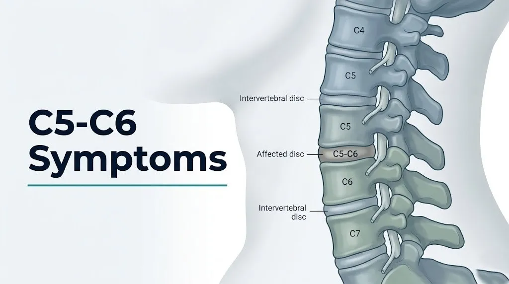
The C5-C6 spinal level consists of the fifth and sixth cervical vertebrae, the intervertebral disc between them, two neural foramina (one on each side) through which the C6 nerve roots exit, and the cervical spinal cord passing through the central canal directly posterior to the disc.
Think of the C5-C6 disc like a jelly donut squeezed between two vertebrae, with nerve tunnels on either side and the spinal cord directly behind it. When the outer ring (annulus fibrosus) tears under pressure, the inner jelly (nucleus pulposus) pushes out. If it pushes to one side, it enters the nerve tunnel and squeezes the C6 nerve root. If it pushes straight back toward the spine, it can compress the spinal cord itself.
The C5-C6 intervertebral disc sits at a biomechanical pivot point. The lower cervical spine (C5-C6, C6-C7) performs a disproportionate share of cervical flexion and extension movements compared to other levels, explaining why this segment accumulates degenerative stress and disc wear faster than most other cervical levels.
The C6 nerve root, after exiting the C5-C6 foramen, contributes fibers to the musculocutaneous nerve (innervating the biceps brachii) and the radial nerve (innervating the brachioradialis and extensor carpi radialis longus). The C6 dermatome covers the lateral forearm, the thumb, and the index finger. This anatomical map directly predicts where a person will feel pain and numbness from C6 nerve root compression.
According to the National Institute of Neurological Disorders and Stroke, cervical disc herniations most frequently occur at C5-C6 and C6-C7, with the posterolateral position (toward the nerve tunnel) being the most anatomically typical direction of herniation, explaining why the C6 nerve root is the most frequently affected root in cervical disc disease.
C5-C6 Neck and Shoulder Pain
Neck and shoulder pain from C5-C6 pathology typically presents as a deep, aching pain in the posterior and lateral cervical region, often radiating to the medial scapular border, the trapezius muscle, and the lateral shoulder, and it frequently worsens with extension or lateral bending toward the affected side.
The neck pain arises from two distinct mechanisms. First, the disc itself contains nociceptive nerve fibers in its outer third (the annulus fibrosus), innervated by the sinuvertebral nerve (also called the recurrent meningeal nerve). When the annulus tears, these nociceptors are activated, producing deep, poorly localized axial neck pain that patients often describe as an aching pressure or stiffness. This is called discogenic pain and does not require nerve root compression to occur.
Second, when the herniated disc material contacts the C6 nerve root or the dorsal root ganglion at the C5-C6 level, the nucleus pulposus releases phospholipase A2, an enzyme that initiates an arachidonic acid inflammatory cascade producing prostaglandin E2 locally. This sensitizes nociceptive C-fibers in the nerve root and the peridiscal tissue, amplifying the pain signal beyond what mechanical pressure alone would produce.
Shoulder pain in C5-C6 disease is frequently misattributed to rotator cuff pathology, bursitis, or shoulder impingement, because the shoulder and upper arm are in the C5 dermatomal territory and the C5 nerve root contributes to the axillary nerve innervating the deltoid muscle. The distinguishing clinical feature: C5-C6 shoulder pain is worsened by neck movement (particularly lateral rotation and extension) and does not change with shoulder-specific provocative tests like the Neer or Hawkins impingement tests.
People with diabetes mellitus have a higher rate of both cervical disc disease and peripheral neuropathy, which complicates symptom attribution. Shoulder pain in a diabetic patient may reflect C5-C6 radiculopathy, diabetic amyotrophy (Bruns-Garland syndrome affecting the brachial plexus), or shoulder pathology, and a board-certified neurologist with EMG testing is often needed to differentiate these conditions.
Key Takeaway: Neck and shoulder pain from C5-C6 disease arises from both discogenic nociceptor activation in the torn annulus fibrosus and from phospholipase A2-driven inflammation sensitizing the nerve root; shoulder pain that worsens with neck movement rather than shoulder movement should prompt consideration of cervical nerve root involvement.
C6 Nerve Root Symptoms: Arm, Forearm, and Hand
C6 nerve root compression at the C5-C6 level produces a specific, reproducible symptom pattern: pain and paresthesias (tingling and burning) in the lateral forearm, extending to the thumb, index finger, and the lateral palm and thenar eminence (the fleshy base of the thumb).
This anatomical specificity is not coincidental. The C6 dermatome precisely maps the skin territory supplied by sensory fibers traveling through the C6 nerve root. When that root is compressed, the brain interprets the nerve’s distorted signal as coming from the skin and tissue in its dermatomal territory, even though the actual compression is happening in the neck. This is the same mechanism as referred pain, where a heart attack produces left arm pain because the brain cannot distinguish the location of an unusual signal along a familiar nerve pathway.
The pain character in C6 radiculopathy is typically electric, shooting, or burning, distinct from the dull ache of the neck and shoulder component. It travels down the arm in a predictable path: from the neck or shoulder, along the lateral forearm, into the thumb and index finger. Patients frequently describe a specific burning or buzzing sensation in the thumb that they cannot explain until imaging reveals the C6 compression.
According to the North American Spine Society Clinical Guidelines, the combination of arm pain in the C6 dermatomal distribution with diminished brachioradialis reflex and weakness of the biceps or wrist extensors has high diagnostic specificity for C6 radiculopathy, with the Spurling test (lateral compression of the cervical spine toward the affected side) having a specificity of approximately 93 percent for cervical radiculopathy when positive.
Pregnant women experiencing thumb and lateral forearm numbness should have both C6 radiculopathy and carpal tunnel syndrome (median nerve compression at the wrist) considered in the differential diagnosis. Carpal tunnel syndrome is common in pregnancy from hormonal fluid retention and produces symptoms overlapping with C6 in the lateral palm, thumb, and index finger. A clinical neurologist can distinguish these with EMG and nerve conduction velocity testing.
C5 Nerve Root Symptoms at the C5-C6 Level
The C5 nerve root can be affected at the C5-C6 level through central or medial disc herniation, foraminal stenosis from uncovertebral joint osteophytes, or anomalous disc anatomy, producing a symptom pattern distinctly different from C6 involvement.
C5 nerve root compression produces pain and sensory changes in the lateral upper arm and shoulder, in the deltoid muscle region extending to approximately the elbow. The C5 dermatome does not extend below the elbow, which is the critical distinguishing feature from C6 involvement. A person who says “my shoulder and upper arm hurt but nothing below my elbow is numb” is describing C5 territory, not C6 territory.
Motor involvement of the C5 nerve root produces weakness of the deltoid muscle, innervated by the axillary nerve (C5-C6, predominantly C5), producing difficulty raising the arm to the side (shoulder abduction). This is tested clinically by asking the patient to hold their arm horizontally while the examiner pushes downward. Deltoid weakness from C5 involvement can be mistaken for rotator cuff pathology or adhesive capsulitis and requires MRI and clinical examination to differentiate.
According to research published in the Journal of Bone and Joint Surgery, isolated C5 nerve root palsy, sometimes called C5 palsy, is a recognized complication of cervical spine surgery but can also occur spontaneously from foraminal stenosis at the C5-C6 level. It presents as sudden deltoid and biceps weakness without significant pain, which can be alarming and is often misdiagnosed as stroke or peripheral nerve injury before the cervical origin is recognized.
Older adults over age 65 are more likely to have C5 nerve root symptoms from foraminal stenosis (bony narrowing of the nerve tunnel) rather than from acute disc herniation, because age-related osteophyte formation at the uncovertebral joints is the dominant pathological mechanism in this age group rather than acute nuclear herniation.
C5-C6 Weakness: Which Muscles Are Affected
C5-C6 nerve root compression produces weakness in specific, predictable muscle groups, and knowing which muscles to test is the most practical way to assess whether nerve function is intact or deteriorating between clinical appointments.
The muscles most affected by C5-C6 pathology, organized by nerve root:
| Nerve Root | Muscle | Function | Clinical Test |
|---|---|---|---|
| C5 (primary) | Deltoid (axillary nerve) | Shoulder abduction | Raise arm to 90 degrees against resistance |
| C5-C6 | Biceps brachii (musculocutaneous nerve) | Elbow flexion and forearm supination | Curl the forearm up against resistance |
| C5-C6 (C6 primary) | Brachioradialis (radial nerve) | Elbow flexion with forearm neutral | Bend elbow with thumb pointing up against resistance |
| C6 (primary) | Extensor carpi radialis longus (radial nerve) | Wrist extension | Extend wrist against downward pressure |
| C6-C7 | Pronator teres | Forearm pronation | Turn palm down against resistance |
A person with C6 nerve root compression will typically notice weakness when opening jars (requiring wrist extension), doing curls at the gym (biceps), or performing any activity requiring sustained grip with wrist stabilization. These functional limitations are often noticed before a clinical examination documents them formally.
The weakness of C5-C6 radiculopathy is a lower motor neuron weakness: it is typically unilateral, may be associated with muscle atrophy in chronic cases, and does not produce spasticity (increased muscle tone). This distinguishes it from the upper motor neuron weakness of myelopathy, which produces bilateral weakness with spasticity below the level of cord compression.
According to the American Academy of Orthopaedic Surgeons, progressive motor weakness (strength decreasing over weeks to months) in any muscle group supplied by the C5 or C6 nerve root is an indication for surgical consultation regardless of pain intensity, because irreversible motor neuron damage can occur if the compression is not relieved.
Manual workers performing repetitive overhead activities with sustained cervical extension face elevated risk of progressive weakness from chronic foraminal narrowing at C5-C6. Any sustained occupation-related position that reproduces arm weakness should be modified immediately pending clinical evaluation.
C5-C6 Numbness and Tingling Patterns
Numbness and tingling from C5-C6 pathology follow the dermatomal distribution of the compressed nerve root with a precision that is one of the most useful diagnostic clues available without any testing equipment.
C6 nerve root numbness produces tingling, numbness, or altered sensation in the lateral (outer) forearm, the dorsal and palmar surface of the thumb, the index finger, and sometimes the lateral palm extending toward the thenar eminence. Patients frequently describe this as a buzzing or electric feeling in the thumb that is worse when the neck is held in certain positions (particularly extension or ipsilateral lateral bending) and may partially or completely relieve when the arm is held overhead (a position that opens the neural foramen and reduces nerve root tension).
C5 nerve root numbness produces sensory changes in the lateral shoulder, the outer upper arm from shoulder to approximately the elbow, and the deltoid patch (a characteristic region of reduced sensation over the lateral deltoid muscle). It does not extend below the elbow. This is the definitive anatomical distinction: below-elbow numbness is C6 or lower; above-elbow-only numbness points to C5.
The position-dependent nature of C5-C6 numbness is diagnostically useful. Symptoms that worsen with neck extension or lateral bending toward the symptomatic side (narrowing the neural foramen) and improve with the arm held overhead (distraction test) are consistent with nerve root origin. Numbness that is constant, position-independent, and most pronounced at the wrist and palm points more toward carpal tunnel syndrome than cervical radiculopathy.
Research published in Spine found that approximately 40 percent of patients with confirmed cervical radiculopathy report their paresthesias as their most bothersome symptom, even more than pain, underscoring the importance of precise dermatomal characterization in clinical assessment.
Athletes who participate in contact sports (football, rugby, wrestling) and develop sudden bilateral upper extremity burning or tingling after a tackle or fall (“burners or stingers”) should be evaluated for both peripheral brachial plexus stretch injury and central C5-C6 cord contusion, as these presentations can be clinically similar acutely.
Key Takeaway: C6 numbness extends from the lateral forearm to the thumb and index finger, while C5 numbness stays in the lateral upper arm and shoulder and never extends below the elbow; this dermatomal distinction is the most reliable clinical feature for identifying which nerve root is compressed without imaging.
C5-C6 Reflex Changes and What They Mean
Reflex changes at the C5-C6 level are among the most objective clinical findings in cervical disc disease, and understanding which reflex corresponds to which nerve root helps explain what your neurologist or orthopedic surgeon found on examination.
| Reflex | Nerve Root | Testing Method | Abnormal Finding | Clinical Significance |
|---|---|---|---|---|
| Biceps reflex | C5 (primary), C6 | Tap the biceps tendon in the antecubital fossa | Diminished or absent response | Suggests C5 nerve root dysfunction |
| Brachioradialis reflex | C6 (primary) | Tap the brachioradialis tendon at the wrist | Diminished or absent response | Highly specific for C6 nerve root dysfunction |
| Triceps reflex | C7 (primary) | Tap the triceps tendon above the olecranon | Diminished or absent response | Suggests C7 nerve root dysfunction, not C5-C6 |
| Hoffmann sign | Upper motor neuron (myelopathy) | Flick the distal phalanx of the middle finger | Thumb and index finger flex | Indicates spinal cord compression (myelopathy) |
The brachioradialis reflex is the most specific reflex for C6 nerve root involvement. When the C6 root is compressed at the C5-C6 level, this reflex is typically diminished or absent on the affected side. A neurologist or orthopedic spine surgeon will routinely compare the reflex response between the two sides, because subtle asymmetry between right and left is more clinically informative than comparing against a population average.
The Hoffmann sign deserves particular attention. When a clinician flicks the distal phalanx of your middle finger and your thumb and index finger flex involuntarily, that is the Hoffmann sign. It is a pathological upper motor neuron reflex indicating dysfunction of the corticospinal tracts in the spinal cord above the level of the hand. In the context of C5-C6 disease, a positive Hoffmann sign suggests that the spinal cord, not just the nerve root, is being compressed. This is a finding that warrants imaging and specialist evaluation regardless of symptom duration.
According to research published in the Journal of Neurosurgery: Spine, the combination of a positive Hoffmann sign with bilateral hand weakness and gait disturbance has high sensitivity for cervical spondylotic myelopathy, and its presence should prompt MRI of the cervical spine within days rather than weeks.
People with rheumatoid arthritis face specific additional cervical reflex examination complexity because atlantoaxial subluxation (C1-C2 instability from rheumatoid pannus) can produce upper motor neuron signs that mimic or coexist with C5-C6 myelopathy. A rheumatologist and spine specialist should both be involved in evaluating cervical symptoms in rheumatoid arthritis patients.
C5-C6 Radiculopathy vs Myelopathy: Key Differences
Cervical radiculopathy from C5-C6 pathology is a nerve root problem producing unilateral arm and hand symptoms, while cervical myelopathy is a spinal cord problem producing symptoms in both arms, the legs, the hands, and potentially the bowel and bladder, and the two conditions require fundamentally different management approaches.
Think of radiculopathy as one specific telephone wire getting pinched: only the circuit it connects gets disrupted, and it stays on that side. Myelopathy is the main telephone exchange being compressed: every circuit it controls gets disrupted, and both sides of the body are affected below the point of compression.
| Feature | C5-C6 Radiculopathy | C5-C6 Myelopathy |
|---|---|---|
| Structure compressed | C5 or C6 nerve root | Cervical spinal cord |
| Symptom side | Unilateral (one arm) | Bilateral (both arms and legs) |
| Pain pattern | Radiating from neck into one arm | Diffuse, often less sharp than radiculopathy |
| Weakness | Specific muscles (biceps, deltoid, wrist extensors) | Diffuse, bilateral, with spasticity |
| Reflexes | Diminished in affected root territory | Diminished at the level; hyperreflexia below |
| Hand symptoms | Numbness in dermatomal pattern | Clumsy, weak hands; difficulty with fine motor tasks |
| Gait | Normal | Shuffling, unsteady, wide-based gait |
| Bowel and bladder | Not affected | May be affected in advanced cases |
| Urgency | Routine to urgent depending on weakness | Urgent to emergency depending on progression rate |
| Hoffmann sign | Negative | Positive |
According to the North American Spine Society, cervical myelopathy is the most prevalent cause of spinal cord dysfunction in adults over age 55 in developed countries, and it is frequently missed or delayed in diagnosis because its early symptoms (hand clumsiness, mild gait unsteadiness) are subtle and attributed to other age-related changes.
Any person with known C5-C6 disc disease who develops bilateral symptoms, hand clumsiness affecting writing or buttoning clothing, or unsteadiness walking should be evaluated by a board-certified neurosurgeon or orthopedic spine surgeon within days, not weeks.
C5-C6 Myelopathy Symptoms and Why They Matter
Cervical spondylotic myelopathy from C5-C6 cord compression produces a specific clinical pattern reflecting which spinal cord tracts are being compressed: the corticospinal tracts (motor), dorsal columns (proprioception and vibration sense), and spinothalamic tracts (pain and temperature below the lesion).
Corticospinal tract compression produces upper motor neuron weakness in the arms and legs: bilateral weakness that is accompanied by spasticity (increased muscle tone), hyperreflexia (exaggerated reflexes below the compression level), and the Hoffmann sign in the hands. The weakness of myelopathy is different from radiculopathy weakness: it tends to be diffuse, bilateral, and accompanied by a stiffness or heaviness in the limbs rather than the focal, flaccid weakness of nerve root compression.
Dorsal column compression produces proprioceptive loss: the inability to sense the position of the feet and hands without looking at them. This is what produces the characteristic wide-based, shuffling gait of cervical myelopathy. The person walks with feet wider apart than normal and may feel unsteady on stairs, on uneven surfaces, or in the dark (where visual compensation for proprioceptive loss is not available). Lhermitte sign (a brief, electric shock-like sensation down the spine and into the limbs when the neck is flexed) reflects dorsal column irritation and is a recognizable myelopathy symptom.
Hand clumsiness is often the earliest myelopathy symptom. Patients notice they are dropping things, have difficulty with fine motor tasks like typing, buttoning shirts, or using keys, or that their handwriting has deteriorated. This reflects compression of the corticospinal fibers controlling hand function combined with proprioceptive loss in the fingers.
According to research published in the Journal of Neurosurgery: Spine, cervical spondylotic myelopathy follows a stepwise deterioration pattern in approximately two-thirds of patients, with periods of stability punctuated by sudden worsening episodes, rather than a smooth progression. This stepwise pattern means that a patient who feels stable may be one minor event (a sudden neck extension, a minor fall) away from acute neurological deterioration.
Elderly patients with cervical spondylotic myelopathy have a higher fall risk due to gait instability and proprioceptive deficits. Any fall in an elderly person with known cervical stenosis warrants evaluation for acute cord injury superimposed on chronic myelopathy.
Key Takeaway: Cervical myelopathy from C5-C6 cord compression produces bilateral weakness, hand clumsiness, and gait unsteadiness through corticospinal tract and dorsal column compression; a positive Hoffmann sign with any bilateral symptoms warrants MRI of the cervical spine within days and neurosurgical consultation.
Causes of C5-C6 Symptoms: Disc Herniation, Stenosis, and Spondylosis
Three distinct pathological processes produce C5-C6 symptoms, and understanding which process applies to you helps set accurate expectations about the clinical course and treatment approach.
Acute disc herniation at C5-C6 occurs when the nucleus pulposus ruptures through a tear in the posterolateral annulus fibrosus, typically after a sudden axial loading event (a fall, a lifting injury, or rapid neck movement) or after cumulative annular fatigue. The herniation is typically sudden or subacute in onset, produces intense radicular pain, and is the predominant mechanism in adults aged 30 to 55. According to research published in Spine, approximately 70 to 80 percent of patients with acute cervical disc herniation experience meaningful clinical improvement within 12 weeks with non-surgical management, making this a condition where watchful waiting combined with physician-directed care is clinically reasonable for most people in the absence of progressive weakness.
Cervical spondylosis at C5-C6 is the age-related degenerative process producing osteophyte formation at the vertebral endplates and uncovertebral joints, disc height loss from chronic dehydration of the nucleus pulposus, and facet joint arthrosis. These changes develop slowly over years or decades, typically producing chronic intermittent symptoms that wax and wane rather than the acute, intense onset of disc herniation. The predominant mechanism shifts from soft disc herniation in younger adults to hard osteophyte-driven foraminal narrowing in adults over 55.
Cervical foraminal stenosis refers to narrowing of the neural foramen through which the C6 nerve root exits, produced by a combination of disc height loss, osteophyte from the uncovertebral joint (joint of Luschka), and facet joint hypertrophy. It produces positional radicular symptoms: worse with neck extension (which closes the foramen further) and relieved with neck flexion or arm elevation overhead (which opens the foramen slightly).
Additional contributing causes of C5-C6 symptoms include ligamentum flavum hypertrophy (the yellow ligament behind the spinal cord thickening with age, reducing central canal diameter), ossification of the posterior longitudinal ligament (OPLL) (a condition more prevalent in East Asian populations that produces diffuse spinal cord compression), and post-traumatic disc injury.
C5-C6 Symptoms by Age and Population
The clinical presentation of C5-C6 symptoms changes substantially based on age, biological sex, and occupational or recreational activity level, and these differences should inform both how symptoms are interpreted and when specialist evaluation is appropriate.
Adults aged 30 to 50 most commonly present with acute or subacute disc herniation producing intense unilateral C6 radiculopathy with arm pain as the dominant symptom. The onset is often linked to a specific exertional event or posture, and the natural history is relatively favorable: the majority of acute herniations improve without surgery as the herniated material dehydrates and contracts over weeks to months.
Adults aged 50 to 70 more commonly present with chronic, bilateral, or positional symptoms from cervical spondylosis and foraminal stenosis rather than acute herniation. Pain may be less intense than in younger patients, but neurological signs (reflex changes, weakness) are often more established. Myelopathy from central canal stenosis becomes increasingly prevalent in this age group as cumulative degenerative changes reduce canal diameter.
Adults over 70 face the highest risk of cervical spondylotic myelopathy presenting with gait instability and hand weakness that may be attributed to peripheral vascular disease, peripheral neuropathy, or general aging rather than cervical cord compression. A clinician who observes progressive gait deterioration in an elderly patient should include cervical MRI in the diagnostic consideration.
Sex-based differences in C5-C6 symptom presentation: research published in Spine documented that women with cervical radiculopathy report higher pain intensity and higher rates of neck-dominant symptoms, while men report arm-dominant symptoms more frequently. Women are also more likely to have cervical radiculopathy co-exist with temporomandibular joint disorder and headache, which can complicate the symptom picture.
Occupational groups at highest C5-C6 risk:
- Hairdressers and surgeons: prolonged cervical flexion
- Construction and manual laborers: repetitive heavy lifting with cervical loading
- Office workers: sustained cervical flexion from screen work
- Overhead athletes (swimmers, baseball pitchers, volleyball players): repetitive cervical extension under load
- Contact sport athletes: acute axial loading injury risk
How C5-C6 Symptoms Are Diagnosed
Diagnosis of C5-C6 pathology begins with a clinical history and physical examination that specifically tests the nerve root levels at risk, then is confirmed with imaging and, when needed, electrodiagnostic testing.
The clinical examination for C5-C6 pathology includes:
- Dermatomal sensory testing: light touch and pin-prick sensation assessed in the C5 territory (lateral upper arm) and C6 territory (lateral forearm, thumb, index finger) compared between sides
- Motor strength testing: deltoid (C5), biceps brachii (C5-C6), brachioradialis (C5-C6), and wrist extensors (C6) graded on the Medical Research Council (MRC) 0 to 5 scale
- Reflex testing: biceps, brachioradialis, and triceps reflexes assessed for asymmetry; Hoffmann sign tested for upper motor neuron involvement
- Provocative testing: Spurling test (lateral compression of the cervical spine toward the symptomatic side reproducing arm pain); shoulder abduction relief test (patient raises arm overhead, which relieves radicular pain if nerve root distraction reduces compression); cervical distraction test (examiner gently lifts the head to open foramina, relieving radicular symptoms)
- Gait and balance assessment: tandem walking, Romberg test (standing with feet together and eyes closed), observation for spasticity or shuffling gait pattern suggesting myelopathy
MRI of the cervical spine is the definitive imaging study. T2-weighted MRI sequences show the disc herniation, neural foraminal narrowing, central canal diameter, and spinal cord signal change. T2 hyperintensity (a bright signal within the spinal cord on T2-weighted MRI) at the C5-C6 level indicates cord compression with edema or ischemia, consistent with myelopathy requiring urgent evaluation.
EMG and nerve conduction velocity (NCV) testing performed by a board-certified neurologist or physiatrist can localize the nerve root involved, distinguish root pathology from peripheral nerve entrapment (such as carpal tunnel syndrome at the wrist), and assess the severity and chronicity of axonal damage.
According to the AAOS OrthoInfo clinical guidelines, MRI is the preferred initial imaging study for suspected cervical radiculopathy with neurological deficit, with CT myelography reserved for patients who cannot undergo MRI or in whom surgical planning requires more detailed bony anatomy visualization.
C5-C6 Treatment Options Overview
Treatment for C5-C6 symptoms is guided by the specific diagnosis (radiculopathy versus myelopathy), the severity of neurological compromise, and the duration of symptoms, with non-surgical management being appropriate for most radiculopathy cases and surgical management required for progressive myelopathy.
| Treatment Approach | What It Addresses | Evidence Basis | Provider |
|---|---|---|---|
| Physician-directed physical therapy | Cervical stabilization, neural mobilization, posture | Supported by multiple randomized controlled trials for radiculopathy | Physical therapist under spine specialist guidance |
| Cervical epidural steroid injection (ESI) | C6 nerve root inflammation reduction | Supported by controlled trials for short-term pain relief; less evidence for long-term outcomes | Interventional pain management physician |
| Cervical collar (short-term) | Immobilization and pain management in acute phase | Supported by clinical guidelines for short-term use only | Primary care physician or spine specialist |
| Oral anti-inflammatory medications | Prostaglandin-mediated nerve root inflammation | Supported by clinical guidelines for symptomatic relief | Primary care physician |
| Anterior cervical discectomy and fusion (ACDF) | Direct decompression of C6 nerve root or spinal cord | Well-established by controlled surgical outcome trials | Board-certified orthopedic spine surgeon or neurosurgeon |
| Cervical disc arthroplasty | Decompression while preserving motion at C5-C6 | Supported by multiple randomized controlled trials vs. ACDF | Board-certified orthopedic spine surgeon or neurosurgeon |
| Posterior cervical foraminotomy | Decompression of neural foramen without fusion | Supported by controlled surgical outcome trials | Board-certified neurosurgeon or orthopedic spine surgeon |
According to a landmark study published in the New England Journal of Medicine (the SPORT trial, which included cervical spine data), patients with confirmed cervical radiculopathy from disc herniation who undergo surgery achieve faster symptom relief compared to non-surgical management, but the long-term outcomes between surgical and non-surgical groups converge over 2 to 4 years for most patients without progressive neurological deficit.
The critical exception: myelopathy does not improve significantly with non-surgical treatment and typically progresses without decompressive surgery. According to the North American Spine Society Clinical Guidelines, surgical decompression for cervical spondylotic myelopathy is recommended for patients with moderate to severe myelopathy or progressive neurological deterioration, based on evidence from multiple prospective trials.
Emergency Symptoms: When C5-C6 Problems Require Immediate Care
Certain symptoms associated with C5-C6 disc disease and cervical stenosis require immediate emergency evaluation. Do not wait for a scheduled appointment if these signs are present.
Call 911 or go to the nearest emergency room immediately if you experience:
- Sudden onset of bilateral arm weakness, leg weakness, or inability to walk: indicates acute cervical cord compression or cervical spinal cord injury requiring emergency neurosurgical evaluation
- Sudden loss of bowel or bladder control in a person with known or suspected cervical stenosis or myelopathy: indicates acute cord compression affecting the sacral parasympathetic pathways through the cord and constitutes a neurological emergency
- Acute neck injury (fall, motor vehicle accident, diving injury) with any new neurological symptoms: indicates possible cervical fracture or acute disc herniation with cord injury; do not move the neck; call 911 immediately
- Rapidly progressive hand weakness or bilateral arm weakness developing over hours to days in a person with known cervical stenosis: indicates acute decompensation of myelopathy requiring emergency surgical evaluation
- Sudden bilateral electric shock sensation throughout the body with neck movement (acute severe Lhermitte sign), particularly after a fall or impact: indicates acute cord compression or injury requiring emergency evaluation
- Loss of sensation below the neck on both sides: indicates acute transverse myelopathy or acute spinal cord injury requiring emergency evaluation
Certain symptoms warrant urgent (same-day or next-day) evaluation rather than emergency room, but should not be ignored:
- Progressive motor weakness in any C5 or C6 innervated muscle over days to weeks
- New gait unsteadiness in a person with known cervical disc disease
- Worsening hand clumsiness affecting daily function
- Positive Hoffmann sign newly identified by a clinician
These emergency presentations can indicate acute cervical spinal cord compression or injury and require emergency medical and neurosurgical assessment, not watchful waiting or urgent care clinic evaluation.
Frequently Asked Questions About C5-C6 Symptoms
What does a C5-C6 pinched nerve feel like?
A pinched C6 nerve root at C5-C6 typically feels like a sharp, burning, or electric pain starting in the neck or shoulder and traveling down the outer forearm into the thumb and index finger.
The pain is often accompanied by tingling or numbness in the lateral forearm and thumb, and may worsen when the neck is tilted or rotated toward the painful side.
Weakness of the biceps when curling the arm or of the wrist extensors when lifting objects is also frequently reported alongside the pain.
Where does C5-C6 pain radiate to?
C5-C6 pain involving the C6 nerve root radiates from the neck down the lateral forearm and into the thumb and index finger.
C5 nerve root involvement at the same level produces pain in the lateral shoulder and upper arm, without extension below the elbow.
The specific pattern of radiation is one of the most reliable clinical indicators for identifying which nerve root is compressed, and a neurologist or spine specialist uses this dermatomal distribution to guide imaging and management decisions.
What muscles are weak with a C5-C6 disc problem?
A C5-C6 disc problem most commonly weakens the biceps brachii (reducing elbow flexion strength), the brachioradialis (reducing forearm flexion with a neutral wrist), and the wrist extensors including the extensor carpi radialis longus.
C5 nerve root involvement at the same level additionally weakens the deltoid muscle, producing difficulty raising the arm to shoulder height.
The American Academy of Orthopaedic Surgeons notes that progressive weakness in these muscle groups, rather than pain alone, is the primary indicator for surgical consultation regardless of symptom duration.
How do I know if my C5-C6 symptoms are serious?
Symptoms that warrant urgent evaluation within days include progressive weakness in the arm or hand, bilateral arm symptoms, gait unsteadiness, hand clumsiness, or bowel and bladder changes.
Symptoms that are unilateral, primarily sensory (pain and numbness without progressive weakness), and present for fewer than 6 weeks can typically be evaluated with a primary care physician and imaging within 1 to 4 weeks without emergency evaluation.
A positive Hoffmann sign, bilateral symptoms, or rapid neurological deterioration should prompt same-day evaluation by a board-certified neurosurgeon or orthopedic spine surgeon.
Can C5-C6 problems cause hand weakness or clumsiness?
Yes, C5-C6 problems can cause hand weakness through two different mechanisms: C6 nerve root compression weakening the wrist extensors and grip in a unilateral, dermatomal pattern, or spinal cord compression causing bilateral hand clumsiness from corticospinal tract dysfunction.
Hand clumsiness affecting both hands simultaneously, particularly with difficulty buttoning shirts, writing, or typing, is more concerning for myelopathy (spinal cord compression) than for simple nerve root involvement.
According to research published in the Journal of Neurosurgery: Spine, bilateral hand clumsiness is one of the earliest and most consistent clinical signs of cervical spondylotic myelopathy and should prompt MRI imaging of the cervical spine.
How long do C5-C6 radiculopathy symptoms last?
Acute C5-C6 radiculopathy from disc herniation typically improves substantially within 6 to 12 weeks in most patients managed non-surgically, based on prospective cohort data reviewed by the North American Spine Society.
Chronic radiculopathy from spondylosis or foraminal stenosis may persist longer, with waxing and waning symptom patterns rather than complete resolution, particularly without addressing contributing factors like posture and occupational exposure.
Symptoms that worsen rather than improve, or that include progressive motor weakness, do not follow the expected natural history and require evaluation by a board-certified orthopedic spine surgeon or neurosurgeon within 1 to 2 weeks.
Closing
C5-C6 disc disease produces a precise, anatomically predictable symptom pattern, and knowing where your pain and numbness are located tells you which nerve root is involved. Lateral forearm and thumb symptoms point to C6; lateral upper arm and shoulder symptoms point to C5. Neither pattern is subtle once you know the dermatomal map.
The most important clinical distinction to make is between radiculopathy (one-sided arm symptoms from nerve root compression) and myelopathy (bilateral symptoms from spinal cord compression). Myelopathy does not improve with time; it progresses. Hand clumsiness, gait unsteadiness, or bilateral arm weakness requires MRI and neurosurgical evaluation within days, not weeks.
If your symptoms include any combination of progressive motor weakness, bilateral hand or arm symptoms, or gait instability, contact a board-certified orthopedic spine surgeon or board-certified neurosurgeon for evaluation. Bring your imaging if you have it, describe which arm symptoms are affected, and specifically mention whether you have noticed any changes in balance, walking, or hand dexterity.