Bronchitis vs Pneumonia Symptoms: Key Differences 2026
The core distinction between bronchitis vs pneumonia symptoms comes down to anatomy: bronchitis inflames the airways (the bronchi and bronchioles), while pneumonia infects the air sacs (the alveoli), and that single anatomical difference produces symptom patterns that overlap in frustrating ways while diverging on the features that matter most for your safety. Both conditions cause a cough, both can cause chest discomfort, and both can make you feel genuinely miserable, but pneumonia carries a risk of respiratory failure that acute bronchitis in a healthy adult does not.
The Centers for Disease Control and Prevention (CDC) reports that pneumonia accounts for more than 1.5 million emergency department visits annually in the United States and remains the leading infectious cause of death in children worldwide. That weight makes the bronchitis/pneumonia distinction a clinical decision, not an academic one.
This article covers every shared and distinguishing symptom between these two conditions, explains the physiological reason each symptom differs, identifies the populations in whom the distinction is hardest to make and most dangerous to miss, and tells you exactly when professional evaluation is non-negotiable.
Bronchitis vs Pneumonia Symptoms: The Key Differences Explained
The single most reliable distinction between bronchitis vs pneumonia symptoms is where in the respiratory tract the disease process is occurring, which directly determines which symptoms are present and how dangerous the illness is.
Bronchitis stays above the alveoli. Pneumonia reaches them. That difference means bronchitis disrupts airflow through the tubes, while pneumonia disrupts gas exchange in the sacs. A person with bronchitis can breathe in enough oxygen; a person with pneumonia may not be able to.
The at-a-glance clinical comparison:
| Feature | Bronchitis | Pneumonia |
|---|---|---|
| Primary anatomical site | Bronchi and bronchioles | Alveoli (air sacs) |
| Cough character | Productive, mucoid | Productive or dry; rust-colored sputum in bacterial |
| Fever | Low-grade or absent | Often high (above 38.5°C/101.3°F) in bacterial |
| Chest pain | Mild, burning, central | Sharp, pleuritic, worse with deep breath |
| Shortness of breath | Mild, on exertion | Present at rest in moderate to severe cases |
| Oxygen saturation | Typically normal (96%+) | May fall below 94% |
| Lung sounds | Wheeze, rhonchi | Crackles (rales), dullness to percussion |
| Chest X-ray | Normal or hyperinflation | Consolidation, infiltrates, possible effusion |
| Systemic illness severity | Mild to moderate | Moderate to severe; sepsis possible |
| Resolves without antibiotics | Yes (most cases) | Bacterial type requires antibiotics |
This table gives the general pattern. The sections that follow explain why each of these differences exists at the cellular and physiological level, and why some cases don’t fit the clean pattern above.
What Is Bronchitis and How Does It Affect the Airways
Acute bronchitis is inflammation of the mucosal lining of the bronchi and larger bronchioles triggered most often by a viral infection, producing symptoms through airway narrowing and excess mucus production rather than alveolar damage.
The mucociliary escalator is the bronchial epithelium’s primary defense: a layer of pseudostratified columnar ciliated cells whose coordinated beating moves a mucus blanket upward toward the pharynx at approximately 1 to 2 cm per minute, trapping and transporting inhaled pathogens outward before they can reach the alveoli. When a virus like rhinovirus, influenza A or B, parainfluenza, or respiratory syncytial virus (RSV) infects the bronchial epithelium, it disrupts this defense through ciliostasis, reducing ciliary beat frequency and impairing mucus clearance.
Simultaneously, the infected epithelium triggers goblet cell hyperplasia: the population of mucus-secreting goblet cells in the bronchial epithelium expands, dramatically increasing mucus output. This excess mucus, impaired clearance, and submucosal edema collectively narrow the bronchial lumen. The mechanical narrowing produces the characteristic expiratory wheeze of bronchitis, while stimulation of bronchial cough receptors (rapidly adapting receptors in the bronchial submucosa) drives the persistent, productive cough.
According to the American Academy of Family Physicians (AAFP), acute bronchitis is caused by viral pathogens in more than 90 percent of cases, with rhinovirus and influenza accounting for the majority. Bacterial causes, primarily Bordetella pertussis (whooping cough), account for less than 10 percent of cases.
The chronic form of bronchitis is a different clinical entity. Chronic bronchitis is defined clinically as productive cough occurring on most days for at least three months in each of two consecutive years, and it represents a component of chronic obstructive pulmonary disease (COPD) driven by long-term smoking-induced bronchial remodeling rather than acute infection.
People with pre-existing asthma are more likely to develop reactive airway disease following acute bronchitis, with prolonged wheeze lasting weeks after the initial infection resolves, because their bronchial smooth muscle has heightened baseline sensitivity to inflammatory triggers.
What Is Pneumonia and How Does It Damage the Lungs
Pneumonia is an infection of the alveoli (the microscopic air sacs where oxygen crosses into the bloodstream) that fills these sacs with inflammatory exudate, directly impairing the gas exchange that keeps blood oxygenated.
Think of the lungs as a vast branching tree. Bronchitis affects the large and medium branches of that tree. Pneumonia reaches the leaves, the 300 to 500 million alveoli that collectively provide approximately 70 square meters of gas exchange surface. When those surfaces fill with fluid, fibrin, bacteria, and dead immune cells, they can no longer do their job.
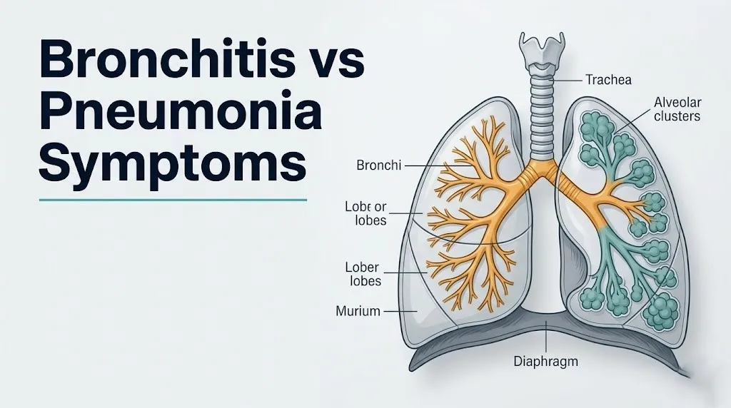
When a pathogen (bacterial, viral, or atypical) overwhelms the alveolar macrophages that patrol the alveolar surface, those macrophages release interleukin-1beta (IL-1beta), interleukin-6 (IL-6), interleukin-8 (IL-8), and tumor necrosis factor-alpha (TNF-alpha). These cytokines recruit neutrophils from pulmonary capillaries into the alveolar space, increase capillary permeability, and trigger protein-rich plasma exudate to flood the alveolar sacs. The alveoli progress through pathological phases: early congestion (capillary engorgement), red hepatization (alveoli packed with red blood cells and fibrin, giving the lung a liver-like consistency), gray hepatization (neutrophil predominance), and finally resolution when the immune system clears the infection.
During active exudate-filling, affected alveoli cannot exchange gas. The blood flowing past them receives no oxygen, creating ventilation-perfusion (V/Q) mismatch: the heart is pumping blood through the lungs, but that blood is passing non-functioning alveoli and returning to circulation still oxygen-poor. This V/Q mismatch produces hypoxemia (low blood oxygen), the physiological mechanism behind the shortness of breath and low pulse oximetry readings that distinguish pneumonia from bronchitis.
The Infectious Diseases Society of America (IDSA) and the American Thoracic Society (ATS), in their 2019 Community-Acquired Pneumonia Clinical Practice Guidelines, identify Streptococcus pneumoniae as the most common bacterial cause of community-acquired pneumonia in adults, followed by Mycoplasma pneumoniae, Haemophilus influenzae, and Legionella pneumophila.
Key Takeaway: Bronchitis inflames the airway tubes (bronchi), producing wheeze and productive cough while gas exchange stays intact; pneumonia infects the air sacs (alveoli), flooding them with exudate and directly impairing oxygen delivery to the blood, which is why pneumonia can drop your oxygen levels in ways bronchitis typically does not.
Symptoms of Bronchitis and Pneumonia That Overlap
Symptoms of bronchitis and pneumonia that overlap exist because both conditions trigger the same bronchial defense mechanisms at the airway level, even though pneumonia’s defining damage occurs deeper in the lung.
The shared symptoms:
- Persistent cough: Both conditions stimulate bronchial cough receptors, either through bronchial mucosal inflammation (bronchitis) or through mucus draining downward from infected alveoli into the bronchi (pneumonia). Cough is present in nearly all bronchitis cases and in approximately 80 to 90 percent of pneumonia cases according to Mayo Clinic clinical data.
- Mucus production: Both can produce sputum, though the character differs (addressed in the next section).
- Chest discomfort: Both conditions produce a sense of chest tightness or heaviness, though through different mechanisms (bronchial inflammation vs. pleural irritation).
- Fatigue and malaise: Both activate the systemic innate immune response. Rising IL-6 from infected tissue travels to the hypothalamus, triggering prostaglandin E2 production through the cyclooxygenase-2 (COX-2) pathway, which produces the fatigue and general malaise that accompany both illnesses.
- Low-grade fever: Both can cause temperature elevation, particularly in the early stages.
- Reduced exercise tolerance: Both conditions make physical exertion feel harder, though the mechanisms differ (airflow limitation in bronchitis vs. reduced oxygen delivery in pneumonia).
The overlap is genuine and physiologically grounded. This is precisely why self-diagnosis is unreliable, and why the features that do differ between the two conditions deserve careful attention.
Pregnant women in their second and third trimester are at elevated risk of having both conditions misclassified, because normal pregnancy physiology produces baseline dyspnea (from diaphragm elevation by the enlarging uterus and increased respiratory drive from progesterone), which can mask the worsening breathlessness that signals pneumonia rather than bronchitis.
Symptoms of Pneumonia vs Bronchitis: The Key Differences
The symptoms of pneumonia vs bronchitis that most reliably distinguish the two conditions in a real-world setting are oxygen saturation, the character of chest pain, the degree of systemic illness, and the findings a clinician hears through a stethoscope.
High fever is more characteristic of pneumonia. A temperature above 38.5°C (101.3°F) alongside a productive cough and chest symptoms should raise clinical suspicion for pneumonia rather than bronchitis, particularly when fever onset is acute (within 24 to 48 hours) rather than gradual.
Pleuritic chest pain is specific to pneumonia. Bronchitis produces a burning or aching central chest discomfort from bronchial inflammation. Pneumonia can produce a sharp, stabbing pain that worsens dramatically with a deep breath or a cough, localized to one side of the chest. This pleuritic pain occurs when the infection reaches or inflames the pleura (the membrane lining the chest wall and lung surface), which contains pain-sensitive nerve fibers.
Oxygen saturation below 94% is a pneumonia finding, not a bronchitis finding. Uncomplicated acute bronchitis in a non-COPD patient does not significantly impair alveolar gas exchange, so pulse oximetry typically reads 96% or above. Any SpO2 at or below 94% at rest suggests alveolar compromise consistent with pneumonia.
The auscultatory difference is the most clinically precise distinguishing feature: bronchitis produces wheeze (a continuous musical sound from air moving through narrowed bronchi) and rhonchi (low-pitched sounds from mucus in the larger airways). Pneumonia produces crackles, also called rales: fine, discontinuous crackling sounds heard in late inspiration as fluid-filled alveoli pop open under inspiratory pressure. Clinicians sometimes describe this sound as similar to rubbing a strand of hair between two fingers near your ear.
Quick Tip:
- Check your temperature: above 38.5°C (101.3°F) with respiratory symptoms is a pneumonia red flag.
- Check your oxygen saturation with a consumer pulse oximeter if available: 94% or below at rest requires same-day professional evaluation.
- Note whether your chest pain worsens with a deep breath: pleuritic pain is not a bronchitis symptom.
- Note the color of your sputum: rust-colored or blood-tinged sputum is characteristic of Streptococcus pneumoniae pneumonia, not bronchitis.
Bronchitis or Pneumonia: How the Cough Differs
The character of the cough is one of the most practically accessible distinguishing features between bronchitis or pneumonia, and it reflects the different anatomical sites of each infection.
In bronchitis, the cough is productive from the earliest days of illness. The excess mucus produced by goblet cell hyperplasia in the bronchial epithelium accumulates in the bronchial lumen and triggers the cough reflex. The sputum in uncomplicated viral bronchitis is initially clear or white, and may turn yellow or green after several days as neutrophils accumulate in the mucus. This color change does not reliably indicate bacterial superinfection, despite the common belief that it does. The AAFP clinical guidelines for acute bronchitis explicitly state that yellow or green sputum color alone should not drive antibiotic prescribing decisions in otherwise healthy adults.
In pneumonia, the cough character is more variable and depends on the causative pathogen. Bacterial pneumonia, especially from Streptococcus pneumoniae, classically produces a productive cough with rust-colored sputum. The rust color results from red blood cells released into the alveolar exudate during the red hepatization phase breaking down into hemosiderin, which gives the sputum its distinctive color. This rust-colored sputum is highly specific for pneumococcal pneumonia and does not occur in bronchitis.
Viral pneumonia (from influenza or SARS-CoV-2, for example) often produces a dry, non-productive cough with more prominent systemic symptoms (high fever, myalgia, headache) than bacterial pneumonia. Atypical pneumonia from Mycoplasma pneumoniae produces a dry, hacking, persistent cough that can be mistaken for bronchitis for days to weeks.
The other cough difference is progression. Bronchitis cough typically peaks in severity around days 3 to 5 and begins improving by days 7 to 10. A cough that starts improving and then worsens, or that is still present and productive beyond two to three weeks, warrants evaluation by a primary care physician to exclude secondary bacterial pneumonia or underlying pulmonary pathology.
Bronchitis vs Pneumonia Fever and Systemic Symptoms
Bronchitis vs pneumonia fever patterns are one of the most practically useful distinguishing features, though the relationship between fever and disease severity is more nuanced than most general health articles acknowledge.
In typical viral acute bronchitis, fever is absent or low-grade, generally below 38.3°C (101°F). The illness feels more like a bad cold with a chest component: fatigue, mild bodyaches, and the dominant symptom of a cough. Systemic symptoms are present but not severe. Most healthy adults with acute bronchitis can maintain their daily activities at a reduced level.
Bacterial pneumonia typically presents with an abrupt-onset, high fever, often 38.5°C to 40°C (101.3°F to 104°F), accompanied by shaking chills (rigors). Rigors occur when the hypothalamus raises its thermostat set point rapidly in response to bacterial pyrogens, triggering involuntary muscle shivering as the body generates heat to reach the new target temperature. This pattern of abrupt fever with rigors is characteristic of pneumococcal pneumonia specifically and is not seen in typical bronchitis.
Systemic symptoms of pneumonia extend beyond the respiratory system. Headache, myalgias, nausea, and confusion (particularly in older adults) can accompany pneumonia because elevated IL-6 and TNF-alpha from the infected lung reach systemic circulation, producing a generalized inflammatory state. When confusion accompanies a respiratory illness, it should be treated as a pneumonia signal requiring same-day medical evaluation regardless of other features.
A critical exception: older adults over age 65 frequently do not mount the expected fever response to pneumonia. This occurs because aging is associated with blunted thermoregulatory response, reduced IL-1 and IL-6 production by macrophages, and impaired hypothalamic febrile response. According to data from the National Heart Lung and Blood Institute (NHLBI), older adults with pneumonia may present with confusion, falls, or functional decline as the primary or only symptom, without fever, without prominent cough, and without obvious respiratory distress. This atypical presentation makes the bronchitis/pneumonia distinction particularly difficult and particularly important in this age group.
Key Takeaway: High fever above 38.5°C (101.3°F) with shaking chills and respiratory symptoms points strongly toward bacterial pneumonia rather than bronchitis, but older adults with pneumonia frequently do not develop fever at all, so confusion or a sudden decline in function in an elderly person with any respiratory symptoms warrants same-day emergency medicine evaluation.
Walking Pneumonia Symptoms vs Bronchitis
Walking pneumonia (most commonly caused by Mycoplasma pneumoniae infection) is the most clinically dangerous mimic of acute bronchitis because it produces a symptom pattern that can convincingly resemble bronchitis for one to three weeks before the true diagnosis becomes apparent.
The term “walking pneumonia” describes what clinicians observe: the patient feels sick but not sick enough to be hospitalized. They are ambulatory, functional, and often attribute their symptoms to a bad chest cold. The distinguishing features are subtle:
| Feature | Walking Pneumonia (Mycoplasma) | Typical Acute Bronchitis |
|---|---|---|
| Onset | Gradual over 1 to 3 weeks | Gradual, typically 3 to 5 days after URI |
| Fever | Low-grade, often below 38.5°C | Low-grade or absent |
| Cough | Dry, persistent, hacking, non-productive | Productive from early in illness |
| Sore throat | Common early symptom | Common early symptom |
| Headache | Prominent | Mild |
| Ear pain | Bullous myringitis in some cases (unique to Mycoplasma) | Absent |
| Chest X-ray | Bilateral patchy infiltrates (may lag behind symptoms) | Normal |
| White blood count | Often normal or only mildly elevated | Normal or mildly elevated |
| Response to antibiotics | Responds to azithromycin, doxycycline (not penicillin) | Does not require antibiotics |
| Duration without treatment | 2 to 4 weeks or longer | 7 to 14 days typically |
The danger of walking pneumonia being mistaken for bronchitis is greatest in school-age children and young adults aged 5 to 40, the populations in whom Mycoplasma pneumoniae most commonly causes infection. According to IDSA/ATS 2019 CAP Guidelines, Mycoplasma pneumoniae accounts for approximately 20 to 30 percent of community-acquired pneumonia cases in ambulatory patients, a proportion that makes it far too common to dismiss as a rare cause.
Clinicians sometimes cannot distinguish walking pneumonia from bronchitis on physical examination alone, because the early chest X-ray in Mycoplasma pneumonia may show minimal findings despite established alveolar involvement, and the stethoscope findings may not yet show the crackles characteristic of more advanced infection. This is the clinical scenario in which serology (Mycoplasma IgM) or a respiratory pathogen panel adds diagnostic certainty.
Bronchitis vs Pneumonia in Children and Older Adults
Bronchitis vs pneumonia in children and older adults is the clinical scenario where standard adult symptom descriptions are the least reliable and where the consequences of misclassification are the most severe.
In children under age 5, the primary clinical indicator of pneumonia is tachypnea (abnormally rapid breathing rate), not cough, fever, or chest pain. The American Academy of Pediatrics (AAP) defines age-specific respiratory rate thresholds for concern:
- Infants under 2 months: respiratory rate above 60 breaths per minute
- Infants 2 to 12 months: respiratory rate above 50 breaths per minute
- Children 1 to 5 years: respiratory rate above 40 breaths per minute
- Children over 5 years: respiratory rate above 30 breaths per minute
Additional signs of severe respiratory illness in children: nasal flaring (widening of the nostrils with each breath, indicating increased respiratory effort), subcostal or intercostal retractions (visible sucking-in of skin at the ribs with each inspiration, indicating the accessory muscles are working hard), and grunting (the child makes an audible grunt with each expiration, using partial vocal cord closure to maintain positive end-expiratory pressure in the alveoli). Any of these signs in a child with respiratory symptoms requires immediate evaluation by a pediatric emergency medicine physician.
In older adults over 65, the presentation of pneumonia is frequently atypical. As noted in the previous section, fever may be absent. The most commonly reported atypical pneumonia presentations in the elderly include new-onset confusion, unexplained falls, sudden inability to perform usual daily activities, and worsening of baseline functional status without obvious respiratory symptoms. Any of these changes in an older adult with any respiratory symptoms at all should prompt same-day evaluation by an emergency medicine physician, not watchful waiting.
Older adults with COPD face a specific additional challenge: they have a baseline of airflow limitation, wheeze, and productive cough from their chronic bronchitis component that can mask the worsening produced by superimposed pneumonia. A COPD patient who notices that their usual cough has become worse, their sputum has changed in character, or their usual rescue inhaler is providing less relief than normal should contact their primary care physician or pulmonologist promptly rather than assuming this is a standard exacerbation.
Bronchitis vs Pneumonia in Immunocompromised Patients
Bronchitis vs pneumonia in immunocompromised patients represents the most challenging clinical distinction because the immune response that produces most of the recognizable symptoms of pneumonia (fever, productive cough, leukocytosis, chest X-ray infiltrates) may be dramatically blunted or absent.
An immunocompromised host is a person whose adaptive or innate immune system is impaired by any of several mechanisms: active HIV infection with CD4 count below 200 cells per microliter, solid organ transplantation with immunosuppressive drug regimens (tacrolimus, mycophenolate, corticosteroids), hematological malignancy with neutropenia (absolute neutrophil count below 500 cells per microliter), high-dose systemic corticosteroid therapy (equivalent to prednisone 20 mg per day or more for more than four weeks), or biologic therapies targeting TNF-alpha or IL-6 for inflammatory conditions.
In these patients, alveolar macrophages and recruited neutrophils cannot mount the normal inflammatory response to alveolar infection. The result is pneumonia without fever in many cases, minimal sputum production (because goblet cell stimulation depends partly on immune mediators), and a chest X-ray that may appear near-normal despite extensive alveolar involvement because the exudate and consolidation that produce radiographic changes depend on a functional inflammatory response.
The opportunistic pathogens that affect immunocompromised patients further complicate the picture. Pneumocystis jirovecii pneumonia (PCP), seen in patients with advanced HIV or on certain immunosuppressive regimens, presents with a gradual onset of dry cough, exertional dyspnea, and low-grade fever that strongly mimics bronchitis for days to weeks before progressing to severe hypoxemia. The chest X-ray in early PCP may be normal; ground-glass bilateral infiltrates appear later. Clinicians use elevated serum LDH (lactate dehydrogenase) and arterial blood gas evidence of hypoxemia disproportionate to clinical appearance as early diagnostic clues.
Any immunocompromised patient with respiratory symptoms of any kind should be evaluated by a primary care physician or infectious disease specialist within 24 hours, with a low threshold for chest X-ray and additional laboratory testing, because the usual symptom thresholds for professional evaluation do not apply to this population.
Key Takeaway: Immunocompromised patients can have pneumonia without fever, without a productive cough, and with a nearly normal chest X-ray, which means the standard symptom comparison between bronchitis and pneumonia does not apply to this population, and any respiratory symptoms in an immunocompromised person require professional evaluation within 24 hours.
How Bronchitis vs Pneumonia Is Diagnosed
Distinguishing bronchitis vs pneumonia definitively requires a physical examination and, in most cases where pneumonia is suspected, imaging, because symptom patterns alone cannot reliably confirm the diagnosis.
The diagnostic workup a primary care physician or emergency medicine physician follows:
- Medical history: duration of symptoms, character and color of sputum, fever pattern and maximum temperature recorded, degree of shortness of breath at rest versus exertion, pleuritic chest pain, any recent sick contacts, vaccination status (influenza vaccine, pneumococcal vaccine), smoking history, and underlying health conditions.
- Vital signs assessment: temperature, heart rate, respiratory rate, blood pressure, and pulse oximetry. These four values alone provide enormous diagnostic information. A respiratory rate above 20 breaths per minute in a resting adult, heart rate above 100, temperature above 38.5°C, or SpO2 at or below 94% all raise pneumonia probability significantly.
- Chest auscultation: the clinician listens with a stethoscope over multiple lung zones bilaterally. Wheeze and rhonchi point toward bronchial disease (bronchitis, asthma, COPD). Crackles (fine, late-inspiratory popping sounds) over a specific lung segment point toward alveolar consolidation. Dullness to percussion over the same area (a hollow sound replaced by a flat sound, like tapping a full box versus an empty one) confirms consolidation in that segment.
- Chest X-ray (posterior-anterior and lateral views): the standard imaging tool for confirming or excluding pneumonia. Bronchitis produces no consolidation on chest X-ray; the X-ray may show mild peribronchial thickening or be entirely normal. Pneumonia produces characteristic consolidation (an area of lung appearing white or opaque on X-ray where air normally provides a dark, air-filled appearance) in a lobar, segmental, or patchy bilateral pattern depending on the causative pathogen.
- Laboratory tests: CBC with differential (leukocytosis with left shift in bacterial pneumonia; lymphocytosis in viral pneumonia), C-reactive protein (CRP, markedly elevated in bacterial pneumonia), procalcitonin (PCT, elevated preferentially in bacterial infections and clinically useful for distinguishing bacterial from viral causes). A 2009 randomized controlled trial published in the New England Journal of Medicine (the ProHOSP trial) demonstrated that procalcitonin-guided antibiotic therapy reduced antibiotic exposure without worsening outcomes in lower respiratory tract infections.
- Severity scoring in confirmed pneumonia: the CURB-65 score (Confusion, Urea above 7 mmol/L, Respiratory rate above 30, Blood pressure below 90/60, age 65 or older) and the Pneumonia Severity Index (PSI/PORT score) both guide hospitalization decisions. A CURB-65 score of 0 to 1 supports outpatient management; a score of 2 warrants consideration of hospital admission; a score of 3 or above warrants hospital admission.
Bronchitis Symptoms vs Pneumonia on Chest X-Ray
The chest X-ray is the most objective single test for separating bronchitis symptoms vs pneumonia, because the anatomical difference between the two conditions produces completely different radiographic findings.
In acute bronchitis, the chest X-ray is normal in the vast majority of cases. Occasionally, mild peribronchial thickening is visible, appearing as slight haziness around the bronchi, but the lung parenchyma (the alveolar tissue) appears dark and normally air-filled. There is no consolidation, no opacity, and no infiltrate. This is the expected and reassuring finding.
In pneumonia, the chest X-ray shows opacification of the affected lung area. Exudate-filled alveoli absorb X-rays instead of allowing them to pass through, producing a white area on the image where normal lung appears dark. The pattern of opacification depends on the pathogen:
- Lobar consolidation: entire lobe appears white and airless; characteristic of Streptococcus pneumoniae (pneumococcal pneumonia); the entire right lower lobe, for example, may appear consolidated with the lobar boundary sharply demarcated
- Segmental consolidation: one or more segments within a lobe; seen in various bacterial pneumonias
- Bilateral patchy infiltrates: scattered ground-glass or fluffy opacities in both lungs; characteristic of viral pneumonia (influenza, SARS-CoV-2) or atypical pneumonia (Mycoplasma, Legionella)
- Pleural effusion: fluid in the pleural space appearing as blunting of the costophrenic angle; can accompany bacterial pneumonia
The important clinical caveat: chest X-ray findings can lag 12 to 24 hours behind the actual development of pneumonia, particularly in patients who are dehydrated (reduced exudate formation temporarily). This is called an “early” or “dry” pneumonia presentation. A patient with clinical findings strongly suggesting pneumonia (high fever, pleuritic pain, crackles on auscultation) may have a near-normal initial X-ray. In this scenario, clinical judgment, laboratory results, and repeat imaging in 24 hours guide management rather than the initial normal X-ray being falsely reassuring.
High-resolution CT of the chest detects alveolar consolidation and interstitial infiltrates that a standard chest X-ray may miss in early or mild pneumonia, and is ordered when clinical suspicion is high despite a normal or equivocal X-ray finding.
Can Bronchitis Turn Into Pneumonia
Bronchitis can progress to pneumonia in specific circumstances, though in otherwise healthy adults this progression is not the expected clinical course.
The mechanism by which bronchitis transitions to pneumonia involves the relationship between bronchial defense impairment and bacterial colonization of the lower respiratory tract. When viral bronchitis disrupts the mucociliary escalator through ciliostasis and goblet cell hyperplasia, the mechanical barrier that normally prevents bacteria from reaching the alveoli is weakened. Bacteria that normally colonize the upper respiratory tract (nasopharynx) without causing disease, including Streptococcus pneumoniae and Haemophilus influenzae, can then descend into the bronchi and, in susceptible individuals, reach the alveoli.
Secondary bacterial pneumonia following viral bronchitis is a well-documented clinical pattern. It is the mechanism behind the deadly pneumonia waves that followed the 1918 influenza pandemic and the elevated bacterial pneumonia burden seen during seasonal influenza epidemics. The CDC has documented that secondary bacterial pneumonia is the leading cause of death in severe influenza, with Staphylococcus aureus (including methicillin-resistant strains) and Streptococcus pneumoniae being the most frequent implicated pathogens.
The clinical signal that bronchitis is progressing to pneumonia is a characteristic pattern sometimes called the “biphasic illness”: the patient begins improving from their initial bronchitis symptoms around days 5 to 7, then experiences a sudden worsening of fever, a return of or increase in productive cough with purulent sputum, increasing shortness of breath, and new pleuritic chest pain. This biphasic pattern with secondary clinical deterioration after apparent initial improvement should prompt immediate evaluation by a primary care physician or emergency medicine physician that day.
The populations at highest risk for this progression include adults over 65, people with COPD, people with diabetes mellitus, people who are immunocompromised, and anyone who has not been vaccinated against influenza or pneumococcus, according to CDC and IDSA risk stratification data.
Bronchitis vs Pneumonia Treatment Differences
The treatment difference between bronchitis vs pneumonia is clinically meaningful and directly affected by the diagnostic distinction, because the most commonly recommended treatment for one condition (antibiotics) is not indicated for the other in most cases.
Acute bronchitis treatment in healthy adults:
Acute viral bronchitis in a healthy, non-elderly adult does not benefit from antibiotic therapy. The AAFP clinical practice guidelines state clearly that antibiotics should not be prescribed for acute bronchitis in otherwise healthy adults. The evidence base for this recommendation includes multiple randomized controlled trials showing no clinically meaningful reduction in duration or severity of acute bronchitis symptoms from antibiotic treatment, while antibiotic use contributes to antimicrobial resistance and carries risks of adverse effects including diarrhea, allergic reactions, and Clostridioides difficile infection. Supportive care (adequate hydration, rest, honey for cough suppression in adults, acetaminophen or ibuprofen for fever and discomfort) represents appropriate management for uncomplicated viral bronchitis.
Pneumonia treatment depends on the causative pathogen:
- Bacterial community-acquired pneumonia (most commonly Streptococcus pneumoniae): treated with antibiotic therapy. The specific agent is selected based on severity, patient risk factors, and local resistance patterns, and is chosen by a physician based on clinical assessment. Antibiotic therapy for bacterial pneumonia is not optional; untreated bacterial pneumonia carries substantial mortality risk.
- Viral pneumonia (influenza): antiviral therapy with oseltamivir is most effective when started within 48 hours of symptom onset in high-risk patients; supportive care for uncomplicated cases in otherwise healthy adults. Antibiotics are not effective for viral pneumonia.
- Atypical pneumonia (Mycoplasma, Legionella, Chlamydophila): requires specific antibiotic classes that penetrate intracellular compartments. Standard beta-lactam antibiotics (penicillin, amoxicillin) do not cover Mycoplasma or Legionella; treatment requires macrolides (azithromycin), doxycycline, or respiratory fluoroquinolones selected by a physician.
- Pneumocystis jirovecii pneumonia (PCP) in immunocompromised patients: requires trimethoprim-sulfamethoxazole, not standard pneumonia antibiotics.
This complexity is exactly why “do I need antibiotics for my chest illness?” is not a question that can be answered without a clinical diagnosis. Pressing a physician for antibiotics for bronchitis contributes to resistance without benefiting the patient. Avoiding antibiotic treatment for bacterial pneumonia out of a general preference to avoid medication can be genuinely dangerous.
Key Takeaway: Antibiotics are not indicated for acute viral bronchitis in healthy adults and will not help you recover faster, but bacterial pneumonia requires antibiotic therapy selected by a physician based on your specific clinical presentation, which is precisely why the diagnostic distinction between these two conditions matters clinically, not just academically.
Emergency Symptoms: When to Call 911 or Go to the ER
Certain symptoms associated with bronchitis and pneumonia require immediate emergency evaluation. Do not wait to see if these resolve on their own.
Call 911 or go to the nearest emergency room immediately if you or someone you are caring for experiences:
- Oxygen saturation (SpO2) at or below 92% on a pulse oximeter at rest (indicates severe hypoxemia requiring supplemental oxygen and immediate clinical assessment for respiratory failure)
- Respiratory rate above 30 breaths per minute at rest in an adult (indicates severe respiratory distress and impending respiratory failure)
- Blue-tinged lips, tongue, or fingertips (central cyanosis, indicating critically low blood oxygen)
- Confusion, disorientation, altered mental status, or new inability to recognize people or follow simple instructions in a person with respiratory symptoms (indicates hypoxemia or sepsis affecting brain perfusion)
- Inability to speak a full sentence without stopping to breathe
- Severe chest pain with difficulty breathing and hemodynamic instability (lightheadedness, rapid or weak pulse, pallor, sweating)
- A child with nasal flaring, subcostal or intercostal retractions, grunting with each breath, or any of the age-specific tachypnea thresholds described earlier in this article
- Any sudden worsening of respiratory symptoms in a person with confirmed pneumonia who was being managed at home (may indicate complications including pleural empyema, lung abscess, or sepsis)
- An older adult over 65 with new confusion and any respiratory symptoms, even without high fever (atypical pneumonia presentation that is a medical emergency)
These presentations can indicate severe pneumonia with impending respiratory failure, septic shock, or hypoxemia-induced end-organ compromise, and they require emergency medical assessment at a hospital emergency department, not an urgent care clinic or a next-day appointment.
Pneumonia vs Bronchitis Symptoms: How to Decide What to Do Next
When you’re trying to decide whether your symptoms represent pneumonia vs bronchitis, the practical decision framework comes down to three measurable factors and one behavioral pattern: temperature, oxygen saturation, the character of chest pain, and what your symptoms are doing over time.
Use this framework to guide your next step:
| Your Situation | Most Likely Action |
|---|---|
| Cough, mild fatigue, clear or yellow sputum, temperature below 38.3°C, SpO2 96%+ , symptoms day 1 to 7 | Monitor at home with supportive care; contact primary care if not improving by day 10 |
| Cough, temperature 38.3°C to 38.5°C, some shortness of breath on exertion but not at rest, SpO2 94% to 95% | Contact primary care physician or urgent care the same day for evaluation |
| Cough, temperature above 38.5°C (101.3°F), pleuritic chest pain, or SpO2 at or below 94% at rest | Evaluate at urgent care or emergency department today; do not drive yourself if short of breath |
| Any of the emergency symptoms listed in the section above | Call 911 or go to the nearest emergency room immediately |
| Older adult (65+) with new confusion, functional decline, or falls alongside any respiratory symptoms | Go to the emergency room immediately, even without fever |
| Child with nasal flaring, grunting, retractions, or age-specific tachypnea | Go to the pediatric emergency department immediately |
| Immunocompromised patient with any new respiratory symptoms | Contact primary care or infectious disease physician within 24 hours |
The single thing this article cannot replace is a physical examination and, where indicated, a chest X-ray. The stethoscope finding of crackles over a specific lung segment, combined with a fever and an area of opacification on chest X-ray, is how bronchitis is ruled out and pneumonia is confirmed. That information requires a clinician.
What this framework gives you is the ability to recognize when your symptom pattern is outside the safe home-monitoring range and when the decision to wait and see could be the wrong one.
Frequently Asked Questions About Bronchitis vs Pneumonia Symptoms
How do I know if I have bronchitis or pneumonia?
You cannot definitively distinguish bronchitis from pneumonia based on symptoms alone; a physical examination and often a chest X-ray are required for a reliable answer.
The clinical features most suggestive of pneumonia rather than bronchitis are high fever above 38.5°C (101.3°F) with shaking chills, oxygen saturation at or below 94%, pleuritic chest pain that worsens with deep breathing, crackles heard through a stethoscope, and consolidation visible on a chest X-ray.
If you have two or more of those features, evaluation by a primary care physician or urgent care clinician the same day is the appropriate next step, not continued home monitoring.
Can you have pneumonia without a fever?
Yes, you can have pneumonia without a fever, and this is particularly common in older adults over 65, immunocompromised individuals, and people on corticosteroid therapy.
In older adults, aging blunts the hypothalamic febrile response through reduced macrophage IL-1 and IL-6 production, meaning the body may not raise its temperature even in response to significant alveolar infection.
Any older adult with new confusion, a sudden decline in functional status, or worsening respiratory symptoms should be evaluated for pneumonia by an emergency medicine physician regardless of whether a fever is present.
Does bronchitis need antibiotics?
Acute bronchitis in otherwise healthy adults does not require antibiotics in more than 90 percent of cases because it is caused by viruses, which do not respond to antibiotic treatment.
The American Academy of Family Physicians clinical guidelines explicitly state that antibiotics should not be prescribed for acute bronchitis in healthy adults, based on multiple randomized controlled trials showing no meaningful benefit in symptom duration or severity.
Antibiotic treatment is appropriate for bacterial causes of bronchitis-like illness, specifically Bordetella pertussis (whooping cough) and Mycoplasma pneumoniae walking pneumonia, both of which require clinical evaluation and specific laboratory testing to identify.
What does pneumonia feel like compared to bronchitis?
Pneumonia typically feels more severe than bronchitis, with a higher fever, more pronounced fatigue, and shortness of breath that may be present even at rest rather than only with exertion.
The chest discomfort in pneumonia is often sharp and localized, worsening dramatically with a deep breath (pleuritic pain), while bronchitis produces a more diffuse burning or aching central chest discomfort from bronchial inflammation.
Many patients with pneumonia describe feeling genuinely unwell in a systemic way (unable to get out of bed, loss of appetite, confusion in older adults) rather than the more localized respiratory misery typical of bronchitis.
Can bronchitis become pneumonia if untreated?
Bronchitis can progress to pneumonia in susceptible individuals when viral-induced impairment of the mucociliary escalator allows bacteria that normally colonize the upper respiratory tract to descend into the alveoli.
The clinical signal is a biphasic illness pattern: initial improvement after days 5 to 7 followed by a sudden return of high fever, increased purulent sputum, worsening shortness of breath, and new pleuritic chest pain.
Anyone experiencing this secondary deterioration pattern should be evaluated by a primary care physician or emergency medicine physician the same day, because secondary bacterial pneumonia following viral bronchitis requires antibiotic treatment.
When should I go to the ER for bronchitis or pneumonia symptoms?
Go to the nearest emergency room immediately if you have oxygen saturation at or below 92%, respiratory rate above 30 breaths per minute at rest, blue-tinged lips or fingertips, confusion, or inability to complete a sentence without stopping to breathe.
Older adults over 65 with any new confusion alongside respiratory symptoms should go to the emergency room immediately even without fever, because pneumonia in this age group frequently presents without the expected high temperature.
Children with nasal flaring, grunting, visible rib retractions with each breath, or respiratory rates above the age-specific thresholds described in the AAP guidelines require immediate evaluation at a pediatric emergency department, not urgent care.
Closing
The distinction between bronchitis and pneumonia is one of the most practically consequential clinical questions in everyday medicine. The features that reliably separate them, temperature, oxygen saturation, the character of chest pain, the lung sounds a clinician hears, and the chest X-ray finding, are accessible through a straightforward medical evaluation.
The framework in this article gives you the clinical vocabulary to recognize when your symptom pattern demands professional assessment today versus when you can reasonably monitor at home. An SpO2 at or below 94%, a fever above 38.5°C with pleuritic chest pain, a child with retractions, or an older adult with new confusion: these are not situations for watchful waiting.
You now know what each symptom means anatomically and why the distinction matters. The next step is straightforward: if your pattern suggests pneumonia, contact a primary care physician or urgent care clinician today. If it suggests one of the emergency scenarios, go to the nearest emergency room immediately.