Erysipelas Symptoms: Causes, Red Flags, and 2026 Care
Erysipelas symptoms include a rapidly spreading, intensely red, warm, swollen skin rash with a sharply raised and well-defined advancing border, accompanied by high fever, chills, and significant pain at the affected site. This is a bacterial infection of the upper layer of the skin that requires same-day medical evaluation: it does not resolve without antibiotic treatment, and in untreated patients it can progress to bacteremia and life-threatening sepsis within days.
According to the Infectious Diseases Society of America (IDSA), erysipelas is a distinct clinical entity from cellulitis, caused primarily by Group A Streptococcus (Streptococcus pyogenes), with the skin of the lower leg as the most common site in adults. The Centers for Disease Control and Prevention reports that Group A Streptococcus causes millions of infections annually in the United States, with skin and soft tissue infections representing a major category.
This article covers each symptom with the anatomical and physiological mechanism behind it, explains how erysipelas differs from cellulitis at a structural level, addresses the specific clinical variants including facial and bullous erysipelas, and provides clear emergency thresholds so you know exactly when to call 911.
Erysipelas Symptoms: What This Infection Looks and Feels Like
Erysipelas symptoms are a rapidly spreading, fiery-red skin rash with a sharply raised, advancing border, intense local pain and warmth, swelling, and systemic symptoms including fever, rigors, and malaise, typically developing within 24 to 48 hours of infection onset.
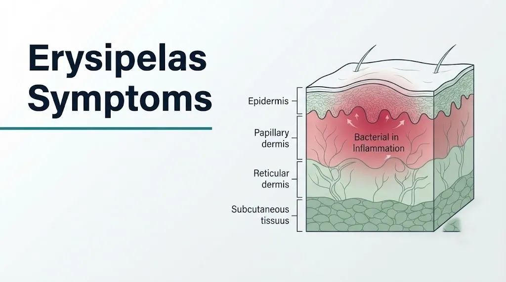
The core symptom cluster of erysipelas reflects infection of the papillary dermis (the upper layer of the dermis) and the superficial lymphatic channels running within it. Bacterial invasion of this specific tissue plane triggers an intense local inflammatory response that produces the visible hallmark signs: brilliant erythema from vasodilation, edema from increased vascular permeability, heat from increased blood flow, and pain from cytokine-mediated nociceptor sensitization.
The defining physical feature that sets erysipelas apart from all other skin infections is the raised, palpable, sharply demarcated border. You can feel it with your fingertip: a definite step up from normal skin to the infected skin edge. This raised border reflects the specific anatomy of the infection, confined within the tissue plane of the papillary dermis, where the boundary with the underlying reticular dermis creates a measurable physical border at the advancing infection edge.
| Erysipelas Symptom | Body Location | What It Feels Like |
|---|---|---|
| Bright red erythema | Affected skin area | Intensely red, orange-red, or crimson |
| Raised advancing border | Edge of erythema | Palpably elevated above surrounding skin |
| Warmth | Entire affected area | Hot to touch, noticeably warmer than adjacent skin |
| Edema (swelling) | Affected area and surrounding tissue | Tight, tense swelling |
| Pain and tenderness | Affected area | Throbbing, burning, or aching; painful to touch |
| Fever | Systemic | 38.5 to 40°C (101.3 to 104°F) |
| Rigors and chills | Systemic | Severe shaking chills preceding or coinciding with fever |
| Malaise and fatigue | Systemic | Profound illness feeling |
Systemic symptoms often begin before the skin changes are fully developed, which is a clinically important feature. A patient may feel intensely unwell with fever and chills for 12 to 24 hours before the skin rash reaches its full visible extent.
What Is Erysipelas and How Does It Develop
Erysipelas is a superficial bacterial skin infection, coded in the International Classification of Diseases as ICD-10: A46, caused primarily by Group A Streptococcus (Streptococcus pyogenes) and characterized by infection of the epidermis and papillary dermis with involvement of the superficial lymphatic channels.
The historical name for erysipelas is “St. Anthony’s fire,” a term that arose from medieval descriptions of the condition’s spreading, flame-like red border and the intense burning pain it produces. The name persisted because the visual impression of spreading fire along the skin surface remains one of the most accurate plain-language descriptions of how erysipelas behaves in its acute phase.
Development follows a predictable sequence. Group A Streptococcus enters through a break in the skin barrier: a small cut, insect bite, skin fissure from tinea pedis (athlete’s foot), intertrigo in a skin fold, a surgical wound, or a venipuncture site. Once inside the papillary dermis, the bacteria deploy specific virulence factors that allow them to spread rapidly and evade immune defenses. The streptococcal M protein on the bacterial surface binds human factor H and blocks deposition of complement factor C3b, effectively making the bacteria invisible to the neutrophils that would normally engulf them. Without effective phagocytosis, the bacteria multiply and spread laterally through the dermis uncontrolled.
Streptokinase then dissolves fibrin barriers by converting plasminogen to plasmin. Hyaluronidase degrades hyaluronic acid in the extracellular matrix. Together, these enzymes clear the biological scaffolding that would otherwise slow lateral spread, allowing the infection front to advance centimeters per hour in severe cases.
The primary causative agent in more than 80 percent of erysipelas cases is Group A Streptococcus, according to microbiological studies cited in the Journal of the American Academy of Dermatology. In a smaller subset, Group C and Group G Streptococcus serve as causative agents. Staphylococcus aureus, including methicillin-resistant Staphylococcus aureus (MRSA), rarely causes classic erysipelas but becomes more relevant in patients with prior antibiotic exposure or healthcare-associated risk factors.
Erysipelas Early Symptoms and Warning Signs
The earliest erysipelas symptoms typically appear as a prodromal phase of fever, chills, malaise, and generalized illness that precedes the visible skin rash by 12 to 24 hours, sometimes misleading patients into thinking they have influenza or another systemic viral illness.
The prodromal systemic symptoms are produced by the early cytokine release from dermal macrophages that have encountered streptococcal antigens. Even before the skin rash becomes visible, interleukin-1 beta (IL-1beta) released from activated macrophages acts on the hypothalamus to trigger prostaglandin E2 synthesis, raising the body’s temperature set point and producing fever. Tumor necrosis factor-alpha (TNF-alpha) simultaneously drives malaise and fatigue at the level of the central nervous system.
Early warning signs specific to erysipelas that distinguish it from a viral prodrome:
- Fever that is sudden in onset and high (typically above 38.5°C / 101.3°F) rather than the gradually rising lower-grade fever of most viral illnesses
- Localized skin tenderness or burning that precedes visible redness, concentrated in the eventual site of the rash
- The redness, when it appears, spreads visibly over hours rather than remaining stable
- The skin becomes warm and then hot to the touch at the affected site even before the full redness develops
- A feeling of skin tightness at the affected site, produced by the early edema forming in the papillary dermis as vascular permeability increases
Patients on corticosteroids or immunosuppressive medications may not develop a high fever despite active erysipelas, because these medications blunt the IL-1beta and TNF-alpha response. In this population, local skin symptoms (spreading redness, warmth, pain) may be the primary early warning signs in the absence of systemic symptoms.
Quick Tip:
- Take your temperature at the first sign of skin redness or warmth in a limb, as fever confirms a systemic infectious process and changes the urgency of evaluation
- Photograph the skin finding and draw a pen line around the visible border of the redness, noting the time; if the redness visibly advances beyond that line within 2 hours, this is rapid spread warranting emergency room evaluation
- Elderly patients may present with confusion or altered mental status as the first sign of erysipelas rather than fever, because their thermoregulatory response is blunted with age
Key Takeaway: Erysipelas begins with systemic fever and chills before the skin rash fully develops, a prodromal pattern that can be mistaken for influenza but is specifically caused by early dermal macrophage IL-1beta and TNF-alpha release from streptococcal antigen recognition.
Erysipelas Skin Symptoms: Redness, Swelling, and the Raised Border
The defining skin symptom of erysipelas is a sharply raised, palpably elevated advancing border surrounding intensely red, edematous, painful skin, an appearance produced by the precise anatomical confinement of the infection to the papillary dermis and superficial lymphatic channels.
Think of the skin’s layers as a layered sandwich. The epidermis is the outermost bread layer. Directly beneath it is the papillary dermis, a thin layer of loose connective tissue riddled with superficial lymphatic capillaries and blood vessel loops. Below that is the reticular dermis, a denser connective tissue layer. The papillary dermis sits on top of the reticular dermis the way a pillow sits on a mattress: with a definable surface boundary between them. When erysipelas infects the papillary dermis, it spreads within that specific layer but cannot easily cross into the reticular dermis below, which has different connective tissue density and different tissue plane characteristics. The result is an infection that spreads laterally and remains in a defined anatomical layer, producing the physically raised advancing edge that is erysipelas’s clinical fingerprint.
The erythema (redness) of erysipelas is characteristically brilliant and uniform within the infected area. It ranges from orange-red to deep crimson and blanches with finger pressure (a blanching erythema, indicating vasodilation rather than hemorrhage into tissue). As the infection advances, the central area of the rash may become less intensely red as acute inflammation shifts to subacute, while the active advancing border remains the brightest and most painful zone.
Edema in erysipelas is produced by two mechanisms: vasodilation increasing blood flow to the area (bringing fluid into the tissue), and increased vascular permeability from IL-6-driven endothelial changes that allow plasma proteins to leak from capillaries into the interstitium. This protein-rich edema produces the characteristic tight, shiny, orange-peel textured surface called peau d’orange, visible in approximately 30 to 40 percent of erysipelas presentations, particularly on the lower leg.
Patients with pre-existing pitting edema from chronic venous insufficiency may find that the erysipelas swelling is harder to visually distinguish from their baseline leg swelling. In this population, the presence of new, escalating warmth and pain localized to one area of the leg, combined with fever, is a more reliable indicator of active erysipelas than the swelling alone.
Erysipelas Fever and Systemic Symptoms
Erysipelas fever is typically abrupt in onset, high-grade (38.5 to 40°C / 101.3 to 104°F), and accompanied by rigors (severe shaking chills), reflecting the systemic release of pro-inflammatory cytokines from dermal macrophage activation by streptococcal antigens.
The systemic component of erysipelas is not simply a side effect of having a skin infection. It is a direct consequence of the inflammatory signaling pathways activated by Group A Streptococcus virulence factors. Streptococcal superantigens, proteins produced by the bacteria, stimulate large numbers of T lymphocytes non-specifically by binding outside the normal antigen-binding site of the T cell receptor. This non-specific T cell activation triggers a massive cytokine release that affects the hypothalamus, liver, bone marrow, and cardiovascular system simultaneously, producing fever, acute phase protein synthesis (CRP, ESR elevation), leukocytosis, and in severe cases, cardiovascular compromise.
According to the IDSA skin and soft tissue infection guidelines (Stevens et al., Clinical Infectious Diseases, 2014), systemic symptoms in erysipelas including fever above 38°C, heart rate above 90 beats per minute, respiratory rate above 24 breaths per minute, or white blood cell count above 12,000 cells per microliter meet criteria for systemic inflammatory response syndrome (SIRS) and represent a clinical threshold requiring hospital-level evaluation.
| Systemic Symptom | Mechanism | Clinical Significance |
|---|---|---|
| Fever 38.5 to 40°C | IL-1beta, prostaglandin E2 acting on hypothalamus | Confirms bacterial infection; above 40°C is a hospital-level sign |
| Rigors and shaking chills | Hypothalamic temperature reset with muscular thermogenesis | Indicates rapid fever rise; common at erysipelas onset |
| Tachycardia above 90 bpm | TNF-alpha cardiovascular effects, fever-driven | Meets SIRS criteria; warrants same-day emergency evaluation |
| Malaise and fatigue | Central nervous system cytokine effects | Present in virtually all active erysipelas cases |
| Nausea and vomiting | Systemic cytokine response | Less common; indicates more severe systemic involvement |
| Altered mental status | Sepsis-related cerebral dysfunction | Emergency: indicates possible sepsis or bacteremia |
Patients with diabetes mellitus may have blunted fever responses due to autonomic neuropathy affecting thermoregulation. A diabetic patient with a spreading leg infection and a temperature of only 37.8°C (100°F) may have the same severity of underlying infection as a non-diabetic patient with 39.5°C (103.1°F), because their fever response is physiologically limited. Physicians evaluating diabetic patients with suspected erysipelas use the skin presentation and laboratory markers rather than temperature alone to assess severity.
Erysipelas on the Leg: Symptoms and Presentation
Erysipelas on the leg is the most common presentation in adults, accounting for approximately 70 to 80 percent of all erysipelas cases according to epidemiological data published in the British Journal of Dermatology, and it most often begins at the lower leg, typically the shin or the area around the ankle.
The lower leg is preferentially affected because it is the most common site for the portal-of-entry skin breaks that allow streptococcal bacteria in. Tinea pedis (athlete’s foot) is a particularly important predisposing factor: the chronic skin maceration and fissuring between the toes created by tinea pedis infection provides an entry point that streptococcal bacteria exploit repeatedly. In patients with recurrent erysipelas of the lower leg, identifying and treating underlying tinea pedis is an essential part of preventing the next episode.
The presentation on the leg characteristically includes:
- Redness that begins at the lower leg and advances proximally toward the knee, with the raised border marking the advancing edge
- Edema that gives the leg a swollen, heavy, tense quality; the peau d’orange texture is frequently visible on the tibial skin
- Severe pain and tenderness on palpation of the entire affected area
- In more advanced presentations, streaks of red along the lymphatic channels of the medial thigh (lymphangitis), indicating that the infection has extended beyond the primary site into the lymphatic drainage vessels
- Enlarged, tender inguinal lymph nodes (inguinal lymphadenitis) reflecting the regional lymph node response to lymphatic bacterial load
- Occasionally, a clearly visible entry wound at the ankle or between the toes, though the entry site is frequently not visible by the time the patient seeks care
Patients who are obese are at elevated risk for lower leg erysipelas because adipose tissue in the lower extremities impairs lymphatic drainage, creating a permanently compromised local immune environment.
Key Takeaway: Erysipelas on the leg most commonly enters through tinea pedis-related skin fissures between the toes, and identifying and treating underlying tinea pedis is an essential step in preventing recurrent leg erysipelas.
Erysipelas on the Face: Symptoms and Specific Risks
Facial erysipelas symptoms include a butterfly-pattern rash across both cheeks and the nose bridge, similar in appearance to lupus erythematosus, with the same sharply raised border, intense redness, edema, and severe pain characteristic of erysipelas elsewhere, but with specific additional risks that make it a more urgent clinical presentation.
The face accounts for approximately 20 percent of erysipelas cases, with the remaining cases distributed across the lower extremities and other body sites. Facial erysipelas most commonly enters through nasal passages, the inner canthus of the eye, or small breaks in the facial skin. The butterfly pattern across both cheeks and the nose occurs because the bacteria spread through the facial lymphatic channels, which drain bilaterally across this anatomical zone.
The specific additional risks of facial erysipelas relate to the anatomy of the facial venous drainage. The facial veins communicate directly with the cavernous sinus, a large venous channel at the base of the brain, via the ophthalmic veins. Septic thrombophlebitis (infected clot formation) in the facial veins can propagate through this communication to the cavernous sinus, producing cavernous sinus thrombosis: a life-threatening intracranial complication with a mortality rate that clinical case series have reported in the range of 20 to 30 percent even with modern treatment.
Warning signs specific to facial erysipelas that indicate possible cavernous sinus thrombosis:
- Sudden severe headache that is new or “worst ever”
- Visual changes including double vision (diplopia) or reduced visual acuity
- Proptosis (protrusion of the eyeball from the orbit)
- Altered mental status or confusion
- High fever with rapid clinical deterioration despite treatment
Any facial erysipelas patient who develops these additional symptoms requires immediate emergency room evaluation without waiting for an outpatient appointment.
Children and infants are disproportionately affected by facial erysipelas compared to adults, and in neonates, Group B Streptococcus rather than Group A Streptococcus may be the causative organism, a distinction that affects antibiotic selection and warrants pediatric emergency evaluation for any neonate with facial erysipelas signs.
Bullous Erysipelas Symptoms
Bullous erysipelas is a variant of erysipelas in which fluid-filled blisters (bullae) form within the area of infection, produced by bacterial toxin-mediated destruction of the connection between the epidermis and the papillary dermis, and it carries a more serious prognosis than non-bullous erysipelas.
The bullae in bullous erysipelas form when streptolysin and other streptococcal exotoxins damage the dermal-epidermal junction, causing the epidermis to separate from the underlying dermis and fill with serous (clear), seropurulent (cloudy), or hemorrhagic (blood-tinged) fluid. Clear fluid bullae indicate earlier-stage blistering with less tissue destruction. Cloudy or hemorrhagic bullae suggest more advanced local tissue damage and a higher risk of progression to necrotizing fasciitis.
According to clinical data published in the Journal of the American Academy of Dermatology, bullous erysipelas is associated with a higher rate of hospitalization, longer treatment duration, and a greater risk of secondary complications compared to non-bullous presentations. Patients with bullous erysipelas are more likely to develop bacteremia and require intravenous antibiotic therapy rather than oral outpatient management.
The bullae can rupture spontaneously, leaving raw, denuded skin that is susceptible to secondary bacterial colonization. The surrounding skin retains the classic erysipelas characteristics: raised advancing border, intense erythema, edema, and warmth.
Patients with pre-existing skin conditions including pemphigus or bullous pemphigoid may have bullous skin changes that are unrelated to infection, creating a potential diagnostic challenge. The combination of fever, rapidly spreading erythema, and new bullae onset within 24 to 48 hours strongly favors erysipelas over a purely dermatological blistering disorder, but laboratory testing and possibly skin biopsy may be needed to confirm the diagnosis in these patients.
Erysipelas vs. Cellulitis: How to Tell Them Apart
Erysipelas and cellulitis are both bacterial skin infections, but erysipelas affects the upper dermis and superficial lymphatics while cellulitis affects the deeper dermis and subcutaneous tissue, and this anatomical difference produces the single most reliable clinical distinguishing feature: erysipelas has a sharply raised, palpable advancing border and cellulitis does not.
Erysipelas stays in the papillary dermis because of the tissue plane boundary between the papillary and reticular dermis. Cellulitis infiltrates the reticular dermis and hypodermis, where no sharp tissue plane boundary exists. The result is that cellulitis spreads diffusely through tissue without producing a raised border; the edge of the redness simply fades gradually into normal skin rather than stepping up abruptly.
| Feature | Erysipelas | Cellulitis |
|---|---|---|
| Skin layer infected | Papillary dermis and superficial lymphatics | Reticular dermis and subcutaneous tissue |
| Border character | Sharply raised, palpably elevated, well-defined | Flat, indistinct, gradually fading |
| Color | Brilliant, uniform orange-red to crimson | Red but less vivid, may be blotchy |
| Common location | Lower leg and face | Lower leg primarily |
| Onset | Very rapid: rash develops fully in 24-48 hours | Can be more gradual |
| Common pathogen | Group A Streptococcus primarily | Streptococcus and Staphylococcus aureus |
| Systemic symptoms | Typically prominent: high fever, rigors | Variable: may be mild or absent |
| Bullae formation | Can occur (bullous erysipelas variant) | Rare |
| Lymphangitis | Common | Less common |
Distinguishing erysipelas from cellulitis matters because the organisms involved differ slightly, the depth of infection affects the choice of antibiotic, and the speed of progression differs. Even for experienced clinicians, the distinction can be difficult in atypical presentations, and the IDSA guidelines acknowledge that clinical differentiation between erysipelas and cellulitis is not always possible on examination alone.
Key Takeaway: The single most reliable physical sign distinguishing erysipelas from cellulitis is the sharply raised, palpably elevated border of erysipelas, produced by the infection’s confinement to the papillary dermis tissue plane, a feature that is absent in cellulitis.
Erysipelas Causes and Risk Factors
Erysipelas is caused by Group A Streptococcus bacteria entering through a break in the skin, and the risk factors that make a person susceptible are any condition that disrupts the skin barrier, impairs local lymphatic function, or weakens the neutrophil-mediated immune response that would otherwise contain bacterial spread.
The CDC surveillance data on Group A Streptococcus documents thousands of invasive cases annually in the United States, with non-invasive skin infections including erysipelas occurring at far higher rates. The bacteria’s ability to exploit any skin entry point makes prevention of skin breaks the primary risk reduction strategy.
Risk factors for developing erysipelas:
- Tinea pedis (athlete’s foot): chronic skin fissuring between toes provides a reliable entry point; present in a large proportion of lower leg erysipelas cases
- Insect bites, cuts, abrasions, and scratches: any break in skin integrity, regardless of how minor
- Chronic venous insufficiency: impaired venous return produces lower leg edema, stretching the skin barrier and impairing local immune surveillance
- Lymphedema: disrupted lymphatic drainage creates a protein-rich fluid accumulation that supports bacterial growth and impairs local immune response
- Obesity: skin folds retain moisture and create friction-related skin breakdown; adipose tissue impairs lymphatic function
- Diabetes mellitus: peripheral neuropathy impairs sensation (masking minor injuries), and hyperglycemia impairs neutrophil function and kills microbial pathogens less effectively
- Prior erysipelas episodes: each episode causes additional lymphatic damage, increasing the risk of the next episode
- Immunosuppressive medications: corticosteroids, biologics, and chemotherapy agents impair neutrophil recruitment and phagocytic capacity
- Alcohol use disorder: associated with impaired neutrophil function and increased skin barrier breakdown
- Surgical wounds and venous catheter sites: disrupted skin integrity in a healthcare setting
Erysipelas in Elderly Patients and Immunocompromised Individuals
Erysipelas in elderly patients and immunocompromised individuals presents with atypical features including blunted fever, milder early skin signs, and a higher rate of rapid deterioration to severe infection, making clinical recognition more challenging and the threshold for aggressive treatment lower than in younger, immunocompetent adults.
The immune system changes measurably with age in a process called immunosenescence: the number and functional capacity of naive T lymphocytes declines, neutrophil chemotaxis and phagocytic efficiency decrease, and the innate immune response to toll-like receptor stimulation by bacterial antigens becomes slower and weaker. The practical result is that an older adult with erysipelas may present with a temperature of only 37.5°C (99.5°F) when an otherwise equivalent younger adult would have a temperature of 39.5°C (103.1°F), because their hypothalamus is producing a weaker IL-1beta-driven temperature response.
According to a study published in Clinical Infectious Diseases, older adults with erysipelas have higher rates of hospitalization, longer hospital stays, and higher rates of bacteremic complications than younger adults, despite initially presenting with less dramatic systemic symptoms. This paradox, less severe initial presentation but worse clinical course, is a consequence of delayed immune response that allows deeper bacterial spread before the host mounts an effective defense.
Immunocompromised patients, including those receiving chemotherapy, biologic agents for autoimmune conditions, or systemic corticosteroids, face a similar risk profile. Their blunted inflammatory response can make erysipelas appear deceptively mild initially while the infection advances aggressively in the deeper tissue. Any immunocompromised patient with a new spreading skin redness, even without high fever, warrants same-day medical evaluation.
Transplant recipients deserve particular attention. They are managed on long-term immunosuppression, often have chronic kidney disease that impairs drug clearance, and have higher rates of MRSA colonization, all of which affect both the presentation and the treatment of erysipelas.
Erysipelas and Lymphedema: Recurrent Infection Symptoms
Patients with lymphedema experience erysipelas at far higher rates than the general population, and each erysipelas episode worsens the underlying lymphedema by damaging additional lymphatic channels, creating a self-reinforcing cycle of recurrent infection and worsening lymphatic dysfunction.
The lymphatic system serves as the clearance pathway for interstitial fluid, proteins, immune cells, and pathogens from the tissue spaces. When lymphatic channels are damaged or absent, this clearance function fails. Protein-rich fluid accumulates in the interstitium, creating a nutrient-rich medium that supports bacterial growth. Local immune surveillance is impaired because the immune cells that would normally patrol this fluid, particularly macrophages and dendritic cells, cannot circulate efficiently without functional lymphatic drainage.
Each erysipelas episode in a lymphedema patient causes direct inflammatory damage to the superficial lymphatic channels at the infection site. Repeated bouts of lymphatic channel inflammation produce fibrosis and permanent scarring of those channels, reducing their functional capacity and worsening the pre-existing lymphedema. The British Journal of Dermatology has published cohort data showing that patients with lower extremity lymphedema who experience one episode of erysipelas have a recurrence rate exceeding 30 percent within the following year.
Recurrent erysipelas in the setting of lymphedema warrants referral to an infectious disease specialist for evaluation of long-term prophylactic antibiotic therapy, which the IDSA guidelines support in patients with three or more episodes per year. The goal of prophylaxis is not just preventing symptomatic infection but slowing the progressive lymphatic damage that each episode produces.
Patients with lymphedema should be specifically advised to inspect the skin of their affected limb daily for early signs of erysipelas, including new localized redness, warmth, or increased firmness, and to seek same-day evaluation at any sign of possible infection rather than monitoring at home.
Key Takeaway: Each erysipelas episode in a patient with lymphedema permanently damages superficial lymphatic channels, worsening the underlying lymphedema and increasing the risk of the next episode, creating a clinical imperative for prophylactic management after three or more recurrences.
Erysipelas Complications and When Symptoms Become Dangerous
Erysipelas complications range from local tissue damage requiring surgical management to life-threatening sepsis and death, and the clinical warning signs that indicate progression toward these serious outcomes include rapid spread, skin color change toward purple or black, fever above 40°C, and altered mental status.
The most feared complication of erysipelas is necrotizing fasciitis, a rapidly progressing bacterial infection that destroys the fascia and subcutaneous tissue and carries a mortality rate of 20 to 40 percent even with emergency surgical debridement, according to data published in the New England Journal of Medicine. Necrotizing fasciitis can begin as what appears to be erysipelas and transition to the deeper, more destructive infection when bacteria breach the reticular dermis and enter the fascial planes. The clinical warning sign that distinguishes early necrotizing fasciitis from erysipelas is pain that is out of proportion to the visible skin findings: the skin surface may look like erysipelas while the deeper tissue destruction is already advancing.
Other serious complications:
- Bacteremia: bacteria entering the bloodstream from the skin infection site. Occurs in approximately 2 to 4 percent of erysipelas cases, more commonly in immunocompromised or elderly patients. Produces sepsis if not rapidly treated.
- Septic arthritis or osteomyelitis: bacterial seeding of adjacent joints or bone from a bacteremic episode
- Post-streptococcal glomerulonephritis: immune complex deposition in renal glomeruli following streptococcal infection, producing hematuria, proteinuria, and in some cases, acute kidney injury
- Cavernous sinus thrombosis (facial erysipelas specifically): described in the facial erysipelas section
- Chronic lymphedema: persistent lymphatic channel damage producing long-term lower extremity swelling even after the infection resolves
- Recurrence: the most common complication; each episode increases risk of the next
Patients receiving anticoagulation therapy (warfarin, direct oral anticoagulants) who develop erysipelas face an elevated risk of spontaneous bleeding into the bullae or surrounding tissue, which can complicate wound management and extend treatment duration.
Erysipelas Diagnosis: What to Expect at the Appointment
Erysipelas is a clinical diagnosis based on the characteristic skin presentation, systemic symptoms, and a compatible infection history. No single laboratory test confirms or rules out erysipelas.
To prepare for a same-day medical evaluation for suspected erysipelas:
- Photograph the affected skin area before leaving home. Include a ruler or coin in the photo for size reference. Draw a pen line around the visible edge of the redness and note the exact time, so the physician can compare it to the current border at the appointment.
- Write down the precise timeline: when you first noticed any fever, chills, or skin discomfort, when the redness first appeared, and how far you estimate it has spread since then.
- Bring a list of all medications, particularly any corticosteroids, immunosuppressive agents, blood thinners, or prior antibiotics taken in the last 3 months.
- Note any recent skin injuries, insect bites, surgical procedures, or signs of tinea pedis at the feet.
- Report any prior episodes of similar skin infection, including when they occurred and how they were treated.
Laboratory tests commonly ordered at the evaluation include a complete blood count (CBC) to identify leukocytosis (elevated white blood cells with band forms), C-reactive protein (CRP) and erythrocyte sedimentation rate (ESR) as inflammatory markers, and a basic metabolic panel to assess kidney function and guide antibiotic dosing. Blood cultures are ordered when bacteremia is clinically suspected (high fever, rigors, hypotension, or signs of SIRS).
Imaging with MRI of the soft tissues may be ordered when necrotizing fasciitis cannot be excluded clinically. MRI can identify deep fascial plane inflammation and gas formation (a sign of anaerobic co-infection) that would not be detectable on clinical examination. A CT scan of the affected area is an alternative when MRI is not immediately available.
Erysipelas Treatment and Symptom Resolution Timeline
Erysipelas is treated with antibiotic therapy targeting Group A Streptococcus, most commonly with penicillin-class antibiotics for outpatient cases or intravenous antibiotics for hospitalized patients, and most uncomplicated cases begin showing visible improvement within 48 to 72 hours of starting appropriate treatment.
The IDSA skin and soft tissue infection guidelines recommend beta-lactam antibiotics (specifically penicillin-class agents) as first-line treatment for non-purulent erysipelas caused by Group A Streptococcus. Patients with penicillin allergy may be treated with alternative agents as determined by their physician based on allergy severity. The specific antibiotic choice, route of administration (oral vs. intravenous), and duration are clinical decisions that belong to the treating physician. No specific antibiotic regimen should be initiated based on online reading.
Symptom resolution follows a predictable timeline in uncomplicated cases:
| Timepoint | Expected Change |
|---|---|
| 24 to 48 hours after starting antibiotics | Fever should begin to reduce; skin spread should slow or stop |
| 48 to 72 hours | Fever resolving; redness may still be present but spreading border should halt |
| 3 to 7 days | Redness beginning to fade from center outward; edema starting to reduce |
| 7 to 14 days | Most skin changes resolved; residual hyperpigmentation (skin darkening) common |
| 2 to 4 weeks | Full skin appearance normalization; lymphedema improvement in patients without pre-existing lymphedema |
Patients should be specifically told by their physician what to watch for as a treatment failure sign. Skin spread beyond the pen line despite 48 hours of treatment, new bullae formation, worsening fever, or developing purple or dark skin discoloration all indicate treatment failure or progression to a more serious infection and require re-evaluation the same day.
Older adults and patients with diabetes may have a slower initial response to antibiotics because of reduced drug distribution in edematous or poorly vascularized tissue, not because the infection is resistant to treatment. This should be discussed with the treating physician before concluding that treatment is failing.
Emergency Symptoms: When to Call 911 or Go to the ER
Certain erysipelas symptoms require immediate emergency evaluation. Do not wait to see if these resolve on their own.
Call 911 or go to the nearest emergency room immediately if you experience:
- Skin color changing from red to purple, grey, or black within the affected area, because this indicates tissue necrosis (death) consistent with necrotizing fasciitis or gangrenous erysipelas requiring emergency surgical evaluation
- Severe pain disproportionate to the visible skin findings, because pain out of proportion is the classic early warning sign of necrotizing fasciitis where deep tissue destruction is advancing faster than surface skin changes
- Fever above 39.4°C (103°F) combined with confusion, altered mental status, or extreme lethargy, because these together indicate possible sepsis or bacteremia requiring intravenous antibiotics and intensive monitoring
- Hypotension (dizziness, lightheadedness, near-fainting) or heart rate above 100 beats per minute at rest combined with a skin infection, because these are cardiovascular signs of sepsis-level systemic response
- Bullae (blisters) with cloudy, dark, or blood-tinged fluid appearing rapidly within an area of spreading redness, because hemorrhagic bullae indicate deep tissue destruction beyond the superficial dermis
- New severe headache, double vision, vision loss, or proptosis (eye bulging) in a patient with facial erysipelas, because these indicate possible cavernous sinus thrombosis, a life-threatening intracranial complication
- Spreading redness that has visibly advanced several centimeters within a two-hour observation period despite the patient being at rest, because this rate of spread indicates uncontrolled bacterial proliferation
These presentations can indicate necrotizing fasciitis, bacteremia with sepsis, cavernous sinus thrombosis, or gangrenous erysipelas and require emergency medical assessment. None of these presentations are appropriate for an urgent care appointment, a telehealth call, or watchful waiting at home.
Frequently Asked Questions About Erysipelas Symptoms
What does erysipelas look like on the skin?
Erysipelas looks like a brilliantly red, swollen, hot, painful area of skin with a sharply raised, palpably elevated advancing border that clearly demarcates it from surrounding normal skin.
The redness is uniform and vivid within the infected zone, and the raised border can be felt with a fingertip as a definite step up from normal to infected skin.
In some cases, the surface has an orange-peel texture (peau d’orange) from dermal edema, and in the bullous variant, fluid-filled blisters develop within the reddened area.
How fast does erysipelas spread?
Erysipelas can spread several centimeters per hour in rapidly progressing cases, driven by streptococcal streptokinase dissolving fibrin barriers and hyaluronidase degrading the extracellular matrix.
Drawing a pen line around the visible border at a specific time and checking it 1 to 2 hours later is a practical way to assess spread rate while awaiting evaluation.
Spread that advances visibly within 2 hours at rest indicates rapidly progressing infection and warrants emergency room evaluation rather than scheduled outpatient care.
Can erysipelas go away on its own without antibiotics?
Erysipelas does not resolve without antibiotic treatment in the vast majority of cases, and delayed treatment significantly increases the risk of bacteremia, sepsis, and necrotizing fasciitis.
The Infectious Diseases Society of America guidelines recommend prompt antibiotic therapy for all erysipelas presentations as the standard of care.
Attempting to manage erysipelas at home without antibiotics places the patient at risk of life-threatening complications that could have been prevented with same-day treatment.
What is the difference between erysipelas and cellulitis?
Erysipelas infects the papillary dermis and superficial lymphatic channels, producing a sharply raised, well-defined advancing border; cellulitis infects the deeper reticular dermis and subcutaneous tissue, producing a flat, indistinct border that fades gradually into normal skin.
The raised, palpable border is the single most reliable clinical feature distinguishing erysipelas from cellulitis on physical examination.
Both conditions require antibiotic treatment and same-day medical evaluation; the distinction affects antibiotic selection and risk assessment, not the urgency of seeking care.
How serious is erysipelas if left untreated?
Untreated erysipelas can progress within days to bacteremia (bacteria in the bloodstream), sepsis, and in rare cases, necrotizing fasciitis, all of which carry a mortality risk.
The New England Journal of Medicine has documented necrotizing fasciitis mortality rates of 20 to 40 percent even with emergency surgical treatment, underscoring why early evaluation prevents catastrophic outcomes.
All suspected erysipelas presentations require same-day evaluation by a primary care physician or urgent care physician; waiting is not clinically appropriate.
When should I go to the emergency room for erysipelas?
Go to the emergency room immediately if you have erysipelas with skin turning purple or black, pain out of proportion to skin findings, fever with confusion, heart rate above 100 at rest, hemorrhagic blisters, or signs of facial involvement with headache or vision changes.
These presentations indicate possible necrotizing fasciitis, sepsis, or cavernous sinus thrombosis, each of which requires emergency rather than outpatient management.
For erysipelas without these signs, same-day evaluation at a primary care office or urgent care clinic is appropriate; do not delay past the same day of symptom recognition.
Act on What the Skin Is Telling You
Erysipelas moves fast. The bacteria spread by design, dissolving the barriers that would normally contain them, and the window between an early treatable infection and a life-threatening complication is shorter than most patients expect.
The most important action is the same whether the infected area is on your leg or your face: seek same-day evaluation. Bring a photograph of the skin taken before you left home, draw a pen line around the border before you go, and tell the physician exactly when you first noticed the fever, where the border was an hour ago, and what medications you take. That information changes the urgency assessment and the treatment decision.
If your skin is changing color toward purple or your pain is escalating faster than the redness, do not drive to urgent care. Call 911 and describe what you are seeing. You have the specific clinical knowledge now to recognize when erysipelas has crossed from urgent to emergency, and that knowledge is what gets you the right care at the right time.