Bruised Ribs Symptoms: Every Sign and Red Flag in 2026
Bruised ribs symptoms include sharp, localized chest wall pain that worsens with breathing, a tender rib area that is painful to touch, muscle spasm around the injury site, difficulty taking a deep breath, and visible bruising or swelling that may appear hours after the impact. These symptoms are produced by injury to the periosteum, the densely innervated fibrous membrane covering the rib’s cortical surface, rather than by the rib bone itself breaking.
According to the American Academy of Orthopaedic Surgeons (AAOS), rib injuries are among the most frequently encountered chest wall injuries in emergency and primary care settings, accounting for a substantial proportion of all blunt chest trauma presentations. The side of the chest where symptoms occur matters clinically: right-sided lower rib bruising overlies the liver and gallbladder, while left-sided lower rib bruising overlies the spleen, and damage to these organs from the same impact that bruised the rib can be life-threatening if missed.
This guide covers the full spectrum of bruised ribs symptoms with the anatomical mechanism behind each one, explains why the right and left sides produce different clinical concerns, identifies the specific symptoms that require emergency evaluation, and tells you when your presentation can be managed at home versus when it needs same-day imaging.
What Is a Bruised Rib and What Causes the Symptoms
A bruised rib, clinically termed a rib contusion (ICD-10 S20.41 for the right side, S20.42 for the left side), is an injury to the soft tissue structures covering the rib, specifically the periosteum and the adjacent intercostal muscles, produced by blunt force that compresses the chest wall without causing a complete structural fracture of the rib cortex.
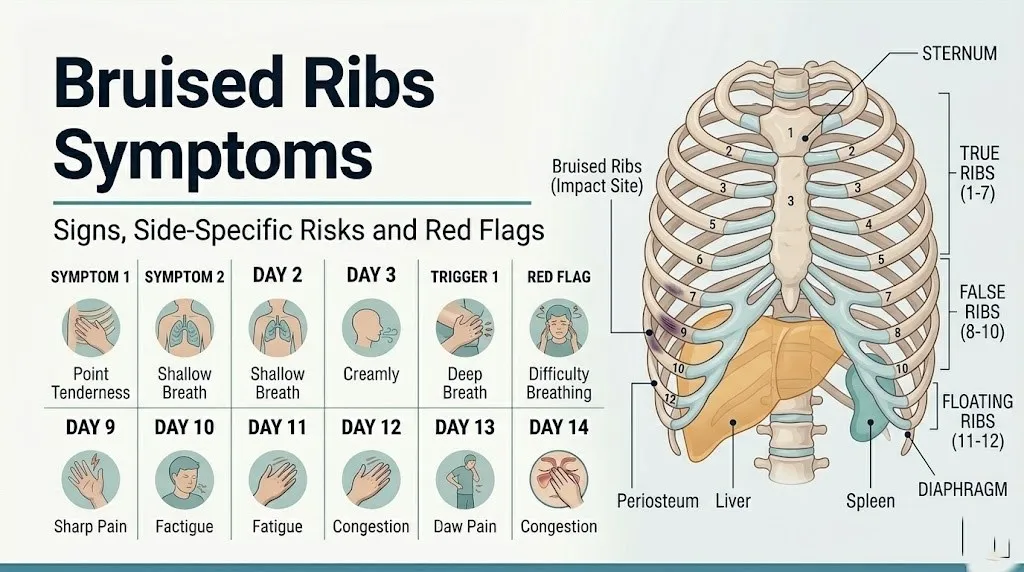
The rib cage consists of twelve paired ribs. True ribs (1-7) attach anteriorly to the sternum via costal cartilage. False ribs (8-10) attach to the costal cartilage of rib 7 rather than directly to the sternum. Floating ribs (11-12) have no anterior cartilaginous attachment. Each rib has a superior border and an inferior border. The inferior border contains the costal groove, which houses the intercostal nerve, artery, and vein running between the internal and innermost intercostal muscle layers.
The periosteum covering each rib is richly supplied with free nerve endings (nociceptors) from the intercostal nerve (the anterior ramus of the corresponding thoracic spinal nerve, T1-T12). These nociceptors are exquisitely sensitive to mechanical deformation and chemical irritation. When blunt force compresses the rib wall, the periosteum is squeezed, stretched, and chemically irritated by the release of prostaglandin E2, bradykinin, histamine, and substance P from damaged cells and activated mast cells.
Causes that produce this injury pattern include sports collisions, falls onto the chest, motor vehicle accidents, direct blows from balls or equipment, and in older adults, even vigorous coughing, which can produce enough rib stress to contuse or fracture ribs with reduced cortical density.
People with osteoporosis or on long-term corticosteroid therapy have ribs with thinner cortex and reduced periosteal integrity, meaning the force threshold for injury is substantially lower than in age-matched individuals with normal bone density. In these populations, bruised rib symptoms may appear after impacts that would be entirely inconsequential in healthy bone.
Bruised Ribs Symptoms: Full Overview
The full spectrum of bruised ribs symptoms encompasses five distinct clinical categories, each produced by a different anatomical mechanism, and each with a different clinical significance level.
The following table presents the complete symptom overview:
| Symptom | Anatomical Source | Physiological Mechanism | Clinical Significance |
|---|---|---|---|
| Sharp, localized chest wall pain | Periosteal nociceptors (intercostal nerve T1-T12) | PGE2 and bradykinin sensitize free nerve endings in injured periosteum | Most consistently present symptom; severity correlates with injury force |
| Pain with breathing (inspiration) | Rib cage expansion stretching injured periosteum | External intercostal muscles elevate ribs during inspiration; motion stresses contusion site | Present in nearly all rib contusions; shallow breathing is a compensatory response |
| Point tenderness on palpation | Periosteal nociceptor hyperalgesia | Substance P-mediated sensitization makes normal touch painful | Highly localizable; finger-point tenderness at the impact site is characteristic |
| Chest wall swelling | Periosteal vessel rupture, local edema | Bradykinin and histamine increase capillary permeability; fluid accumulates in injured tissue | Localizes injury site; may not be visible if over muscular area |
| Ecchymosis (visible bruising) | Subperiosteal and intercostal vessel rupture | Blood tracks through fascial planes to skin surface; may take 12-48 hours to appear | Later-appearing sign; absence does not exclude rib contusion |
| Intercostal muscle spasm | Spinal reflex arc via T1-T12 segments | Intercostal nerve afferents trigger motor neuron activation; involuntary protective muscle contraction | Amplifies pain and reduces respiratory excursion |
| Difficulty taking a deep breath | Splinting and pain-limited inspiration | Reflex intercostal spasm reduces rib cage expansion to minimize periosteal stretch | Requires monitoring; prolonged splinting raises pneumonia risk |
People taking nonsteroidal anti-inflammatory drugs (NSAIDs) for pre-existing conditions before the injury may experience partial suppression of the PGE2-mediated pain component, potentially underestimating injury severity. This matters particularly in older adults, where pain may be less severe than the actual degree of tissue damage warrants.
Rib Pain with Breathing as a Bruised Rib Symptom
Pain with breathing is the most reliably present and diagnostically characteristic symptom of a bruised rib, occurring because every inhalation requires the external intercostal muscles to elevate the rib cage, mechanically stretching and stressing the injured periosteum with each breath.
Normal quiet breathing requires the diaphragm to descend and the chest wall to expand outward as the external intercostal muscles contract. When a rib is bruised, this mandatory expansion of the chest wall applies a repetitive stretching force to the inflamed periosteum at the injury site. The periosteal nociceptors, already sensitized by prostaglandin E2 and bradykinin from the initial injury, respond to this stretch with a sharp, stabbing pain signal conducted along the intercostal nerve to the thoracic spinal cord and upward to the sensory cortex.
The body’s automatic protective response is to reduce the depth of each breath, producing the characteristic shallow, guarded breathing pattern of rib contusion. This response reduces the mechanical stress on the injury site with each breath cycle but comes with a significant downside: reduced tidal volume means less alveolar ventilation per breath, and over time, the smaller breaths fail to adequately expand the lung bases, creating microatelectasis (small areas of lung collapse) that can serve as a nidus for pneumonia.
According to research published in the Journal of Trauma and Acute Care Surgery, pneumonia following rib injuries is a recognized complication that disproportionately affects adults over 65, who have reduced pulmonary reserve and less effective cough mechanics. In older adults with bruised or fractured ribs, the pain-limited shallow breathing pattern substantially elevates pneumonia risk within the first week of injury.
People with pre-existing chronic obstructive pulmonary disease (COPD) or asthma have reduced baseline respiratory reserve. Even a moderate reduction in tidal volume from rib contusion splinting can cause clinically apparent respiratory distress in these patients, warranting earlier and more aggressive pain management evaluation by a primary care physician or pulmonologist.
Key Takeaway: Pain with every breath from a bruised rib is not just discomfort; the shallow breathing it produces reduces lung expansion and raises pneumonia risk, particularly in adults over 65, making adequate pain control and guided deep breathing exercises a clinical priority, not optional.
Pain with Coughing, Sneezing, and Movement in Bruised Ribs
Pain with coughing, sneezing, laughing, and twisting movements is produced by sudden increases in intrapleural pressure transmitted through the chest wall to the injured periosteum, creating sharp, intense pain spikes that are often described as far more severe than the baseline breathing pain.
During a cough or sneeze, the diaphragm and expiratory muscles contract forcefully and suddenly, generating a rapid spike in intrapleural pressure. This pressure wave is transmitted outward through the lung tissue, across the pleural space, and into the chest wall. At the site of periosteal injury, this sudden pressure change compresses and torques the already-sensitized periosteum, activating nociceptors far more intensely than the slow, rhythmic stretch of quiet breathing.
Rotational torso movements (twisting, bending sideways) produce a different but equally painful mechanical stress. The serratus anterior muscle (which attaches to the lateral surfaces of ribs 1-8 and the medial border of the scapula) and the external oblique muscle (which attaches to the outer surfaces of ribs 5-12) both create shearing forces across multiple ribs during torso rotation. When even one of these ribs has a bruised periosteum, this shearing load directly stresses the injury site.
To describe movement-related pain accurately at a medical evaluation:
- Note which specific movements most reliably reproduce the pain (twisting, bending, coughing, lying on the affected side)
- Rate pain on the Visual Analog Scale from 0-10 during the triggering movement and at rest separately
- Record whether pain is localized precisely to one spot or spreads around the chest wall toward the front or back
- Note the time since injury and whether this type of pain was present immediately or developed over hours
- Record any associated symptoms that occur during the painful movement (dizziness, shortness of breath, nausea)
Athletes who must continue training while managing rib contusion symptoms should recognize that rotational sports (tennis, golf, rowing, baseball) apply repeated serratus anterior and oblique force across the rib injury site with every movement, substantially prolonging recovery compared to sports with minimal rotational loading.
Chest Wall Tenderness and Swelling from a Bruised Rib
Chest wall tenderness over the impact site is produced by periosteal hyperalgesia, a state in which the normal touch threshold of the skin and underlying tissue has been dramatically lowered by the inflammatory mediators released at the injury site, making even light contact painful.
The mechanism behind this hypersensitivity is precise. Substance P and calcitonin gene-related peptide (CGRP), both released from the peripheral terminals of activated intercostal nerve nociceptors, create a state of neurogenic inflammation in the periosteum and surrounding soft tissue. These neuropeptides cause local vasodilation and increase vascular permeability, amplifying the local inflammatory response. Simultaneously, prostaglandin E2 directly lowers the activation threshold of nociceptors in the injured tissue, meaning stimuli that would normally register as non-painful (light touch, clothing contact, even pressure from a seatbelt during sitting) now generate pain signals.
Swelling at the injury site results from increased capillary permeability driven by bradykinin and histamine, allowing protein-rich fluid to accumulate in the extravascular tissue space (edema). This edema can produce visible puffiness over the affected rib area, though visibility depends on the amount of overlying muscle and subcutaneous tissue. In muscular individuals or in injuries over the posterior rib cage where the erector spinae muscle group overlies the ribs, swelling may not be visible even when present.
Mayo Clinic clinical guidance on rib injuries notes that localized point tenderness reproducible by pressing directly on the affected rib is one of the most reliable physical examination findings for rib contusion and is used by clinicians to identify the injury site before imaging is ordered.
Postmenopausal women with reduced subcutaneous tissue density over the rib cage may develop more pronounced visible swelling and bruising from the same force than younger adults, because reduced skin turgor allows fluid accumulation to appear more rapidly at the surface.
Muscle Spasm and Splinting as Bruised Rib Symptoms
Muscle spasm and splinting in bruised ribs occur when the spinal reflex arc at the corresponding thoracic spinal cord level (T1-T12) causes involuntary contraction of the external and internal intercostal muscles adjacent to the injured rib, a protective mechanism that limits rib movement but produces its own pain and respiratory consequences.
Think of the spinal reflex arc in rib injury the same way you think of touching a hot stove. The pain signal from the burned finger travels to the spinal cord, where an interneuron immediately activates the motor neurons that withdraw the hand, without waiting for the brain to consciously decide. In rib contusion, the intercostal nerve carries pain signals from the periosteum into the thoracic spinal cord, where interneurons reflexively activate the adjacent intercostal motor neurons, producing involuntary contraction. This is splinting: the muscles lock the injured rib segment in place to prevent further movement.
The clinical presentation of intercostal muscle spasm in rib contusion is a tightness or cramp-like sensation in the chest wall that can be distinctly different in character from the sharp periosteal pain. Some patients describe it as a band-like tightness around the chest at the level of the injured rib. The spasm itself can be tender on deep palpation of the intercostal space between ribs, and this tenderness in the intercostal space (rather than directly on the rib) can help a clinician identify which intercostal level is most affected.
The National Institute of Arthritis and Musculoskeletal and Skin Diseases (NIAMS) notes that musculoskeletal protective splinting, while physiologically appropriate, creates a secondary functional problem when it persists because sustained muscle contraction itself becomes a pain source through ischemia of the contracting muscle fibers, adding a myofascial pain component to the primary periosteal injury pain.
People with pre-existing intercostal neuralgia or thoracic herpes zoster (shingles) may have baseline intercostal nerve irritability that amplifies the splinting response after rib trauma, producing more severe and prolonged muscle spasm than would occur in the same injury in a person without these conditions.
Key Takeaway: Intercostal muscle spasm around a bruised rib is a spinal reflex, not a separate injury; it adds a band-like aching quality to the sharper periosteal pain and can make the affected area feel generally tight and restricted, both of which are expected features of rib contusion rather than signs of a fracture.
Bruised Rib Symptoms Right Side: Organ Involvement and Red Flags
Bruised rib symptoms on the right side carry specific clinical concern for the underlying liver (right lobe under ribs 7-10) and gallbladder (fundus under ribs 9-10), because the same blunt force that contuses the right lower ribs can lacerate or contuse these organs through the chest wall.
The liver is the largest solid abdominal organ and occupies most of the right hypochondriac region of the abdominal cavity, protected anteriorly by ribs 7-10. It is highly vascular, receiving approximately 25 percent of cardiac output through the portal vein and hepatic artery. A blow forceful enough to bruise the right lower ribs (7-10) transmits force directly into the liver parenchyma. Minor hepatic contusions may produce right upper quadrant abdominal pain that initially appears hours after the rib injury. More severe hepatic lacerations produce progressive abdominal pain, hypotension, and tachycardia from internal hemorrhage.
The right-sided lower rib bruising pattern also produces a specific referred pain symptom when the adjacent parietal peritoneum (the lining of the abdominal cavity) becomes irritated by blood or fluid from a hepatic injury: pain referred to the right shoulder tip via the right phrenic nerve (C3-C5). This shoulder tip pain appearing after right lower rib trauma is a red flag for intraabdominal injury.
Key right-sided bruised rib red flags requiring emergency evaluation:
- Right upper quadrant abdominal pain developing hours after right lower rib trauma
- Right shoulder tip pain appearing after the injury (phrenic nerve referred pain from peritoneal irritation)
- Nausea, vomiting, or abdominal guarding after right rib trauma
- Jaundice (yellowing of the skin or eyes) appearing one to two days after rib injury
- Dizziness, rapid heart rate, or fainting after right lower rib trauma
Older adults with hepatomegaly (enlarged liver from fatty liver disease, heart failure, or alcohol use) have a liver that extends further below the right costal margin than normal, making it even more vulnerable to right lower rib trauma. A hepatologist or emergency medicine physician should evaluate any older adult with right lower rib contusion and concurrent right upper quadrant symptoms.
Bruised Rib Symptoms Left Side: Spleen Risk and Warning Signs
Bruised rib symptoms on the left side require specific attention to the spleen, which lies under the left ribs 9-11 and is the most frequently injured solid abdominal organ following left lower rib trauma, with splenic laceration carrying a risk of life-threatening intraabdominal hemorrhage.
The spleen is a highly vascular organ, weighing approximately 150 to 200 grams in adults, located in the left hypochondriac region and protected by ribs 9-11. It sits adjacent to the left hemidiaphragm and behind the stomach. Unlike the liver, which has significant connective tissue capsular strength, the splenic capsule is relatively thin and fragile. Blunt force to the left lower ribs transmits energy directly to the splenic parenchyma. Splenic contusions can bleed slowly (a pattern called delayed splenic rupture) with the person appearing well for 24 to 72 hours after injury before sudden hemodynamic deterioration from hemorrhage into the peritoneal cavity.
The classic warning sign of splenic injury after left lower rib trauma is Kehr’s sign: pain referred to the left shoulder tip. This occurs because blood from a splenic laceration pools beneath the left hemidiaphragm, irritating the parietal peritoneum in contact with the diaphragm. The left phrenic nerve (C3-C5), which innervates this peritoneal surface, refers the pain signal to the left shoulder tip via its dermatomes.
Left-sided bruised rib red flags requiring same-day emergency evaluation:
- Left shoulder tip pain appearing after left lower rib trauma (Kehr’s sign)
- Abdominal pain, guarding, or rigidity after left lower rib injury
- Dizziness, fainting, or rapid heart rate appearing hours after the injury
- Abdominal pain that is worsening rather than improving at 24-48 hours
- Pain that is out of proportion to what would be expected from a rib bruise alone
Young athletes who sustain left lower rib trauma during contact sports (rugby, football, ice hockey, martial arts) and who initially appear well must be monitored specifically for delayed splenic rupture symptoms over the 48 to 72 hours following the injury, per clinical guidance from the American College of Emergency Physicians (ACEP).
Key Takeaway: Left shoulder tip pain (Kehr’s sign) appearing after left lower rib trauma is a red flag for splenic injury, not a muscular strain; it requires same-day emergency evaluation regardless of how well the person feels at the time it appears.
Bruised Rib Back Pain and Referred Pain Patterns
Back pain from a bruised rib occurs through two distinct mechanisms: direct injury to the costovertebral joint (where the rib head articulates with the vertebral body and transverse process of the corresponding thoracic vertebra) and referred pain carried along the intercostal nerve from the anterior or lateral rib injury site around the chest wall to the posterior thorax.
The intercostal nerve travels in the costal groove on the inferior surface of each rib, running from the spine anteriorly around the chest wall to terminate in the anterior cutaneous branches at the front of the chest. Because this nerve supplies sensory innervation to the entire circumferential strip of chest wall at its level, injury to the anterior or lateral rib can produce pain that radiates posteriorly toward the spine along the path of the nerve. This is referred pain along the intercostal nerve distribution, not a separate injury to the back.
Simultaneously, each rib articulates with two points on the spine: the costovertebral joint (where the rib head meets the vertebral body) and the costotransverse joint (where the rib tubercle meets the transverse process). Blunt rib trauma can stress these posterior articular joints, producing deep posterior thoracic pain adjacent to the spine at the level of the injured rib. This joint pain is distinct from the anterior periosteal pain and from intercostal nerve referred pain in character: it is typically described as a deep, aching, joint-like pain reproduced by pressing on the costovertebral joint area.
Cleveland Clinic clinical resources on rib injuries note that patients frequently describe their rib pain as wrapping around from the front to the back, which reflects the anatomical reality that the intercostal nerve spans this entire territory rather than any additional structural injury.
People with pre-existing thoracic spine conditions including thoracic disc herniation, degenerative disc disease at T7-T12 levels, or thoracic spondylosis may have pre-sensitized intercostal nerve roots at the affected levels, producing more extensive and severe back pain from a rib contusion than would occur in a person with normal thoracic spine anatomy.
Early Versus Later Stage Bruised Rib Symptoms
Bruised rib symptoms progress through a predictable timeline, with the character and severity of each symptom changing meaningfully from the first hours post-injury through the subacute recovery phase, and recognizing this progression tells you whether your recovery is on track or whether something has changed that warrants re-evaluation.
In the first 24 to 48 hours (acute phase), the primary symptom is intense, sharp, localized pain at the impact site that is worst with any movement or breathing. The inflammatory cascade is at its peak during this window, with maximal prostaglandin E2 and bradykinin production from activated COX-2 enzymes in the injured periosteum. Visible bruising (ecchymosis) is often absent in the first 12 to 24 hours because blood requires time to track through fascial planes from the subperiosteal space to the skin surface. Swelling appears more quickly because vascular permeability changes occur within minutes of injury.
From 48 hours to about two weeks (subacute phase), the inflammatory mediator burden begins to decline as macrophages clear cellular debris and begin the tissue repair process. Pain severity should be decreasing gradually during this phase, though it remains present with deep breathing and activity. Bruising typically becomes visible and may spread beyond the original impact site as blood continues to track through the tissue. The IL-1β and TNF-α from macrophages sustain a lower-level inflammatory response that transitions the injury toward healing.
| Phase | Timing | Expected Symptoms | Warning Signs Suggesting Worsening |
|---|---|---|---|
| Acute | 0-48 hours | Severe localized pain, pain with all movement and breathing, swelling | Fever, shortness of breath at rest, abdominal pain |
| Subacute | 2-14 days | Improving pain, visible bruising appears and spreads, still painful with deep breath | Pain not improving or worsening, new cough, fever |
| Recovery | 2-6 weeks | Gradual pain reduction, improving breathing depth | Persistent severe pain beyond 4 weeks, new neurological symptoms |
Pregnant women in the second and third trimester heal rib contusions on the same timeline as non-pregnant adults in terms of tissue biology, but the elevated diaphragm position (which moves up to 4 cm superiorly in late pregnancy) alters the force distribution across the lower ribs and may produce more intense early symptoms from the same impact force than would occur outside of pregnancy.
Bruised Rib Symptoms at Night and with Rest
Nighttime worsening of bruised rib symptoms is a predictable and physiologically explained feature of rib contusion that affects the majority of patients during the acute and early subacute phases, driven by the loss of the postural and muscular support that reduces rib cage stress during daytime activities.
During the day, upright posture and the gentle tonic contraction of the erector spinae and thoracic paraspinal muscles provide a degree of thoracic cage stabilization that reduces motion at the injured rib site. At night, supine or side-lying positions change the gravitational load on the chest wall and remove this postural support. The weight of the chest wall itself rests differently on the ribs depending on sleep position, and the injured periosteum receives different pressure patterns with each repositioning.
Lying directly on the injured side is typically the most painful sleep position because it applies direct compressive pressure to the injured periosteum and intercostal structures. Lying on the opposite side stretches the chest wall on the injured side as the contralateral shoulder drops, creating a lateral bending force across the rib cage that stresses the injury. Even supine positioning can be painful because the chest wall expansion with each breath occurs without the stabilizing resistance of gravity-assisted chest wall compression that walking and sitting provide.
Quick Tip:
- Sleeping semi-reclined at approximately 30 to 45 degrees with pillow support reduces chest wall stress compared to fully supine positioning during the acute phase
- A pillow pressed gently against the injured side when coughing provides counter-pressure that reduces periosteal stress during forced expiration
- People with pre-existing sleep apnea using CPAP devices should notify their sleep medicine provider about rib contusion, because the positive airway pressure may need adjustment if chest wall pain is interfering with comfortable device use during sleep
Key Takeaway: Nighttime worsening of bruised rib pain is an expected physiological feature of rib contusion, not a sign of fracture; however, if pain is severe enough that you cannot lie in any position without distress after 48 hours, this warrants evaluation by a primary care physician to exclude a fracture or developing complication.
Bruised Rib Versus Broken Rib Symptoms: How to Tell the Difference
Distinguishing a bruised rib from a broken (fractured) rib based on symptoms alone is frequently impossible, because both injuries share the same primary pain mechanism (periosteal nociceptor activation) and produce nearly identical symptom patterns including pain with breathing, point tenderness, and visible bruising.
The periosteum is the pain-generating structure in both injuries. A rib fracture is a structural discontinuity in the rib cortex, but this fracture itself does not directly produce additional pain pathways that a bruise doesn’t also engage; the fractured periosteum generates pain through the same PGE2 and bradykinin cascade as a contused periosteum. This is why patients with hairline rib fractures consistently report symptom intensity indistinguishable from those with pure contusions.
The features that most reliably suggest fracture over bruise, though not definitively without imaging, are:
- A palpable crunching or clicking sensation (crepitus) when pressing on the rib or during breathing, indicating movement of fractured bone ends
- Pain that is severe enough to significantly limit breathing at rest, not just with deep breaths
- Visible deformity of the rib cage contour
- Paradoxical chest wall movement (a section of the chest wall moves inward during inspiration rather than outward), which indicates flail chest, a medical emergency from multiple adjacent rib fractures
| Feature | Bruised Rib | Fractured Rib |
|---|---|---|
| Pain character | Sharp, localized, breathing-related | Same, often more severe |
| Point tenderness | Present directly over injury | Present; may be exquisitely severe |
| Crepitus on palpation | Absent | May be present |
| Pain at rest | Mild to moderate | Moderate to severe |
| Visible deformity | Absent | May be present |
| Respiratory mechanics | Shallow breathing but normal movement | May show paradoxical movement if flail |
| X-ray appearance | Normal rib cortex | Cortical discontinuity visible (sometimes) |
| CT appearance | Normal rib cortex | Fracture line clearly visible |
| Confirmation | Clinical diagnosis; no imaging required if mild | X-ray or CT required to confirm |
The AAOS states that chest X-rays detect only approximately 50 percent of rib fractures, because many fractures are hairline cortical cracks not visible on plain radiographs. CT scanning detects rib fractures with much higher sensitivity and should be ordered when fracture is clinically suspected but X-ray is negative.
Older adults over 65 with osteoporosis should receive CT chest imaging after any significant rib trauma, because their reduced bone density means fractures occur at lower impact thresholds and may not produce symptom patterns that clinically suggest fracture over bruise.
Bruised Rib Symptoms in Older Adults and People on Anticoagulants
Bruised rib symptoms in adults over 65 and in people on anticoagulant therapy require specific clinical modification because both populations face substantially elevated risks of complications from rib injuries that appear clinically mild by standard symptom assessment.
Older adults have a convergence of anatomical and physiological changes that alter the bruised rib clinical picture. Rib cortical bone loses density progressively with age through reduced osteoblast activity and increased osteoclast activity in the bone remodeling cycle, a process accelerated by postmenopausal estrogen withdrawal in women. This reduced cortical thickness means ribs that absorb impact through elastic deformation in younger adults instead crack, making occult fractures far more likely at the forces that produce only contusion in younger individuals. Pulmonary reserve also declines with age: reduced forced vital capacity, impaired mucociliary clearance, and weakened cough mechanics make older adults with rib injuries highly susceptible to pneumonia from the pain-limited breathing pattern.
Research published in the Journal of Trauma and Acute Care Surgery has documented that each additional rib fracture in adults over 65 is associated with a stepwise increase in pneumonia risk and mortality, and that clinical outcomes in this age group are substantially worse than in younger adults with equivalent injuries. While this specific research focused on fractures, the same physiological vulnerabilities (pulmonary reserve, bone density) apply to contusions that may have concurrent occult fractures.
People on anticoagulant medications (warfarin, apixaban, rivaroxaban, dabigatran, and enoxaparin) face elevated hemothorax risk after rib trauma. The intercostal vessels running in the costal groove can bleed significantly when disrupted; in a person with normal coagulation, this bleeding is self-limited by clot formation. In an anticoagulated person, this bleeding may continue, accumulating in the pleural space and producing a hemothorax. Initial symptoms of hemothorax may be indistinguishable from rib contusion alone, making imaging evaluation appropriate for any anticoagulated person with rib trauma regardless of symptom severity.
Bruised Rib Symptoms in Athletes and Pregnant Women
Athletes and pregnant women represent two populations in which bruised rib symptoms present with distinct clinical features or require modified assessment and management approaches.
Athletes, particularly those in contact sports (football, rugby, ice hockey, wrestling), repetitive overhead sports (swimming, rowing, baseball pitching), and high-impact sports (cycling, equestrian), experience rib contusions frequently but also face two specific additional considerations: the possibility of a pre-existing rib stress fracture (a repetitive loading injury most common in rowers, golfers, and throwing athletes) that may have been asymptomatic before the trauma, and the professional pressure to return to sport before the injury has adequately healed.
Stress fractures of the ribs produce a different pain pattern than acute contusion: the pain develops gradually over weeks rather than immediately after impact, is localized to the same rib areas as contusion, and is worsened by the specific repetitive loading movement of the athlete’s sport. A rower with left posterolateral rib pain that has been gradually worsening over weeks and is now suddenly worse after a fall has a presentation that could represent a new contusion on top of a pre-existing stress fracture, and requires X-ray and clinical evaluation before return to rowing.
Third-trimester pregnant women have specific anatomical changes that alter the bruised rib clinical picture. The uterus expands superiorly, pushing the diaphragm upward by up to 4 centimeters and the lower ribs outward (the thoracic cage diameter increases). This means the floating ribs (11-12) and the lower false ribs (8-10) are in slightly different anatomical positions than in a non-pregnant person, and the underlying organs (particularly the spleen on the left, which is also slightly displaced) are in different locations relative to the rib landmarks used for clinical assessment.
Any rib trauma in a pregnant woman should prompt evaluation by both an emergency medicine physician and the patient’s obstetrician-gynecologist, because the force assessment and organ injury risk differs from the non-pregnant case.
When Bruised Rib Symptoms Need Emergency Evaluation
Certain bruised rib symptoms and post-rib-trauma presentations require same-day emergency evaluation. Do not wait to see if these resolve on their own.
Call 911 or go to the nearest emergency room immediately if you experience after rib trauma:
- Worsening shortness of breath or difficulty breathing at rest (may indicate pneumothorax, where air enters the pleural space, or hemothorax, where blood accumulates in the pleural space, compressing the lung)
- Left shoulder tip pain appearing after left lower rib trauma (Kehr’s sign, indicating possible splenic laceration and intraabdominal hemorrhage)
- Right shoulder tip pain appearing after right lower rib trauma (phrenic nerve referred pain from hepatic injury or peritoneal irritation)
- Abdominal pain, guarding, or rigidity appearing after rib trauma (organ injury until proven otherwise)
- Dizziness, fainting, or a rapid heart rate appearing after rib trauma (possible internal hemorrhage from hemothorax or organ laceration)
- Coughing up blood after rib trauma (pulmonary laceration or significant hemoptysis requiring immediate evaluation)
- A section of the chest wall moving inward during inhalation (paradoxical chest wall movement indicating flail chest from multiple rib fractures)
- Any rib trauma in a person over 65, on anticoagulants, or with a known bleeding disorder (imaging evaluation is appropriate regardless of symptom severity)
- Fever developing within 48 hours of rib trauma (possible pneumonia from splinting-related atelectasis or hemothorax infection)
These presentations can indicate pneumothorax, hemothorax, splenic laceration, hepatic laceration, or flail chest, all of which are life-threatening emergencies requiring immediate imaging and clinical assessment, not a wait-and-see approach at home.
Diagnosis and Recovery Timeline for Bruised Rib Symptoms
The diagnosis of a bruised rib is primarily clinical, meaning it is based on the history of trauma, the pattern of symptoms, and the physical examination findings, with imaging reserved for situations where fracture, pneumothorax, hemothorax, or organ injury needs to be excluded.
A clinician evaluating suspected rib contusion will perform: a detailed history of the mechanism of injury and the exact symptom pattern; palpation of each rib along its length to identify the point of maximum tenderness; auscultation of both lung fields to assess for reduced breath sounds (pneumothorax) or dullness (hemothorax); assessment of respiratory mechanics; and abdominal examination for organ injury signs.
Diagnostic tests ordered based on clinical picture:
| Investigation | What It Detects | When It Is Ordered |
|---|---|---|
| Chest X-ray | Rib fractures (50% sensitivity), pneumothorax, hemothorax | All significant rib trauma presentations |
| CT chest | Rib fractures (high sensitivity), pneumothorax, hemothorax, pulmonary contusion | Negative X-ray with ongoing clinical concern; elderly patients; high-energy mechanisms |
| FAST ultrasound | Free intraabdominal fluid (organ injury), pericardial fluid | In emergency settings after any significant chest or abdominal trauma |
| CT abdomen/pelvis with contrast | Hepatic, splenic, or renal laceration | Any rib trauma with abdominal pain or signs of organ injury |
The recovery timeline for an uncomplicated bruised rib in a healthy adult follows a well-established pattern. Acute pain and severe movement limitation typically begins improving by day 5 to 7 as the peak inflammatory phase resolves. Most patients report pain severity reduced to manageable levels by two to three weeks. Full resolution of all symptoms, including the ability to take a deep breath without pain, typically occurs between three and six weeks from injury.
According to clinical guidelines referenced by Cleveland Clinic, the recovery timeline for a bruised rib is three to six weeks for uncomplicated cases in otherwise healthy adults. Rib fractures without displacement typically take six to eight weeks. Any deviation from the expected improvement trajectory (pain worsening after the first 48 hours, new symptoms appearing after initial improvement, persistent severe pain beyond four weeks) warrants re-evaluation by a primary care physician.
Key Takeaway: A bruised rib that is genuinely improving each week, even if slowly, is following the expected recovery trajectory; it is the rib pain that worsens after the first 48 hours, or that fails to improve at all by two weeks, that needs a clinical re-evaluation and possible imaging.
Frequently Asked Questions About Bruised Ribs Symptoms
What does a bruised rib feel like?
A bruised rib feels like a sharp, localized pain at the injury site that is consistently worsened by breathing, coughing, sneezing, or twisting movements of the torso.
The pain is produced by injury to the periosteum, the densely innervated fibrous membrane covering the rib surface, which becomes hyperalgesia from inflammatory mediators including prostaglandin E2 and bradykinin.
Point tenderness directly over the injured rib, meaning pain when pressing precisely on the rib at the impact site, is the most consistently present and diagnostically characteristic physical finding.
How do I know if my rib is bruised or broken?
Distinguishing a bruised rib from a broken rib on symptoms alone is clinically unreliable, because both injuries involve periosteal damage and produce very similar pain patterns with breathing, tenderness, and movement.
A crunching or clicking sensation (crepitus) when pressing on the rib, severe pain at rest rather than only with movement, or a section of the chest wall moving abnormally during breathing suggest fracture rather than contusion.
Chest X-ray detects approximately 50 percent of rib fractures, and CT imaging with much higher sensitivity is appropriate when fracture is suspected but X-ray is negative, particularly in adults over 65.
How long do bruised rib symptoms last?
Bruised rib symptoms in otherwise healthy adults typically begin improving within five to seven days and resolve fully within three to six weeks.
The acute inflammatory phase with the most severe pain peaks in the first 48 hours and then gradually declines as the COX-2-mediated prostaglandin E2 production decreases and macrophages begin clearing damaged tissue.
Bruised rib symptoms persisting beyond four weeks without measurable improvement should be evaluated by a primary care physician to exclude an undetected rib fracture or a developing complication.
Can bruised ribs cause back pain?
Yes, bruised ribs can cause back pain through two mechanisms: referred pain carried along the intercostal nerve (the anterior ramus of the thoracic spinal nerve T1-T12) from the anterior or lateral rib injury site around the chest wall to the posterior thorax, and direct stress to the costovertebral and costotransverse joints where each rib articulates with the thoracic spine.
The referred pain pattern from an intercostal nerve follows the nerve’s circumferential course around the chest wall, producing pain that wraps from the front or side of the chest toward the spine at the level of the injured rib.
Back pain that is significantly worsening, associated with leg weakness or numbness, or located deep and near the spine without a clear referred pain pattern should be evaluated by a primary care physician to exclude thoracic spine injury.
Should I go to the ER for bruised rib symptoms?
You should go to the emergency room immediately for rib trauma if you develop shortness of breath worsening at rest, left or right shoulder tip pain appearing after lower rib trauma, abdominal pain or guarding, dizziness, fainting, or coughing up blood.
Any rib trauma in an adult over 65 or in a person on anticoagulant medications warrants same-day emergency evaluation and imaging regardless of initial symptom severity.
Mild, localized rib pain after minor trauma in an otherwise healthy young to middle-aged adult, with no respiratory difficulty at rest and no abdominal symptoms, can be initially evaluated by a primary care physician rather than requiring emergency department care.
What are the warning signs that a bruised rib is getting worse?
Warning signs that a bruised rib is getting worse include worsening shortness of breath, pain that is increasing rather than gradually improving after the first 48 hours, new abdominal pain developing after the initial injury, fever appearing within the first week, and pain that is severe enough to prevent any comfortable rest position.
These features can indicate developing pneumothorax, hemothorax, pneumonia from inadequate breathing, or organ injury from the original trauma that was not immediately apparent.
Any worsening of rib symptoms after an initial improvement period should be evaluated by a primary care physician or emergency medicine physician the same day, bringing a description of the original injury mechanism, the initial symptom pattern, and the specific changes that have occurred.
Closing
Bruised rib symptoms follow a specific anatomical logic. The periosteum generates the pain. The intercostal nerve determines where it travels. The protective muscle spasm reduces your breathing depth. Knowing this tells you what to expect and what to watch for.
The most important clinical fact to act on: the side of the chest where symptoms occur matters. Left lower rib bruising and a subsequent shoulder tip pain on the left, or right lower rib bruising with shoulder tip pain on the right and upper abdominal discomfort, are not additional muscle soreness. They are signals from injured organs that need same-day imaging.
If your symptoms are improving gradually each week, the biology is working. If you’re not improving at two weeks, or if any new symptom appears after an initial period of stability, a primary care physician can determine whether the diagnosis is still a simple contusion or whether imaging needs to be revisited.