What Causes Optical Migraines: All Causes and Red Flags
What causes optical migraines is a question with two distinct answers depending on whether you’re experiencing a visual aura that affects both eyes (migraine with visual aura, also called a classical or optical migraine) or a temporary loss of vision in one eye only (retinal migraine): the first is caused by a self-propagating wave of electrical suppression across the visual cortex called cortical spreading depression, and the second is caused by vasospasm of the small arteries supplying the retina. These are not the same event, they don’t carry the same risks, and in some cases, one of them is a medical emergency.
According to the National Institute of Neurological Disorders and Stroke (NINDS), migraine affects approximately 12 percent of the population, with visual disturbances occurring in roughly 25 to 30 percent of all migraine sufferers. The distinction between a benign cortical event and a vascular event affecting the retina or brain is one that every person who experiences optical migraine symptoms genuinely needs to understand.
This guide covers the complete cause picture: the neurochemistry behind cortical spreading depression, the vascular mechanism behind retinal migraine, every named trigger and the specific physiological pathway by which it fires the system, the populations most at risk, and the specific situations where optical migraine symptoms require emergency evaluation rather than watchful waiting.
What Are Optical Migraines and How Do They Differ from Regular Migraines
An optical migraine is a lay term describing either migraine with visual aura or retinal migraine, two conditions that produce visual disturbances through entirely different physiological mechanisms and carry different clinical implications.
A regular migraine without aura produces head pain through activation of the trigeminovascular system, specifically the release of inflammatory neuropeptides from trigeminal nerve terminals surrounding the meningeal blood vessels, without any preceding visual disturbance. An optical migraine adds a visual component: either bilateral shimmering lights, zigzag patterns, or expanding blind spots (the cortical aura), or monocular vision loss affecting one eye only (the retinal variant).
The clinical terminology matters for your evaluation. The International Headache Society (IHS) in its International Classification of Headache Disorders, Third Edition (ICHD-3) defines migraine with aura (ICD-10 G43.109) as a migraine with fully reversible neurological symptoms that develop gradually over five or more minutes and last between five and sixty minutes. Retinal migraine (ICD-10 G43.81) is specifically defined as repeated attacks of monocular visual disturbance lasting less than sixty minutes, directly confirmed by examination during the attack or by the patient’s own drawing of a monocular field defect.
The key practical distinction is which eye is affected. Cover each eye alternately during an episode. If the visual disturbance is present in both eyes when each is covered separately, the event is binocular and originates in the visual cortex. If covering one eye makes it disappear, the event is monocular and originates in that eye’s retinal circulation.
People over 50 experiencing a first-ever episode of monocular vision loss should not assume it is a benign retinal migraine. A board-certified ophthalmologist or emergency medicine physician should evaluate the episode the same day to exclude central retinal artery occlusion (CRAO) or transient ischemic attack (TIA) before a retinal migraine diagnosis is assigned.
What Causes Optical Migraines: The Primary Physiological Mechanisms
Optical migraines are caused by a genetically predisposed, hyperexcitable nervous system or an unstable retinal vascular system that, when activated by specific triggers, produces either a wave of suppressed electrical activity across the visual cortex or a vasospasm of the retinal blood supply.
These underlying causes are structural and physiological, not simply behavioral. A person who gets optical migraines has a brain or vascular system that is predisposed to these events. The triggers discussed in later sections activate this predisposed system; they are not the primary cause in isolation.
Two distinct mechanisms account for all optical migraine episodes:
| Mechanism | Condition | Primary Structure | Visual Symptom | Laterality |
|---|---|---|---|---|
| Cortical spreading depression (CSD) | Migraine with visual aura | Primary visual cortex (V1, occipital lobe) | Shimmering arc, zigzag lines, expanding scotoma | Binocular (both eyes) |
| Retinal vasospasm | Retinal migraine | Central retinal artery, cilioretinal arteries | Dimming, graying, or complete vision loss | Monocular (one eye only) |
The genetic basis of optical migraine susceptibility is well-established. Research published in Cephalalgia (the journal of the International Headache Society) has documented specific gene variants associated with elevated cortical hyperexcitability in migraine families, including variants affecting ion channel function in cortical neurons, supporting the concept that the threshold for CSD initiation is genetically determined.
Women who are pregnant and experience a first-time optical migraine episode should seek same-day evaluation from their obstetrician-gynecologist, because preeclampsia can produce visual disturbances through a mechanism involving cerebral vasospasm and cortical edema that must be distinguished from benign migraine aura.
Cortical Spreading Depression as the Cause of Visual Aura in Optical Migraines
Cortical spreading depression (CSD) causes the visual aura of optical migraines by generating a self-propagating wave of neuronal depolarization followed by electrical suppression that travels across the primary visual cortex (V1) in the occipital lobe at a rate of approximately 3 to 5 millimeters per minute.
Think of CSD like a slow-motion power outage moving across a city’s electrical grid. First, a surge of activity lights up every circuit simultaneously. Then the grid shuts down section by section as the surge passes through. In your visual cortex, that initial surge produces the bright, shimmering, or pulsating lights of the visual aura. As each section of cortex enters the suppression phase, the corresponding region of your visual field goes dark, producing the expanding scotoma (blind spot).
The biochemistry behind CSD is well-characterized by controlled neuroimaging and electrophysiology research. Cortical neurons release potassium ions (K+) and glutamate into the extracellular space when they reach a depolarization threshold. The elevated extracellular K+ and glutamate activate adjacent neurons through NMDA (N-methyl-D-aspartate) receptor stimulation, spreading the depolarization to neighboring cortex. Behind the wave front, the affected neurons enter a prolonged refractory period of electrical suppression lasting several minutes.
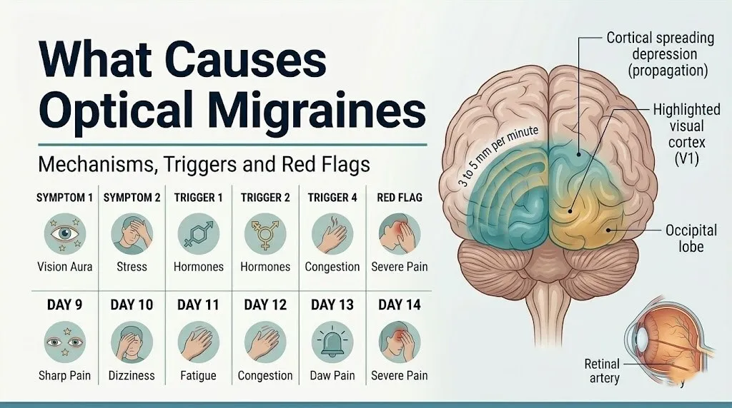
Because V1 is organized retinotopically (meaning each region of the cortex corresponds to a specific region of the visual field), the CSD wave’s progress across the occipital cortex maps directly onto the expanding pattern of the visual aura. The characteristic crescent shape of the scintillating scotoma, which typically starts centrally near fixation and expands toward the peripheral visual field, reflects the CSD wave moving from the posterior occipital cortex (which represents central vision) anteriorly toward cortex representing peripheral vision.
According to the National Institute of Neurological Disorders and Stroke (NINDS), CSD also activates perivascular trigeminal nerve fibers in the cortical meninges, triggering the release of calcitonin gene-related peptide (CGRP) and substance P. This trigeminovascular activation is what converts the visual aura into the subsequent headache phase in people whose optical migraine is followed by pain.
Adolescents with a first-time visual aura episode should be evaluated by a primary care physician before the diagnosis of optical migraine is confirmed, because occipital lobe seizures (benign occipital epilepsy of childhood, including Panayiotopoulos syndrome) can produce visual auras that superficially resemble migraine aura but require different management.
Key Takeaway: CSD is not a vascular event; it is a neurochemical wave propagating across the occipital cortex at 3-5 mm/minute, and the binocular, gradually expanding zigzag arc that expands over 15-30 minutes is its visual signature, distinguishing it from the sudden, monocular vision loss of retinal migraine or TIA.
Retinal Vasospasm as the Cause of Retinal Migraines
Retinal migraine is caused by vasospasm of the central retinal artery, the cilioretinal arteries, or the short posterior ciliary arteries, which reduces blood flow to the retina and produces a temporary, monocular reduction or loss of vision.
The central retinal artery is a branch of the ophthalmic artery, which in turn is the first intracranial branch of the internal carotid artery. It enters the optic nerve approximately 10 millimeters behind the globe and divides into superior and inferior branches supplying the inner layers of the retina. The cilioretinal arteries, present in approximately 20 to 35 percent of people, branch from the short posterior ciliary arteries and supply a segment of the papillomacular bundle (the central visual pathway of the retina). Vasospasm of any of these vessels reduces retinal perfusion to the territory they supply, producing the monocular visual symptom.
Retinal vasospasm differs mechanistically from CSD because it involves abnormal vascular smooth muscle contraction rather than cortical neuronal activity. The vascular smooth muscle surrounding the retinal arterioles contains 5-HT2 serotonin receptors that, when activated, produce contraction. Serotonin fluctuations associated with the migraine cascade may trigger this contraction. Nitric oxide (NO) dysregulation and endothelin-1 release from vascular endothelium may contribute to abnormal vasomotor reactivity in susceptible individuals.
Mayo Clinic clinical guidance on retinal migraine states that the visual loss typically affects the entire monocular field or a segment of it, is described as dimming, graying, or complete blackout of one eye, and is fully reversible within sixty minutes. Any monocular visual loss lasting more than sixty minutes should be treated as a central retinal artery occlusion until proven otherwise by ophthalmoscopic examination.
Men over 50 with hypertension, hyperlipidemia, or a smoking history who experience monocular vision loss have a substantially higher probability of CRAO or TIA than of retinal migraine, because their vascular risk profile alters the probability of an embolic rather than a vasospastic mechanism. These individuals should proceed to emergency evaluation rather than self-attributing the event to a known migraine pattern.
The Trigeminovascular System and Optical Migraine Pain
The trigeminovascular system is the neural pathway that converts a cortical spreading depression event or a retinal vasospasm event into headache pain, by activating nociceptors (pain receptors) on the trigeminal nerve (cranial nerve V) that surround the meningeal blood vessels of the brain.
The trigeminal nerve’s ophthalmic branch (V1) innervates the meningeal blood vessels with sensory fibers that detect stretch, inflammation, and chemical irritation. CSD activates these perivascular fibers directly, triggering them to release calcitonin gene-related peptide (CGRP) and substance P from their terminals into the dural vessel wall. This produces neurogenic inflammation: vasodilation of the dural vessels, plasma protein leakage, and local prostaglandin release. The inflammatory signals travel back along the trigeminal pathway to the trigeminal nucleus caudalis in the brainstem, which relays the pain signal to the thalamus and then to the cerebral cortex for pain perception.
CGRP is now established as the primary mediator of migraine pain. Research published in Cephalalgia demonstrated that intravenous CGRP administration reliably triggers migraine attacks in susceptible individuals, and that CGRP levels in the external jugular vein are elevated during spontaneous migraine attacks. This finding underpins the development of CGRP-targeting therapies used by neurologists in migraine prevention.
The trigeminal nucleus caudalis also receives input from the upper cervical spine (C1-C2 nerve roots), which is why some people with optical migraines experience neck stiffness or pain at the base of the skull as part of the migraine episode.
People with trigeminal neuralgia or prior trigeminal nerve injury have altered trigeminovascular baseline sensitivity, which may modify the pain component of their optical migraine episodes in ways that make standard clinical descriptions less applicable to their experience.
Hormonal Causes of Optical Migraines
Hormonal fluctuations, specifically the withdrawal of estrogen (17-beta estradiol) produced by the ovarian follicles and corpus luteum, cause optical migraines by lowering the threshold at which the visual cortex reaches the depolarization level required to initiate cortical spreading depression.
Estrogen exerts modulatory effects on cortical excitability through multiple mechanisms. High estrogen levels upregulate serotonin receptor expression in the cortex and raphe nuclei, stabilizing serotonergic tone and raising the CSD initiation threshold. They also modulate NMDA receptor sensitivity, reducing the ease with which glutamate-mediated depolarization cascades initiate. When estrogen drops sharply, as it does in the days before menstruation, during the placebo pill week of a combined oral contraceptive cycle, or in the perimenopausal transition, this stabilizing effect is withdrawn, and cortical excitability increases.
This mechanism explains why migraine, including the visual aura variant, is approximately three times more prevalent in women than in men throughout the reproductive years, converging toward equal prevalence after menopause, according to epidemiological data cited by the American Migraine Foundation. The subset of women whose migraines occur specifically in the two days before and the first two days of menstruation (perimenstrual or menstrual migraine) are responding to the most precipitous estrogen drop of the hormonal cycle.
To document a hormonal pattern for your gynecologist or neurologist before an appointment:
- Keep a daily headache and visual aura diary for at least two full menstrual cycles
- Record the date, time, and duration of each episode alongside the day of your menstrual cycle
- Note whether episodes occur specifically in days 27-29 and 1-2 of a 28-day cycle (the perimenstrual window)
- Document any relationship between starting, stopping, or changing hormonal contraception and episode frequency
- Bring a record of all current hormonal medications to the appointment, including dosage and formulation
Postmenopausal women starting hormone replacement therapy (estrogen-containing preparations) may experience new-onset or worsening optical migraines if the estrogen delivery method produces fluctuating rather than stable estrogen levels, with transdermal (patch or gel) delivery generally producing more stable levels than oral formulations.
Key Takeaway: If your optical migraines cluster in the two to three days before your period, the cause is estrogen withdrawal, not a random trigger, and this pattern is specifically addressable through discussions with a gynecologist or headache neurologist about cycle-stabilizing strategies.
Stress, Sleep Deprivation, and Optical Migraine Causes
Psychological stress and sleep deprivation cause optical migraines through two distinct physiological pathways: stress activates the hypothalamic-pituitary-adrenal (HPA) axis to release cortisol, which alters cortical excitability thresholds, and sleep deprivation increases extracellular adenosine accumulation in the brain, sensitizing the cortical environment to CSD initiation.
During sustained psychological stress, the HPA axis drives cortisol release from the adrenal cortex. Cortisol has documented effects on glutamate receptor sensitivity in the cerebral cortex, with animal research showing that elevated cortisol lowers the extracellular potassium concentration required to initiate CSD. Clinically, this explains why optical migraines frequently occur not during the peak of a stressful period but during the “letdown” phase afterward, when acute cortisol elevation drops rapidly, destabilizing a cortex that had been sensitized by the stress exposure.
Sleep deprivation raises extracellular adenosine concentrations throughout the brain through impaired glymphatic clearance and reduced ATP reutilization. Adenosine at the A1 receptor normally provides inhibitory tone that modulates neuronal excitability. When adenosine accumulates beyond normal levels due to sleep debt, and then drops rapidly upon recovery sleep, the oscillating adenosine levels create periods of elevated cortical excitability that can cross the CSD threshold.
The practical implication of the stress-letdown mechanism is specific: people who get optical migraines on weekends, the first day of vacation, or the day after a major work deadline are experiencing the “letdown migraine” pattern. This is caused by rapid normalization of cortisol after a sustained stress elevation, not by any specific weekend behavioral change.
People with generalized anxiety disorder (GAD) or post-traumatic stress disorder (PTSD) have chronically dysregulated HPA axis activity, producing more frequent cortisol fluctuations that may explain why these populations have higher migraine prevalence, per research published in Headache: The Journal of Head and Face Pain.
Dietary and Caffeine Causes of Optical Migraines
Certain dietary components cause optical migraines by altering neurotransmitter levels, cerebrovascular tone, or cortical excitability through named biochemical mechanisms, though the evidence quality varies substantially between individual dietary triggers.
Caffeine acts as an adenosine receptor antagonist, blocking the inhibitory A1 and A2A adenosine receptors throughout the brain. Regular caffeine consumption causes upregulation of adenosine receptors to compensate for this blockade. When caffeine is suddenly withdrawn, the elevated number of adenosine receptors is exposed to normal adenosine levels, producing a disproportionate inhibitory effect on vascular smooth muscle tone and triggering rebound cerebrovascular dilation. This rebound vasodilation was historically thought to be the primary headache mechanism, though current research places greater emphasis on the resulting changes in cortical excitability through adenosinergic pathways. Caffeine withdrawal is among the most well-established dietary migraine triggers in the clinical literature, supported by controlled challenge studies.
Tyramine, a biogenic amine found in aged cheeses, cured meats, fermented foods, and red wine, triggers norepinephrine release from peripheral sympathetic nerve terminals. This produces transient changes in cerebrovascular tone that may lower the CSD initiation threshold in susceptible individuals. The tyramine-migraine association is supported by observational data and patient-reported trigger diaries, though controlled dietary challenge studies have produced inconsistent results.
Alcohol, particularly red wine, contains tyramine, histamine, and sulfites. Red wine is consistently among the most frequently reported dietary migraine triggers in patient surveys, though the specific active component has not been definitively isolated in controlled research. Ethanol itself also produces direct cerebrovascular effects through nitric oxide-mediated vasodilation.
Monosodium glutamate (MSG) contains glutamate, the primary excitatory neurotransmitter that drives CSD propagation. Whether dietary MSG increases extracellular brain glutamate concentrations sufficiently to lower the CSD threshold is debated; controlled blinded challenge studies have not consistently confirmed MSG as a reliable migraine trigger across all subjects.
Older adults who develop new dietary trigger sensitivities for optical migraines alongside weight loss, fatigue, or night sweats should be evaluated by a primary care physician to exclude secondary causes such as giant cell arteritis (temporal arteritis), which can produce visual disturbances through a vascular mechanism and requires urgent diagnosis.
Dehydration and Sensory Triggers as Optical Migraine Causes
Dehydration causes optical migraines by altering plasma osmolality, which the hypothalamus detects through specialized osmoreceptor neurons, triggering a cascade of neuroendocrine and vascular regulatory changes that lower the cortical depolarization threshold.
The hypothalamus contains osmoreceptive neurons in the organum vasculosum of the lamina terminalis (OVLT) and the subfornical organ that continuously monitor blood osmolality. When plasma osmolality rises due to inadequate fluid intake, these neurons trigger antidiuretic hormone (ADH, vasopressin) release from the posterior pituitary and activate autonomic pathways that affect cerebrovascular tone. Animal research has shown that hyperosmolar states reduce the extracellular potassium threshold for CSD initiation. The hypothalamus is also the proposed primary migraine generator (the “migraine generator hypothesis” supported by PET imaging studies published in Cephalalgia), meaning that any input disturbing hypothalamic function, including osmotic stress, may prime the system for a migraine episode.
Bright light and repetitive visual patterns cause optical migraines through photic stimulation of the visual cortex. Pattern glare from high-contrast repetitive stimuli (striped patterns, flickering screens, fluorescent lighting) drives synchronized oscillatory activity in V1. In individuals with cortical hyperexcitability, this synchronized cortical activity may cross the threshold for CSD initiation. Research in this area has demonstrated elevated visual cortex excitability in migraine patients compared to controls, measured by transcranial magnetic stimulation (TMS) techniques.
Strong odors, including perfumes and chemical solvents, cause optical migraine episodes in some individuals through activation of the olfactory bulb projections to the entorhinal cortex and limbic system, which have direct connections to the hypothalamic migraine generator. This mechanism is less well-characterized mechanistically than hormonal or caffeine triggers.
Children and adolescents are particularly vulnerable to photic and pattern-glare triggers for visual aura because their developing visual cortex has higher baseline excitability than the adult cortex, a phenomenon documented in visual evoked potential studies comparing pediatric migraine patients to controls.
Key Takeaway: Dehydration lowers the CSD threshold through hypothalamic osmoreceptor activation, making consistent daily fluid intake a genuinely physiologically relevant preventive measure, not just general health advice.
Why Optical Migraines Happen Without Headache
Optical migraines happen without headache in a substantial proportion of people because cortical spreading depression can occur and resolve without fully activating the trigeminovascular cascade that generates pain, leaving only the visual aura as the clinical manifestation.
The trigeminovascular cascade that produces migraine headache requires CSD to activate perivascular trigeminal fibers sufficiently to initiate the CGRP and substance P release that causes the sustained dural vasodilation and neurogenic inflammation responsible for pain. This activation threshold is not always reached. When CSD propagates through V1 and activates only a portion of the meningeal nociceptor network, or when the trigeminal nucleus caudalis does not receive sufficient input to generate a pain signal, the person experiences only the visual aura.
The clinical term for this presentation is migraine aura without headache, also called a silent migraine or acephalgic migraine. The ICHD-3 recognizes this as a distinct migraine subtype. According to the American Migraine Foundation, migraine aura without headache is more prevalent in men and in older adults, and its incidence increases with age as the headache threshold rises.
This age-related shift is clinically important. A person in their 50s or 60s who begins experiencing new visual aura episodes without headache is more likely to be experiencing acephalgic migraine if they have a prior migraine history, but statistically more likely to be experiencing TIA if they have cardiovascular risk factors and no prior migraine diagnosis. This distinction cannot be made on symptoms alone.
Men over 50 with new-onset visual disturbances without headache and no prior migraine history should be evaluated by an emergency medicine physician or primary care physician within 24 hours, because the absence of headache does not reduce the clinical concern for TIA in this population.
Optical Migraine Causes in Women, During Pregnancy, and on Oral Contraceptives
In women, the causes of optical migraines are predominantly hormonal, specifically driven by the cyclical withdrawal of estrogen during the menstrual cycle, but are substantially modified by pregnancy and oral contraceptive use in ways that change both the frequency of episodes and the clinical risk assessment.
During pregnancy, estrogen levels rise steadily throughout the first trimester and reach their highest sustained levels in the second and third trimesters. This sustained high-estrogen state typically stabilizes cortical excitability, and research published in Headache: The Journal of Head and Face Pain has found that approximately 50 to 80 percent of women with a prior history of migraine experience improvement in migraine frequency during the second and third trimesters. However, women who experience new-onset optical migraine or aura during pregnancy, particularly in the third trimester, require urgent evaluation by their obstetrician-gynecologist to rule out preeclampsia, which produces visual disturbances through cerebral vasospasm and posterior reversible encephalopathy syndrome (PRES), not through CSD.
Combined oral contraceptives (COCs), which contain both synthetic estrogen and progestogen, create a cyclical estrogen withdrawal during the placebo (pill-free) week that mirrors the perimenstrual estrogen drop and triggers optical migraines in susceptible women. Women with migraine with aura who use combined oral contraceptives face an elevated risk of ischemic stroke compared to the general population, a risk that is multiplicative with smoking. The American Academy of Neurology and the ICHD-3 both address this risk, and women with frequent migraine with visual aura who smoke should discuss alternative contraception with their gynecologist.
Progestogen-only contraceptives (the mini-pill, the hormonal IUD, the implant) do not produce the cyclical estrogen withdrawal of COCs and are generally considered a preferable option for women with migraine with aura, though individual hormonal responses vary.
Optical Migraine Causes in Men and Older Adults
Men experience optical migraines less frequently than women during the reproductive years, but when they occur in men, the causes are more likely to be vascular in nature rather than hormonal, and the differential diagnosis with TIA is more clinically pressing.
In men, the absence of cyclical hormonal fluctuation means that the primary underlying cause of optical migraine susceptibility is cortical hyperexcitability from genetic factors, without the estrogen-withdrawal amplification that drives so many episodes in women. Triggers in men are proportionally more likely to be behavioral: irregular sleep, high stress loads, alcohol, and caffeine withdrawal, with no hormonal override.
The retinal migraine variant is proportionally more common in men over 40 with cardiovascular risk factors. The vascular mechanism that causes retinal vasospasm in younger people can shift toward an embolic mechanism in older men with atherosclerosis, hypertension, or atrial fibrillation. A 55-year-old man with hypertension, elevated LDL cholesterol, and a ten-pack-year smoking history who experiences monocular vision loss for twenty minutes is not a straightforward retinal migraine case. He requires same-day evaluation to exclude an embolus from a carotid plaque or a cardiac source as the cause of the retinal ischemia.
According to research published in Ophthalmology (the journal of the American Academy of Ophthalmology), monocular transient vision loss in older adults with vascular risk factors has a clinically meaningful rate of carotid artery stenosis and cardiac embolic sources identified on workup, making the “wait and see if it recurs” approach inappropriate in this population.
Quick Tip:
- Men over 40 experiencing a first episode of monocular vision loss should be evaluated the same day, not reassured that it is likely a migraine
- A carotid Doppler ultrasound and cardiac evaluation are appropriate components of the workup for monocular visual loss in any man with cardiovascular risk factors
- Men with younger-onset optical migraines (under 35) and no cardiovascular risk factors have a clinical picture much closer to women with migraine with aura and can be managed accordingly
Risk Factors That Make Optical Migraines More Likely
The risk factors for optical migraines amplify the underlying physiological susceptibility (cortical hyperexcitability or unstable retinal vasculature) and increase the probability that an individual episode will be triggered in a given person.
Well-established risk factors, based on epidemiological and clinical research:
- Female sex during reproductive years: Threefold higher prevalence than in men, driven by estrogen fluctuation mechanism
- Family history of migraine with aura: First-degree relatives of affected individuals have substantially elevated risk, reflecting the genetic basis of cortical hyperexcitability
- Prior history of motion sickness or vertigo: Associated with heightened vestibular-cortical sensitivity that overlaps with migraine susceptibility
- History of depression or anxiety: Associated with altered serotonergic tone, which modulates trigeminovascular activation threshold
- Irregular sleep patterns: Shift workers and people with highly variable sleep schedules have elevated migraine frequency, through the adenosine and HPA axis mechanisms described above
- High caffeine intake followed by withdrawal: Dose-dependent relationship; higher habitual caffeine intake creates a larger withdrawal effect
- Patent foramen ovale (PFO): An unclosed atrial septal communication present in approximately 25 percent of the general population; associated with elevated migraine with aura prevalence in multiple cohort studies, possibly through paradoxical platelet emboli or vasoactive substance passage from venous to arterial circulation
- Antiphospholipid syndrome: Produces a prothrombotic state that elevates retinal migraine risk and the risk of misdiagnosis as retinal migraine when the true mechanism is thrombosis
According to the American Migraine Foundation, migraine with aura carries a small but measurably elevated risk of ischemic stroke compared to the population without migraine, particularly in women under 45 who smoke and use combined oral contraceptives. This does not mean that optical migraines are routinely dangerous; it means that specific risk factor combinations warrant discussion with a neurologist about prevention.
People with established systemic lupus erythematosus (SLE) have an elevated rate of ocular migraine-like visual episodes that may reflect cerebral vasculitis rather than benign CSD, warranting rheumatologic evaluation of any new visual disturbance in this population.
Key Takeaway: A patent foramen ovale is present in approximately 25 percent of adults and is associated with elevated migraine with aura prevalence; if your neurologist has not addressed PFO in the context of your optical migraine workup, it is a reasonable question to raise at your next appointment.
Can Optical Migraines Be Caused by Something Serious
Optical migraines can, in some cases, be caused by or represent a serious underlying condition, specifically when the visual disturbance is monocular, sudden, prolonged, or accompanied by neurological symptoms that are inconsistent with the typical CSD pattern.
The conditions that can produce visual disturbances resembling an optical migraine but representing a serious underlying cause include:
- Transient ischemic attack (TIA): An embolic or thrombotic event temporarily reducing cerebral or retinal blood flow; produces visual disturbances that can be indistinguishable from optical migraine by symptom description alone but carry a high short-term risk of completed stroke (approximately 10 to 15 percent risk of stroke within 90 days of a TIA, per data from multiple prospective studies)
- Central retinal artery occlusion (CRAO): Permanent or near-permanent loss of vision in one eye from complete arterial occlusion; may begin with a transient episode that is misattributed to retinal migraine before completing
- Amaurosis fugax: Transient monocular blindness from retinal ischemia, a recognized TIA equivalent; caused by emboli from the internal carotid artery or heart, not vasospasm
- Posterior reversible encephalopathy syndrome (PRES): Produces visual disturbances and headache through cerebral vasospasm and edema; associated with severe hypertension, preeclampsia, and immunosuppressive medications
- Occipital lobe tumor or arteriovenous malformation (AVM): Can produce recurrent visual disturbances resembling migraine aura; the characteristic feature that suggests structural pathology is aura that is always in the same visual field location (hemianopic) rather than expanding bilaterally
- Giant cell arteritis (temporal arteritis): Granulomatous inflammation of the temporal arteries and posterior ciliary arteries in adults over 50; produces monocular vision loss from anterior ischemic optic neuropathy; erythrocyte sedimentation rate (ESR) and C-reactive protein (CRP) are typically markedly elevated
The ICHD-3 diagnostic criteria for migraine with aura and retinal migraine require that structural causes have been excluded before the diagnosis is assigned. A diagnosis of optical migraine in a patient who has never had imaging or formal neurological evaluation is a clinical assumption, not a confirmed diagnosis.
Optical Migraine Versus TIA and Retinal Emergency: How to Tell the Difference
Distinguishing an optical migraine from a transient ischemic attack (TIA) or a retinal emergency is the most clinically important differentiation in this topic, because the management decisions, urgency, and downstream risks differ entirely.
The following features distinguish these conditions:
| Feature | Optical Migraine (Visual Aura) | Retinal Migraine (Benign Vasospasm) | TIA / CRAO / Amaurosis Fugax |
|---|---|---|---|
| Laterality | Binocular (both eyes) | Monocular (one eye) | Monocular (one eye) |
| Onset speed | Gradual, over 5-20 minutes | Gradual or rapid | Often sudden (seconds) |
| Visual pattern | Expanding scintillating arc, zigzag | Dimming, graying, complete monocular loss | Curtain dropping, complete monocular blackout |
| Duration | 5-60 minutes (ICHD-3 criterion) | Under 60 minutes | Minutes (TIA) to permanent (CRAO) |
| Headache following | Often (not always) | Mild or absent | Absent |
| Positive visual phenomena | Shimmering, zigzag, colored arcs | Rare | Rare |
| Age of first episode | Often under 40 | Any age | More common over 50 |
| Cardiovascular risk factors | Not typically elevated | Elevated risk in retinal migraine | Elevated; embolic source common |
| Neurological symptoms | Absent | Absent | May include unilateral weakness, speech difficulty |
| Resolution | Complete, fully reversible | Complete | May be incomplete or permanent |
The single most important distinguishing question: Is the visual disturbance in one eye or both? Covering each eye alternately takes 10 seconds and provides the most important initial discriminating information available without any medical equipment.
According to the American Academy of Ophthalmology (AAO), any monocular visual disturbance in a person over 50 with cardiovascular risk factors warrants urgent ophthalmologic evaluation within 24 hours. Monocular vision loss lasting more than 60 minutes in any person warrants emergency evaluation.
Emergency Symptoms: When to Call 911 or Go to the ER
Certain visual symptoms associated with optical migraines can represent a life-threatening or vision-threatening emergency. Do not wait to see if these resolve on their own.
Call 911 or go to the nearest emergency room immediately if you experience:
- Complete loss of vision in one eye lasting more than 60 minutes: May indicate central retinal artery occlusion, which has a narrow treatment window (within hours) for vision restoration
- Visual disturbance accompanied by facial drooping, arm weakness, or speech difficulty: These additional neurological signs indicate a stroke, not a migraine
- A first-ever visual aura with the worst headache of your life: Sudden severe headache with visual disturbance may indicate subarachnoid hemorrhage from a ruptured aneurysm
- Visual disturbance with sudden severe neck stiffness and fever: Indicates possible meningitis with neurological involvement
- Monocular vision loss in a person with severe hypertension or known preeclampsia: May indicate posterior reversible encephalopathy syndrome (PRES), which requires emergency blood pressure management
- Visual symptoms that do not fully resolve within 60 minutes in any person without an established migraine diagnosis
- Any visual disturbance occurring after a head injury: Do not attribute post-trauma visual symptoms to migraine without medical evaluation
These presentations can indicate ischemic stroke, subarachnoid hemorrhage, central retinal artery occlusion, or posterior reversible encephalopathy syndrome, all of which require emergency evaluation and cannot be assessed at an urgent care clinic or resolved by lying down in a dark room.
Diagnosis and When to See a Doctor for Optical Migraines
Optical migraines are diagnosed through a combination of detailed clinical history, neurological examination, and targeted investigations to exclude the serious causes described above, rather than through a single definitive test.
The clinical history is the most important diagnostic tool. Your clinician will be looking for: the visual symptom pattern (binocular vs. monocular, gradual vs. sudden, positive phenomena vs. negative), the duration, the relationship to headache, associated neurological symptoms, family migraine history, cardiovascular risk factors, and any trigger patterns you have identified.
Investigations typically ordered for a new presentation of optical migraine symptoms:
| Investigation | What It Detects | When It Is Ordered |
|---|---|---|
| MRI brain (with and without contrast) | Occipital lesions, AVM, tumor, prior infarction | First episode of visual aura at any age |
| MRA (magnetic resonance angiography) | Intracranial aneurysm, arterial stenosis | Suspected vascular cause, atypical aura features |
| Carotid Doppler ultrasound | Carotid artery stenosis, embolic source | Any monocular visual loss, especially in older adults |
| Echocardiogram | Cardiac embolic source, patent foramen ovale | Retinal migraine, monocular visual loss, cryptogenic |
| ESR and CRP | Giant cell arteritis | Any person over 50 with new monocular visual loss |
| Ophthalmoscopy | Retinal artery occlusion, papilledema, retinal detachment | Any monocular visual disturbance |
| CT scan brain | Intracranial hemorrhage | When MRI not immediately available in emergency |
For an established diagnosis of optical migraine, the appropriate specialist is a board-certified neurologist with subspecialty training in headache medicine, who can provide evidence-based migraine prevention strategies. For retinal migraine evaluation, a board-certified ophthalmologist or neuro-ophthalmologist should be involved to assess the retinal vasculature directly. Primary care physicians and general practitioners can initiate the workup and refer appropriately.
People who have had an established optical migraine diagnosis for years but experience a change in the character, frequency, or associated features of their episodes should return to their neurologist rather than assuming the new presentation is the same condition.
Key Takeaway: A first episode of optical migraine symptoms in any person without an established migraine diagnosis warrants a brain MRI and clinical evaluation to exclude structural causes before the condition is attributed to migraine, regardless of how classic the visual symptom pattern appears.
Frequently Asked Questions About Optical Migraine Causes
What causes optical migraines to happen suddenly?
Optical migraines happen suddenly when a specific trigger raises cortical excitability to the threshold needed to initiate cortical spreading depression in predisposed individuals.
The most common acute triggers are emotional stress, sleep disruption, caffeine withdrawal, hormonal fluctuation near menstruation, bright light exposure, and dehydration, all of which lower the depolarization threshold of the visual cortex.
A first-time episode with no prior migraine history that appears suddenly warrants evaluation by a primary care physician or neurologist to exclude structural causes before it is attributed to optical migraine.
Can stress alone cause an optical migraine?
Stress can cause an optical migraine by elevating cortisol through hypothalamic-pituitary-adrenal axis activation, which lowers the cortical depolarization threshold for cortical spreading depression.
The mechanism is particularly active during the post-stress “letdown” phase, when rapid cortisol normalization destabilizes a cortex sensitized by the preceding stress exposure, which is why many people experience optical migraines the day after a stressful event rather than during it.
Chronic stress from generalized anxiety disorder or PTSD creates ongoing HPA axis dysregulation that may explain the higher migraine prevalence in these populations, per research in Headache: The Journal of Head and Face Pain.
Are optical migraines dangerous or a sign of something serious?
Optical migraines with visual aura (binocular, gradually expanding zigzag pattern) are generally benign in people with an established migraine diagnosis and no cardiovascular risk factors.
Monocular visual disturbances (affecting one eye only), visual symptoms lasting more than 60 minutes, or visual symptoms accompanied by neurological signs such as weakness or speech difficulty are not reliably benign and require same-day medical evaluation.
Women with migraine with aura who smoke and use combined oral contraceptives have a measurably elevated ischemic stroke risk that warrants a discussion with their neurologist and gynecologist about contraceptive alternatives.
What is the difference between an optical migraine and a TIA?
An optical migraine produces binocular visual disturbances (both eyes simultaneously) that develop gradually over 5 to 20 minutes, are often followed by headache, and fully resolve within 60 minutes.
A TIA more commonly produces monocular vision loss (one eye only) or neurological symptoms including facial drooping, arm weakness, or speech difficulty alongside the visual disturbance.
The distinction requires clinical evaluation: visual symptoms in one eye only in a person with cardiovascular risk factors should be treated as a TIA until proven otherwise, and Call 911 or go to the emergency room immediately if neurological symptoms accompany any visual disturbance.
Why do I get optical migraines but no headache?
Optical migraines without headache, called acephalgic or silent migraines, occur when cortical spreading depression propagates through the visual cortex but does not fully activate the trigeminovascular cascade needed to generate pain.
This pattern is more prevalent in men and becomes more common with age, as the headache threshold rises while the susceptibility to CSD may persist.
According to the American Migraine Foundation, new-onset visual aura without headache in adults over 50 with cardiovascular risk factors should be evaluated by an emergency medicine physician or primary care physician the same day to exclude TIA, even when the visual pattern itself appears benign.
What type of doctor should I see for optical migraines?
A board-certified neurologist with subspecialty training in headache medicine is the appropriate specialist for managing recurring optical migraines with confirmed diagnosis.
For a first episode, a primary care physician can initiate the evaluation and refer to neurology and ophthalmology as appropriate.
Any monocular vision loss warrants same-day evaluation by a board-certified ophthalmologist or neuro-ophthalmologist, and if accompanied by neurological symptoms, an emergency medicine physician at an emergency department.
Closing
The causes of optical migraines are mechanistically specific, not mysterious. Cortical spreading depression is a well-characterized neurochemical wave. Retinal vasospasm is a defined vascular event. Estrogen withdrawal, caffeine withdrawal, sleep disruption, and stress all act through named physiological pathways on a cortex or vascular system that is genetically predisposed to these episodes.
Understanding which cause is most likely in your situation changes what you can do about it. A perimenstrual pattern points toward a discussion with a gynecologist or headache neurologist about hormonal stabilization. A caffeine-withdrawal pattern points toward gradual dose reduction rather than cold-turkey cessation. A stress-letdown pattern points toward sleep and stress regulation strategies that prevent the cortisol oscillation rather than just managing the episode.
What you cannot do is self-manage monocular vision loss, visual symptoms accompanied by neurological signs, or a first-ever episode without an established diagnosis. If your visual disturbances are new, worsening, longer-lasting, or accompanied by any neurological change, the right step is medical evaluation by a primary care physician within 24 to 48 hours, or by emergency services immediately if the episode is ongoing.