Red Eyes Causes: What Makes Your Eyes Turn Bloodshot
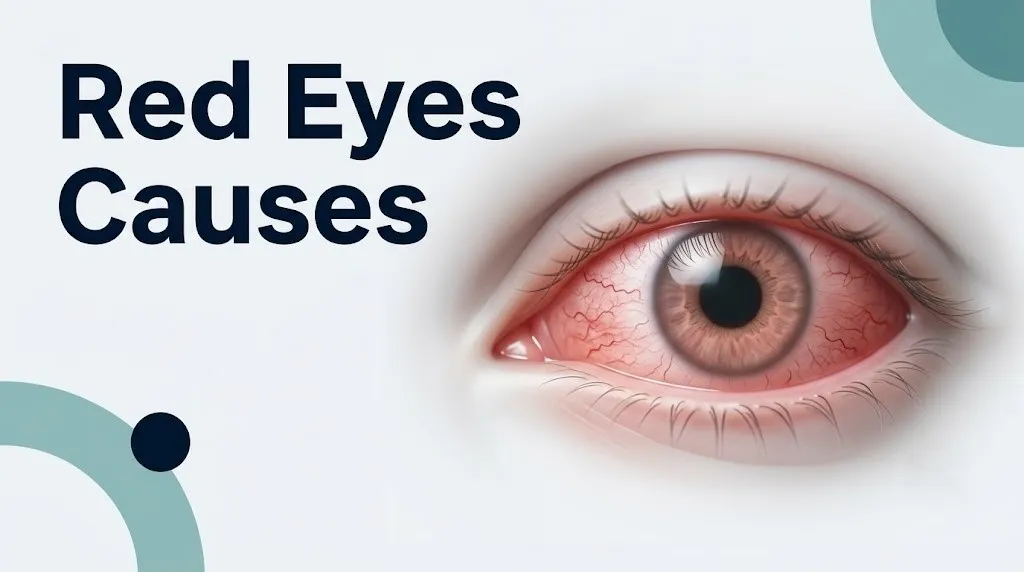
Red eyes causes range from completely harmless, self-resolving irritation to genuine ocular emergencies requiring immediate evaluation. The common thread across all of them is the same: blood vessels within the bulbar conjunctiva and episcleral plexus dilate, making the normally near-invisible capillary network visible as red or pink discoloration across the white of the eye.
According to the National Eye Institute, tens of millions of Americans experience some form of red eye presentation every year, with allergic conjunctivitis alone affecting an estimated 20 percent of the U.S. population. Red eye is one of the most frequent reasons people seek both emergency ophthalmic and primary care consultations. That frequency makes it easy to dismiss, and that dismissal is the single biggest clinical mistake.
This article covers the specific physiological mechanism behind each named cause, from the most common and benign to the rare but serious. It also provides a clear framework for deciding whether your situation needs home monitoring, a same-day eye appointment, or an emergency room visit.
Red Eyes Causes: Why Blood Vessels in the Eye Dilate
Red eyes causes share one anatomical endpoint: dilation of the blood vessels in the bulbar conjunctiva and the episcleral vascular plexus, the layers of fine blood vessels covering the white of the eye.
The conjunctiva is a transparent mucous membrane. It lines the inner surface of the eyelids and folds back to cover the anterior sclera. Under normal conditions, its blood vessels are so narrow that the eye appears white. When any irritant, infection, inflammatory signal, or pressure change reaches that tissue, vessels dilate rapidly to increase blood flow, producing the familiar redness.
The pathway from trigger to visible redness differs by cause. In allergic reactions, IgE-armed mast cells within the conjunctival stroma degranulate and release histamine, which acts directly on H1 receptors on conjunctival endothelial cells, causing rapid vasodilation and increased vascular permeability. In bacterial infections, prostaglandins and interleukin-1 beta drive inflammatory vasodilation through the cyclooxygenase-2 pathway. In dry eye disease, hyperosmotic tear film triggers a stress-signaling cascade in the conjunctival epithelium that activates MAPK inflammatory pathways, producing secondary vessel dilation. In acute angle-closure glaucoma, mechanically elevated intraocular pressure compresses the limbal vasculature and ciliary vessels, producing a distinct deep redness around the corneal margin called ciliary injection.
Understanding which pathway is active in your situation is what tells you whether a cold cloth, an antihistamine eye drop, or a 911 call is the right response.
Quick Tip:
- Redness scattered across the entire white of the eye (diffuse injection) usually points to allergic, viral, or dry-eye causes.
- Redness concentrated in a ring around the corneal edge (ciliary or pericorneal injection) is a more serious sign and warrants same-day evaluation.
- A bright red, sharply defined patch on the white of the eye that does not change with blinking is characteristic of subconjunctival hemorrhage, not infection.
What Causes Red Bloodshot Eyes: The Main Mechanisms
What causes red bloodshot eyes is one of four primary physiological mechanisms: immune-inflammatory activation, mechanical irritation, vascular pressure change, or toxic vasodilation.
Each mechanism recruits different mediators and produces a somewhat different clinical picture. Immune-inflammatory causes (allergies, conjunctivitis, uveitis) produce redness alongside discharge, tearing, or light sensitivity because the same inflammatory cascade that dilates vessels also increases mucus production and stimulates sensory nerve endings in the cornea and conjunctiva. Mechanical causes (contact lens wear, foreign body, corneal abrasion) produce redness localized to the area of irritation, often with a scratching sensation rather than true pain.
Vascular pressure changes, as in acute angle-closure glaucoma, produce a specific, severe, deep-seated eye pain alongside redness because the trabecular meshwork has lost its ability to drain aqueous humor, and pressure builds rapidly within the anterior chamber. Toxic vasodilation, as with cannabis, alcohol, or certain medications, produces bilateral, painless redness because the vasodilating compound reaches episcleral vessels through the systemic circulation rather than through local tissue inflammation.
| Mechanism | Named Mediator | Typical Pattern | Pain Level |
|---|---|---|---|
| IgE-mediated allergy | Histamine, leukotriene C4 | Bilateral, diffuse, itchy | Mild to none |
| Bacterial/viral infection | Prostaglandins, IL-1 beta | Unilateral or bilateral, discharge | Mild |
| Dry eye hyperosmolarity | MAPK pathway activation | Diffuse, chronic, worsens in afternoon | Mild irritation |
| Mechanical irritation | Substance P, bradykinin | Localized, sudden onset | Mild to moderate |
| Elevated IOP | Physical vessel compression | Pericorneal, ciliary injection | Severe |
| Toxic/systemic vasodilation | CB1 receptor activation (cannabis) | Bilateral, diffuse, no discharge | None |
Allergic Conjunctivitis and Red Eyes
Allergic conjunctivitis is the leading cause of red eyes worldwide, producing bilateral redness through an IgE-mediated mast cell response in the conjunctival stroma.
When an allergen such as pollen, pet dander, dust mite protein, or mold spore contacts the conjunctival surface, it binds to IgE antibodies already attached to resident mast cells. This triggers degranulation: mast cells rapidly release histamine, tryptase, leukotriene C4, and prostaglandin D2. Histamine acts within seconds on H1 receptors on conjunctival vascular endothelial cells, causing vasodilation and increased vascular permeability. The result is rapid-onset bilateral redness, intense itching (the most distinguishing symptom), tearing, and eyelid swelling.
A study published in JAMA Ophthalmology in 2022 found that itching is the single symptom that most reliably distinguishes allergic conjunctivitis from other causes of red eye. Pain, photophobia, or visual changes are not features of allergic conjunctivitis and, if present alongside redness, should prompt evaluation for a different diagnosis.
Seasonal allergic conjunctivitis flares with specific pollen seasons, while perennial allergic conjunctivitis persists year-round due to indoor allergens. A more severe form called vernal keratoconjunctivitis affects primarily young males under 25 and can damage the cornea if untreated. Children and adolescents with atopic dermatitis carry a higher risk of all allergic conjunctivitis subtypes due to systemic Th2-skewed immune activity. People using antihistamine tablets for nasal allergies may partially suppress conjunctival histamine responses but often experience worsened dry-eye redness because systemic antihistamines also suppress lacrimal gland secretion.
Key red eye symptoms specific to allergic conjunctivitis:
- Intense bilateral itching, often described as unbearable or irresistible
- Watery, clear discharge without crusting
- Eyelid swelling (chemosis), particularly on waking
- Seasonal or exposure-linked pattern: symptoms worsen near animals, in high-pollen weather, or after vacuuming
- No reduction in visual clarity (vision is not blurred)
Bacterial and Viral Conjunctivitis Causes
Bacterial and viral conjunctivitis cause red eyes through distinct but overlapping inflammatory pathways that both converge on prostaglandin-mediated conjunctival vessel dilation.
Bacterial conjunctivitis is most commonly caused by Staphylococcus aureus in adults, Haemophilus influenzae and Streptococcus pneumoniae in children, and Neisseria gonorrhoeae or Chlamydia trachomatis in sexually active adults and newborns. When bacteria colonize the conjunctival epithelium, they trigger toll-like receptor activation and the release of pro-inflammatory cytokines including interleukin-1 beta and tumor necrosis factor-alpha. These cytokines upregulate cyclooxygenase-2 (COX-2) in the conjunctival stroma, driving prostaglandin E2 synthesis and vascular dilation. The defining clinical feature is mucopurulent discharge: thick, yellow-green, and copious enough to cause eyelids to stick together on waking.
Viral conjunctivitis, most often caused by adenovirus (accounting for 65 to 90 percent of viral conjunctivitis cases, according to the American Academy of Ophthalmology), produces redness through a similar cytokine cascade but with watery rather than purulent discharge. Viral conjunctivitis is highly contagious, spreads through direct contact and contaminated surfaces, and typically begins in one eye before spreading to the other within 24 to 48 hours. It is self-limiting in most cases, resolving within one to two weeks without antibiotic treatment.
Herpes simplex virus (HSV) keratitis is a distinct and more serious viral cause. It typically affects one eye and produces a dendritic ulcer pattern visible on fluorescein staining. Unlike adenoviral conjunctivitis, HSV keratitis can scar the cornea and requires antiviral treatment. People who are immunocompromised, whether from HIV, organ transplant immunosuppression, or systemic corticosteroid use, face a higher risk of severe HSV ocular involvement.
| Cause | Discharge Type | Unilateral or Bilateral | Contagious | Treatment Needed |
|---|---|---|---|---|
| Bacterial (Staph, Strep) | Thick, yellow-green | Often starts unilateral | Moderately | Antibiotic drops |
| Bacterial (Gonorrhea) | Profuse, purulent | Bilateral | Yes | Systemic antibiotics urgently |
| Adenoviral | Watery, clear | Starts unilateral, spreads | Highly | Supportive only |
| Herpes simplex | Watery, may have crusting | Unilateral | Low | Antiviral medication |
Key Takeaway: Itching is the hallmark of allergic conjunctivitis; thick yellow-green discharge points to bacterial infection; watery discharge with high contagiousness suggests adenoviral conjunctivitis, which is self-limiting but requires handwashing precautions to prevent spread.
Dry Eye Syndrome and Red Eyes
Dry eye syndrome causes red eyes by generating a hyperosmotic, pro-inflammatory tear film environment that activates stress-signaling pathways in conjunctival epithelial cells, ultimately recruiting inflammatory mediators that dilate surface blood vessels.
The lacrimal functional unit coordinates tear film production. It includes the lacrimal gland (producing the aqueous tear layer), the meibomian glands in the eyelid margins (producing the lipid layer that prevents evaporation), and the conjunctival goblet cells (producing mucin that anchors tears to the corneal surface). When any component fails, tear film osmolarity rises. Hyperosmotic tears activate MAPK inflammatory signaling in conjunctival epithelial cells, which releases matrix metalloproteinase-9 (MMP-9), interleukin-1 beta, and tumor necrosis factor-alpha. These mediators damage goblet cells further, worsen tear instability, and dilate conjunctival vessels in a self-perpetuating inflammatory loop.
The DEWS II Report, published in The Ocular Surface in 2017 and developed by a panel of 150 international researchers, defines dry eye disease as a chronic, multifactorial disease of the ocular surface characterized by tear film instability, hyperosmolarity, ocular surface inflammation, and neurosensory abnormalities. Redness is one of the cardinal clinical signs because of this inflammatory surface cascade.
Postmenopausal women experience disproportionately higher rates of dry eye disease compared to men of the same age. The reason is androgen-dependent regulation of meibomian gland secretion: androgens (primarily testosterone and its derivative dihydrotestosterone) stimulate lipid production in meibomian glands. After menopause, androgen levels fall, reducing the lipid layer of the tear film and accelerating evaporative dry eye. The National Eye Institute reports that women account for approximately 65 percent of all dry eye diagnoses, with postmenopausal status being the most consistent demographic risk factor.
Quick Tip:
- Red eyes that worsen over the course of the day, especially in air-conditioned or heated environments, suggest evaporative dry eye.
- Redness that improves after blinking or briefly closing the eyes supports a tear film instability mechanism.
- Red eyes in a person taking antihistamines, diuretics, beta-blockers, antidepressants, or isotretinoin may reflect medication-induced lacrimal gland suppression rather than primary dry eye.
Subconjunctival Hemorrhage Causes
Subconjunctival hemorrhage causes a bright, sharply defined red patch on the white of the eye by rupturing one or more small blood vessels within the bulbar conjunctiva, allowing blood to pool between the conjunctiva and the underlying sclera.
The appearance is dramatic: a vivid, bright-red or dark-red patch with clearly defined borders, often noticed suddenly when looking in a mirror. Despite the alarming look, most subconjunctival hemorrhages are painless and do not affect vision. The blood is trapped beneath the transparent conjunctiva, unable to drain, and reabsorbs naturally over one to two weeks as the tissue breaks down hemoglobin through local enzymatic activity.
Common triggers include physical straining (coughing, sneezing, vomiting, heavy lifting, straining during bowel movements), direct eye trauma, Valsalva maneuver during intense exercise, and vigorous eye rubbing. These actions cause a transient spike in venous pressure that overwhelms the fragile conjunctival capillaries. High blood pressure (hypertension) is associated with recurrent subconjunctival hemorrhage in some patients, because chronically elevated arterial pressure weakens vessel walls over time. According to a study published in the British Journal of Ophthalmology, anticoagulant medications such as warfarin, aspirin at therapeutic doses, and newer oral anticoagulants (NOACs like apixaban and rivaroxaban) increase the risk of subconjunctival hemorrhage without increasing the size or clinical significance of the bleed.
People on blood thinners who notice a subconjunctival hemorrhage should not adjust their anticoagulant dose without speaking to the prescribing physician. The hemorrhage itself does not require treatment, but a pattern of recurrent unprovoked hemorrhages warrants evaluation for hypertension, a bleeding disorder, or over-anticoagulation by a primary care physician or hematologist.
Blepharitis and Red Eye Causes
Blepharitis causes red eyes through chronic inflammation of the eyelid margins, which disrupts meibomian gland function and produces a toxic, inflammatory tear film that bathes the conjunctival surface and dilates its blood vessels.
The eyelid margins host the openings of the meibomian glands, which number approximately 25 to 40 per eyelid. In blepharitis, these gland openings become blocked by thickened secretions, debris, or bacterial biofilm, most commonly from Staphylococcus aureus colonization at the lid margin. The bacteria produce lipases that break down meibomian lipids into irritating free fatty acids. These compounds cause direct chemical irritation of the conjunctival epithelium, triggering a low-grade but persistent inflammatory response: upregulation of interleukin-6, interleukin-8, and MMP-9, vessel dilation, and chronic surface redness.
Ocular rosacea is a closely related and frequently underdiagnosed condition. Rosacea affects sebaceous glands throughout the face, and when it involves the eyelid margin glands, it produces a form of blepharitis with particularly severe meibomian gland dysfunction. The American Academy of Ophthalmology notes that up to 58 percent of patients with facial rosacea have ocular involvement, making chronic red eyes one of the presenting features of an underlying dermatological diagnosis.
Middle-aged adults and older adults experience blepharitis more frequently than younger patients, partly because meibomian gland secretion becomes more viscous with age and the eyelid skin loses elasticity, reducing the mechanical pumping action of blinking that normally helps express meibomian secretions. Contact lens wearers also face higher rates of blepharitis-associated red eyes because lens wear alters the microbiome of the ocular surface and physically disrupts tear film distribution across the lid margin.
Key Takeaway: Subconjunctival hemorrhage looks alarming but is typically painless and self-resolving; blepharitis and ocular rosacea cause chronic, low-grade red eyes that require consistent lid hygiene and often professional management rather than eye drops alone.
Anterior Uveitis Causes
Anterior uveitis causes red eyes through immune-mediated inflammation of the uveal tract in the anterior segment of the eye: specifically the iris and the ciliary body, producing a distinct pattern of deep, aching eye pain, light sensitivity, and pericorneal redness that does not respond to antihistamine drops.
The uvea is the pigmented, highly vascular middle layer of the eye. The anterior uvea includes the iris and ciliary body. When inflammation strikes this tissue, white blood cells and inflammatory mediators flood the anterior chamber of the eye. Bradykinin and prostaglandins produced by activated uveal macrophages and T-cells cause intense dilation of the deep episcleral vessels in a ring around the cornea. This specific redness pattern, pericorneal or ciliary injection, is a key distinguishing feature from more superficial conjunctival causes of red eye.
According to research published in the British Journal of Ophthalmology, the HLA-B27 antigen is associated with approximately 50 percent of cases of acute anterior uveitis in Western populations. Conditions carrying the HLA-B27 genetic marker include ankylosing spondylitis, reactive arthritis (formerly Reiter syndrome), psoriatic arthritis, and inflammatory bowel disease. A person who develops a first episode of acute anterior uveitis and has lower back stiffness, joint pain, or bowel symptoms should be referred to a rheumatologist for evaluation of an underlying HLA-B27-associated spondyloarthropathy.
Other causes of anterior uveitis include herpes simplex virus, cytomegalovirus in immunocompromised patients, sarcoidosis, and Behcet disease. Anterior uveitis left untreated risks posterior synechiae (adhesions between the iris and lens), cataract formation, and secondary glaucoma. A unilateral red eye with photophobia and deep eye ache requires same-day evaluation by an ophthalmologist, not an urgent care center.
Contact Lens Red Eye Causes
Contact lens-related red eyes occur because contact lens wear disrupts the ocular surface environment in multiple ways simultaneously, triggering inflammatory pathways that dilate conjunctival vessels within hours of lens insertion.
Modern soft contact lenses sit directly on the cornea, reducing oxygen transmissibility to the corneal epithelium. The cornea is avascular by design: it gets its oxygen from the tear film and directly from the atmosphere. When oxygen delivery is reduced by lens wear, particularly with extended-wear or overnight lenses, the cornea signals for new blood vessel growth at its limbal margin, a process called corneal neovascularization. Limbal blood vessels also dilate in response to the relative hypoxia, producing the characteristic redness around the iris border seen in habitual contact lens overwear.
Contact lens-associated red eye (CLARE) is a specific syndrome characterized by acute, unilateral redness with mild discomfort that typically occurs after overnight lens wear and resolves after lens removal. It results from colonization of the lens surface by gram-negative bacteria (most commonly Pseudomonas aeruginosa), whose lipopolysaccharide components provoke complement activation and inflammatory cell recruitment to the corneal and conjunctival surfaces.
The most serious contact lens-related cause of red eye is microbial keratitis, a corneal infection that can progress to corneal scarring or perforation within hours. Risk factors include overnight wear, swimming while wearing lenses, tap water rinsing of lenses, and continued lens wear after symptoms begin. Microbial keratitis presents as a red eye with moderate to severe pain, photophobia, and a visible white or grey spot on the cornea.
The American Academy of Ophthalmology recommends immediate lens removal and same-day evaluation by an eye care provider if a contact lens wearer experiences any of the following:
- Sudden increase in redness during or after lens wear
- Pain that persists after removing the lens
- Blurred vision that does not clear
- A visible white spot on the cornea
- Increasing light sensitivity
Digital Eye Strain and Red Eyes
Digital eye strain causes red eyes by dramatically reducing blink rate during screen use, allowing the tear film to break up and expose the corneal surface, triggering the same osmotic-stress inflammatory cascade that drives dry eye disease.
The average blink rate during alert activity off screens is 15 to 20 blinks per minute. Multiple studies, including research published in JAMA Ophthalmology, have confirmed that blink rate falls to six to eight blinks per minute during focused screen work. This reduction, combined with the common habit of holding screens below eye level (which increases the exposed surface area of the eye), accelerates tear film evaporation. The corneal and conjunctival epithelium, now bathed in a hyperosmotic tear film, activates MMP-9 and interleukin-1 beta signaling, which dilates surface vessels and produces the familiar end-of-workday redness, burning, and tired-eye feeling.
The 20-20-20 rule is the most widely cited clinical strategy for reducing digital eye strain: every 20 minutes, look at something at least 20 feet away for at least 20 seconds. This forces a full blink cycle, redistributes tear film, and temporarily reduces accommodative strain on the ciliary muscle. While this rule is based primarily on expert consensus and observational data rather than randomized controlled trial evidence, it is supported by the underlying physiology of blink rate and tear film dynamics.
Adults who work in low-humidity office environments with overhead heating or air conditioning face a compounding risk: low ambient humidity accelerates tear film evaporation independently of blink rate. Contact lens wearers using screens for more than six hours daily carry the highest cumulative risk for digital-eye-strain-related red eyes and corneal surface inflammation. Taking scheduled breaks every 30 to 60 minutes and using preservative-free lubricating eye drops rated for contact lens compatibility can reduce symptoms without requiring a prescription.
Key Takeaway: Anterior uveitis is one of the most underrecognized serious causes of red eyes, presenting with deep aching pain, photophobia, and pericorneal redness. Any unilateral red eye with these features needs same-day ophthalmology evaluation, not eye drops from a pharmacy shelf.
Acute Angle-Closure Glaucoma Red Eye
Acute angle-closure glaucoma causes red eyes in the context of a true ocular emergency: intraocular pressure spikes rapidly to 40 to 80 mmHg (normal range 10 to 21 mmHg) when the drainage angle between the iris and the cornea closes abruptly, blocking aqueous humor from exiting through the trabecular meshwork.
Think of the anterior chamber of the eye like a sealed room with one small drainage vent. Under normal conditions, the ciliary body continuously produces aqueous humor, and the trabecular meshwork continuously drains it, maintaining stable pressure. In angle-closure glaucoma, the iris physically blocks the drainage angle, like a door swinging shut over the vent. Pressure builds rapidly, compresses the blood vessels around the corneal margin, and produces the emergency combination of severe eye pain, halos around lights, blurred vision, headache, nausea, and vomiting alongside a visibly red eye with a mid-dilated, non-reactive pupil.
This presentation is a medical emergency. Every hour of elevated intraocular pressure destroys retinal ganglion cells in the optic nerve. Permanent vision loss can begin within hours. The American Academy of Ophthalmology categorizes acute primary angle-closure as a sight-threatening emergency requiring immediate intervention, typically with intravenous acetazolamide, topical pressure-lowering agents, and laser peripheral iridotomy once the acute attack is controlled.
Risk factors for acute angle-closure glaucoma include: hyperopia (far-sightedness, which correlates with a shallower anterior chamber), age over 60, female sex (women have anatomically shallower anterior chambers on average), East Asian ancestry (higher prevalence of anatomically narrow angles), and certain medications including antihistamines, antidepressants, and nasal decongestants that cause pupillary dilation and can precipitate an attack.
Call 911 or go to the nearest emergency room immediately if you or someone near you develops sudden severe eye pain, halos around lights, blurred vision, nausea, or vomiting with a red eye. This is not a symptom to watch overnight.
Red Eyes Causes With No Pain
Red eyes with no pain narrow the differential diagnosis considerably, pointing toward causes that do not involve corneal nerve activation or significant tissue inflammation.
The cornea is the most densely innervated tissue in the human body, with nerve terminal density approximately 300 to 600 times greater than skin. Causes of red eye that involve the cornea or iris almost invariably produce at least some degree of discomfort, photophobia, or foreign body sensation. Painless red eyes point toward the bulbar conjunctiva or the superficial episcleral vessels as the primary site of involvement.
The most common painless red eye causes include:
- Subconjunctival hemorrhage: bright red, sharply bordered, no pain, no discharge, no vision change. Resolves in seven to fourteen days.
- Allergic conjunctivitis: bilateral, diffuse redness with intense itching. Itching, not pain, is the sensory signature.
- Viral conjunctivitis: watery discharge, mild grittiness rather than pain, highly contagious.
- Episcleritis: localized, often sectoral redness with mild tenderness on palpation. Usually self-limiting within two to three weeks. More common in adults under 40.
- Cannabis or alcohol use: bilateral, diffuse, painless redness from systemic vasodilation via CB1 receptor activation in episcleral vessels or direct alcohol-induced vasodilation.
- Dry eye disease: diffuse redness with burning or stinging rather than sharp pain, typically worsening through the day.
- Digital eye strain: similar diffuse redness with fatigue and burning sensation rather than pain.
People taking systemic blood thinners who notice painless, non-blinking red areas on the eye should mention this to their prescribing physician at the next visit, even though the presentation itself is benign. Older adults on warfarin or direct oral anticoagulants have a higher rate of spontaneous subconjunctival hemorrhage, and a cluster of events may prompt a review of medication dose or blood pressure management.
One Red Eye Causes
One red eye, meaning unilateral redness affecting only a single eye rather than both, shifts the diagnostic probability toward localized causes: infection, inflammation of the uvea, contact lens complication, foreign body, corneal pathology, or episcleritis.
Bilateral red eye, by contrast, points toward systemic or surface-wide causes: allergens, viral conjunctivitis spread to both eyes, dry eye, or systemic vasodilation from medications or substances. When redness stays firmly in one eye for more than 24 hours with no contralateral spread, localized pathology is the most likely explanation.
The most clinically important unilateral red eye causes, ranked by urgency:
| Cause | Typical Additional Features | Urgency Level |
|---|---|---|
| Anterior uveitis | Deep ache, photophobia, no discharge | Same-day ophthalmology |
| Microbial keratitis | Severe pain, white corneal spot, contact lens user | Emergency same-day |
| Acute angle-closure glaucoma | Severe pain, halos, nausea | Call 911 / ER now |
| Herpes simplex keratitis | Dendritic pattern on fluorescein staining | Same-day ophthalmology |
| Episcleritis | Localized sectoral redness, mild discomfort | Non-urgent evaluation |
| Subconjunctival hemorrhage | Bright red patch, painless, no discharge | Monitor at home |
| Foreign body | Scratching sensation, tearing, localized | Urgent care or ER |
| Bacterial conjunctivitis (early) | Mild discharge, may spread to second eye | Primary care or optometrist |
A unilateral red eye with any degree of pain, photophobia, or reduced visual clarity should be evaluated by an optometrist or ophthalmologist the same day. These features together, specifically pain plus photophobia plus one red eye, represent the clinical triad most associated with anterior uveitis and should not be attributed to “pink eye” without professional examination.
Key Takeaway: A painless bright-red patch on the white of one eye is almost always a subconjunctival hemorrhage, which resolves without treatment; a unilateral red eye with pain, light sensitivity, and blurred vision requires same-day evaluation for anterior uveitis, corneal infection, or glaucoma.
Red Eyes in the Morning: Causes
Red eyes specifically in the morning, meaning redness that is worst on waking and improves over the first hour of the day, point toward causes that are amplified by reduced tear film activity, reduced blink rate, or allergen exposure during sleep.
During sleep, the eyes are closed, blink rate drops to zero, and tear film dynamics shift. The lacrimal gland produces significantly less aqueous tears during sleep, and the eyelid’s compressive contact with the eye surface temporarily increases the local concentration of inflammatory cytokines and meibomian secretions. People who sleep in environments with pets, dust mite-heavy bedding, or indoor mold are also exposed to allergens throughout the night at close range.
The most common causes of morning-predominant red eyes:
- Nocturnal lagophthalmos: incomplete eyelid closure during sleep, exposing the corneal and conjunctival surface to air and causing surface drying and secondary inflammation. Often associated with thyroid eye disease, facial nerve palsy, or anatomical factors.
- Allergic conjunctivitis from bedding allergens: dust mite proteins peak in pillowcases and mattresses. Symptoms are worst on waking and improve once the person moves away from the sleeping surface.
- Meibomian gland dysfunction and evaporative dry eye: overnight lid closure and reduced blinking allow meibomian plugs to thicken. The first blinks of the morning express this thickened material across the tear film, causing temporary irritation and redness.
- Floppy eyelid syndrome: a condition most common in men with obstructive sleep apnea (OSA) and obesity, in which the upper eyelid cartilage (tarsal plate) becomes rubbery and hypermobile. During sleep, the eyelid everts spontaneously, exposing the superior palpebral conjunctiva to friction against the pillow, causing papillary conjunctivitis and morning redness.
Men with obesity and known or suspected OSA who report chronic morning red eyes warrant evaluation for floppy eyelid syndrome by an ophthalmologist, as the condition is closely linked to systemic hypoxia from sleep apnea and requires treatment of both the ocular and systemic condition simultaneously.
Red Eyes Causes in Children and Infants
Red eyes causes in children and infants include all the causes seen in adults plus several pediatric-specific causes that require different diagnostic thresholds and, in some cases, much more urgent responses.
The Centers for Disease Control and Prevention estimates that bacterial conjunctivitis is the most common ocular infection in school-age children, and schools are a major transmission environment due to hand-to-eye contact behavior. The typical bacterial pathogens in children older than one month are Haemophilus influenzae biotype III (non-typeable) and Streptococcus pneumoniae. These produce copious mucopurulent discharge, conjunctival redness, and lid swelling, and respond to antibiotic drops or ointment within two to three days.
In children under 28 days of age, red eyes with discharge represent a distinct and potentially serious emergency. Ophthalmia neonatorum (neonatal conjunctivitis) can be caused by Neisseria gonorrhoeae transmitted during vaginal delivery. Gonococcal ophthalmia neonatorum presents within 24 to 72 hours of birth with bilateral profuse purulent discharge and severe eyelid swelling. Without prompt systemic antibiotic treatment, it can cause corneal perforation and permanent blindness. Chlamydia trachomatis ophthalmia neonatorum presents later, typically between five and fourteen days after birth, with mucopurulent discharge. It is more common than gonococcal ophthalmia neonatorum and is associated with pulmonary chlamydial infection in the infant.
Any red eye with discharge in an infant under 28 days of age requires same-day evaluation by a pediatric ophthalmologist or a pediatrician who can arrange ophthalmology consultation. This is not a condition for watchful waiting at home.
Toddlers and school-age children also experience allergic conjunctivitis, particularly those with atopic dermatitis or asthma. Viral conjunctivitis in daycare and school settings spreads rapidly. In older children and adolescents, anterior uveitis associated with juvenile idiopathic arthritis (JIA) is notable because it is often entirely silent, producing no redness, pain, or discharge in its early stages. Children with JIA require regular scheduled ophthalmology screening because their uveitis can cause severe vision loss before any red eye symptom appears.
Emergency Symptoms: When to Call 911 or Go to the ER
Certain presentations of red eyes require immediate emergency evaluation. Do not wait to see if these resolve on their own.
Call 911 or go to the nearest emergency room immediately if you experience:
- Sudden severe eye pain with halos around lights, blurred vision, nausea, or vomiting (these together indicate possible acute angle-closure glaucoma, which can cause permanent vision loss within hours)
- Sudden complete or partial loss of vision in one or both eyes alongside redness (possible retinal artery occlusion, retinal detachment, or severe uveitis)
- Red eye following significant trauma to the eye or face, particularly if the eye appears deformed, has a visible laceration, or there is concern about a penetrating injury
- Red eye with proptosis (the eye appearing to push forward out of the socket), fever, and severe pain around the orbit (possible orbital cellulitis, a life-threatening infection that can spread to the brain)
- Red eye in a newborn under 28 days old with purulent discharge: go to the emergency room or call the pediatrician immediately for same-day evaluation
- Rapidly worsening red eye in a contact lens wearer with severe pain and a white spot on the cornea (possible microbial keratitis with risk of corneal perforation)
These presentations can indicate sight-threatening or life-threatening conditions including acute angle-closure glaucoma, orbital cellulitis, corneal perforation, and gonococcal ophthalmia neonatorum. They require emergency medical assessment, not an urgent care appointment or a phone call to the pharmacy.
Key Takeaway: Acute angle-closure glaucoma presents with sudden severe eye pain, halos around lights, and nausea. It is a medical emergency requiring 911 or an emergency room visit because intraocular pressure can destroy optic nerve fibers within hours, causing permanent vision loss.
When to See a Doctor for Red Eyes
Knowing when to see a doctor for red eyes depends on the specific combination of symptoms present, not on the redness alone.
Many causes of red eyes are self-limiting and do not require professional evaluation. A subconjunctival hemorrhage in a person not on blood thinners with no pain or vision change can be monitored at home and will resolve in one to two weeks. Mild allergic conjunctivitis during known pollen season, bilateral and itchy without pain or discharge, can be managed with over-the-counter antihistamine eye drops (such as olopatadine or ketotifen) while avoiding the trigger allergen. Digital eye strain redness that improves with screen breaks and lubricating drops does not require an appointment.
The following presentations do require professional evaluation:
- Red eye that has not improved within seven days with conservative home management
- Any red eye with pain, even mild pain, particularly if located deep within the eye rather than on the surface
- Red eye with photophobia (light sensitivity), even without severe pain
- Red eye with any reduction in visual clarity, halos, or double vision
- Red eye with yellow-green discharge that is thick and copious
- Red eye in a contact lens wearer: always remove lenses and contact an optometrist or ophthalmologist the same day if redness is accompanied by any discomfort
- Recurrent red eye in the same eye, particularly if associated with joint pain or bowel symptoms (possible systemic HLA-B27-associated disease)
- Red eye in any infant or young child under one year of age with discharge
For straightforward presentations without pain or vision change, a licensed optometrist is the appropriate first contact for red eye evaluation in most communities. An optometrist can perform slit-lamp examination, fluorescein staining, and tonometry to differentiate the most common causes. If the clinical picture suggests anterior uveitis, herpes simplex keratitis, angle-closure glaucoma, or scleritis, the optometrist will refer to an ophthalmologist immediately.
A primary care physician is appropriate for initial evaluation of suspected bacterial conjunctivitis, particularly in children, and for assessing systemic causes or medications contributing to red eyes. An ophthalmologist (board-certified specialist in medical and surgical eye disease) is the appropriate provider for any complex, painful, or recurrent red eye presentation and for all cases where an emergency diagnosis cannot be ruled out in a non-emergency setting.
To prepare for your appointment:
- Note exactly when the redness started and whether it began in one eye or both.
- Record any associated symptoms: pain, itching, burning, discharge color and amount, vision changes, light sensitivity, and headache.
- List all current medications, eye drops, and contact lens products, including frequency of lens wear and whether lenses were slept in.
- Bring your contact lens case and solution to the appointment if you wear lenses.
- Note any recent illness, tick exposure, sexually transmitted infection history, or known allergies.
- Tell the provider if you have any systemic conditions including rosacea, ankylosing spondylitis, IBD, psoriatic arthritis, or a history of cold sores.
Frequently Asked Questions About Red Eyes Causes
What is the most common cause of red eyes?
Allergic conjunctivitis is the most common cause of red eyes globally, affecting an estimated 20 percent of the population in countries with high allergen exposure.
It produces bilateral redness through IgE-mediated mast cell degranulation in the conjunctival stroma, releasing histamine that dilates conjunctival blood vessels within seconds of allergen contact.
According to the American Academy of Ophthalmology, itching is the most distinguishing feature: if redness is accompanied by intense itching rather than pain, allergic conjunctivitis is the most likely diagnosis.
Can red eyes go away on their own without treatment?
Many causes of red eyes resolve without treatment, including subconjunctival hemorrhage (one to two weeks), mild viral conjunctivitis (one to two weeks), episcleritis (two to three weeks), and digital eye strain redness (within hours of resting the eyes).
Causes that require treatment and do not reliably resolve on their own include bacterial conjunctivitis, anterior uveitis, microbial keratitis, and acute angle-closure glaucoma.
Any red eye with pain, vision change, or photophobia should be evaluated professionally rather than waited out.
What causes one eye to be red and not the other?
A single red eye is more likely to reflect a localized cause such as a foreign body, corneal abrasion, anterior uveitis, subconjunctival hemorrhage, episcleritis, or contact lens complication than a bilateral systemic cause.
The most urgent unilateral causes are anterior uveitis (presenting with deep aching pain and photophobia) and microbial keratitis (presenting with severe pain and a white spot on the cornea in a contact lens wearer).
A unilateral red eye with any pain or vision change requires same-day evaluation by an optometrist or ophthalmologist.
Are red eyes in the morning a sign of something serious?
Morning-predominant red eyes are most often caused by meibomian gland dysfunction, nocturnal allergen exposure from bedding, or nocturnal lagophthalmos, all of which are manageable rather than dangerous.
An exception is floppy eyelid syndrome in men with obesity and sleep apnea, which causes morning papillary conjunctivitis and chronic redness, and signals the need for sleep apnea evaluation alongside ophthalmology care.
If morning redness is accompanied by pain, photophobia, or vision changes on waking, same-day evaluation by an eye care provider is warranted.
What causes red eyes in babies and newborns?
Red eyes in a newborn under 28 days old with any discharge should be evaluated the same day by a pediatrician or emergency physician because the most serious cause, gonococcal ophthalmia neonatorum caused by Neisseria gonorrhoeae, can cause corneal perforation and blindness within days without systemic antibiotic treatment.
Chlamydia trachomatis ophthalmia neonatorum is more common and typically appears five to fourteen days after birth with mucopurulent discharge and requires antibiotic treatment for both the eye and any concurrent pulmonary chlamydial infection.
In older infants and toddlers, bacterial conjunctivitis from Haemophilus influenzae or Streptococcus pneumoniae is the most common cause and responds to topical antibiotic treatment within two to three days.
When should I go to the emergency room for red eyes?
Go to the emergency room immediately for red eyes accompanied by sudden severe eye pain, halos around lights, blurred vision, nausea, or vomiting, which are the hallmark signs of acute angle-closure glaucoma, an emergency that can cause permanent vision loss within hours.
Other emergency-level red eye presentations include a red eye after significant eye trauma, proptosis with fever suggesting orbital cellulitis, and profuse purulent discharge in a newborn under 28 days old.
Call 911 for sudden vision loss with red eye, or when the person cannot safely be transported without emergency services.
Red eyes are one of the most common reasons people seek medical care. Most cases are benign and self-resolving. The clinical reality, though, is that a small but meaningful percentage of red eyes represent sight-threatening or life-threatening emergencies, and the difference is not always obvious without knowing what to look for.
The key is symptom context. Pain, photophobia, halos around lights, blurred vision, or any presentation in a newborn with discharge are the red flags that move any case from watchful waiting to same-day professional evaluation or emergency room care. Painless bilateral redness with itching after pollen exposure can reasonably be managed with antihistamine eye drops. Sudden unilateral severe eye pain with nausea cannot.
If you are uncertain whether your red eye warrants a call to an eye care provider today, apply this single clinical rule: if there is any pain, any photophobia, any change in vision, or any involvement of a newborn or contact lens wearer with discomfort, call an optometrist or ophthalmologist today. For sudden severe pain with halos and nausea, skip the call and go directly to the nearest emergency room.