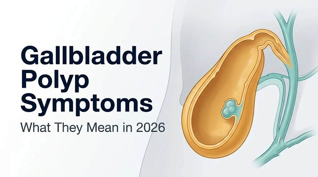
Gallbladder Polyp Symptoms: What They Mean in 2026
Gallbladder polyp symptoms are often absent entirely, but when they do occur, they point to specific disruptions in biliary mechanics that deserve careful clinical evaluation rather than reassurance alone. Understanding what these symptoms are, why they happen, and how they differ from other gallbladder problems gives you the clearest picture of what a polyp finding actually means for your health.
According to the National Institute of Diabetes and Digestive and Kidney Diseases (NIDDK), gallbladder disease broadly affects millions of Americans, and incidentally discovered gallbladder polyps are becoming more frequent as abdominal ultrasound use increases across routine and diagnostic imaging. A 2020 systematic review published in Gut estimated that gallbladder polyps are detected in approximately 0.3% to 9.5% of the general population undergoing abdominal ultrasound, with most patients having no idea the polyp existed before their scan.
This article covers what gallbladder polyps are anatomically, the full clinical picture of symptoms when they occur, why most polyps produce no symptoms at all, how polyp type and size change the risk equation, and the specific thresholds that move this from “watch and wait” to urgent medical action.
What Are Gallbladder Polyps
A gallbladder polyp is a focal projection of tissue growing from the inner wall of the gallbladder into its hollow lumen. The gallbladder is a small, pear-shaped organ nestled in the gallbladder fossa on the inferior surface of the right hepatic lobe. Its primary job is to store and concentrate bile produced by the liver, releasing it into the common bile duct and then the duodenum via the sphincter of Oddi when a meal arrives.
Polyps form when cells in the gallbladder mucosa (the columnar epithelial lining) or deeper wall layers proliferate abnormally or when cholesterol and other lipids accumulate in foam cells within that mucosal layer. The result is a growth that protrudes into the lumen. Most gallbladder polyps are small (under 10mm), sessile or pedunculated (stalk-based), and discovered incidentally rather than through symptomatic investigation.
The clinical importance of a gallbladder polyp depends almost entirely on its type, size, and the characteristics of the person who has it. A 4mm cholesterol deposit with a stalk carries essentially no cancer risk. A 12mm sessile lesion in a 65-year-old with primary sclerosing cholangitis carries a substantially different clinical weight.
Think of the gallbladder like a small balloon that fills with bile between meals and squeezes to release it when you eat. A polyp growing from its wall is like a bump on the inside of that balloon. Small bumps change nothing. Larger bumps, or bumps near the outlet, can interfere with the squeeze.
Key clinical fact: The American College of Gastroenterology (ACG) distinguishes between polypoid lesions of the gallbladder that are non-neoplastic (the majority) and those that are true neoplasms requiring active management, a distinction that drives every decision from surveillance interval to surgery timing.
How Common Are Gallbladder Polyps
Gallbladder polyps are found in roughly 0.3% to 9.5% of the general population, with the wide range reflecting differences in imaging technology, study population, and geographic region. A large population-based study published in Gut in 2016, analyzing over 50,000 individuals in Denmark, found a prevalence of approximately 4.5% in adults undergoing abdominal ultrasound, making gallbladder polyps a relatively frequent incidental finding in clinical practice.
The vast majority of detected polyps are benign. Research published in the British Journal of Surgery consistently shows that approximately 95% of gallbladder polyps are non-neoplastic cholesterol pseudopolyps or adenomyomatosis lesions. True neoplastic polyps (adenomas and early carcinomas) account for roughly 4% to 5% of all gallbladder polypoid lesions.
Detection rates have risen noticeably over the past two decades. The widespread adoption of high-resolution transabdominal ultrasound for evaluating abdominal complaints means more polyps are found at smaller sizes, in younger patients, before any symptoms develop. This incidental discovery pattern means many people receiving this diagnosis are understandably confused about what it actually implies for their health.
Prevalence appears slightly higher in East Asian populations compared to European and North American populations in several epidemiological studies, though researchers have not yet identified a definitive biological mechanism for this geographic variation. Within any population, age over 50 and male biological sex correlate with higher rates of neoplastic polyp type specifically, not with overall polyp prevalence.
Gallbladder Polyp Symptoms
The direct clinical answer is this: most gallbladder polyps produce no symptoms whatsoever, but when gallbladder polyp symptoms do occur, they are typically felt as a dull to cramping ache in the right upper quadrant (RUQ) of the abdomen, sometimes accompanied by nausea, bloating, or intermittent discomfort that worsens after high-fat meals.
The symptom pattern, when present, is generated by the same biliary mechanics responsible for gallstone symptoms. The cystic duct connecting the gallbladder neck to the common bile duct is a narrow passage. When a polyp grows near the gallbladder neck or reaches a size that contributes to intermittent flow disruption, the pressure generated by cholecystokinin (CCK)-triggered gallbladder smooth muscle contraction against that partial obstruction activates visceral afferent pain fibers running along T8 to T9 spinal pathways. The result is a cramping or squeezing sensation in the RUQ.
Here is a full inventory of recognized gallbladder polyp symptoms when they occur:
| Symptom | Mechanism | Clinical Significance |
|---|---|---|
| Right upper quadrant pain (dull to cramping) | Increased biliary pressure against cystic duct during contraction | Most common symptomatic presentation |
| Pain after fatty meals | CCK release triggers gallbladder contraction against restricted outflow | Temporally linked to dietary fat intake |
| Nausea without vomiting | Visceral afferent nerve activation; delayed gastric emptying from biliary dysfunction | Frequently accompanies RUQ discomfort |
| Right shoulder or scapular pain | Referred pain via phrenic nerve (C3-C5) and right subscapular afferents | Often misattributed to musculoskeletal cause |
| Bloating or upper abdominal fullness | Altered biliary motility affecting gastric emptying | Nonspecific; easily confused with other GI conditions |
| Jaundice (rare) | Larger polyp or associated stone causing common bile duct obstruction | Requires urgent evaluation |
| Fever and rigors (rare) | Secondary infection, acute cholecystitis complicating a polyp | Emergency presentation |
Women over 40 may experience RUQ symptoms that are initially attributed to musculoskeletal causes or irritable bowel syndrome (IBS) before biliary pathology is considered, which can delay correct diagnosis. Any right-sided upper abdominal pain lasting more than 30 minutes that occurs consistently after meals warrants evaluation by a primary care physician with abdominal ultrasound ordered.
Do Gallbladder Polyps Cause Pain
Gallbladder polyps can cause pain, but whether they do depends on the polyp’s size, location within the gallbladder, and whether it creates any degree of obstruction to the cystic duct or gallbladder neck during the contraction cycle.
The gallbladder contracts in response to cholecystokinin, a peptide hormone secreted by I-cells in the duodenal mucosa when dietary fat and protein enter the small intestine. This contraction pushes bile from the gallbladder through the cystic duct into the common bile duct. Under normal circumstances, this process is painless. When a polyp partially blocks or stiffens the gallbladder outlet, the pressure generated during contraction rises above the threshold that activates visceral nociceptors in the gallbladder wall’s visceral peritoneum, producing pain.
Small polyps under 5mm in a mid-body location rarely produce this effect. Larger polyps or those at the gallbladder neck have a higher chance of contributing to biliary pressure elevation. The pain is typically described as a dull pressure or cramping sensation rather than a sharp stabbing pain, and it typically builds over 15 to 30 minutes after eating before gradually easing.
It’s worth noting that many patients with confirmed gallbladder polyps who report RUQ pain may be experiencing symptoms from concurrent gallstones or biliary dyskinesia rather than from the polyp itself. A 2019 retrospective analysis published in the Journal of Gastrointestinal Surgery found that polyp-related symptoms are difficult to isolate clinically because polyps and gallstones frequently coexist. Imaging and clinical correlation are both necessary before attributing pain to a polyp specifically.
Populations where pain evaluation is more complex:
- Older adults over 65 may have attenuated visceral pain responses, presenting with milder or more vague discomfort despite significant pathology
- People taking chronic nonsteroidal anti-inflammatory drugs (NSAIDs) may have reduced symptom intensity from the anti-prostaglandin effect, masking escalating biliary pressure symptoms
- Pregnant women experience shifts in biliary motility from elevated progesterone, which relaxes gallbladder smooth muscle and reduces contraction force, potentially altering the typical symptom pattern
Symptoms of Gallbladder Polyps After Eating
Gallbladder polyp symptoms after eating occur because food intake, particularly fat-containing meals, is the primary trigger for gallbladder contraction via CCK release, and any polyp contributing to biliary flow resistance becomes symptomatic only during that active contraction phase.
The sequence is reliable. A meal containing fat or protein reaches the duodenum within 20 to 40 minutes of consumption. Duodenal I-cells respond by secreting CCK into the bloodstream. CCK binds to CCK-A receptors on gallbladder smooth muscle, triggering sustained contraction. If a polyp creates any resistance at the gallbladder neck or cystic duct junction, pain develops during this contraction window. Symptoms typically appear 20 to 60 minutes after eating and may last anywhere from 20 minutes to a few hours before resolving as the gallbladder relaxes.
Foods that reliably trigger this response include:
- Full-fat dairy products (cream, cheese, butter)
- Fried foods and oils
- Fatty cuts of meat and processed meats
- Pastries and baked goods with high fat content
- Avocado and nuts in large quantities (both contain significant dietary fat)
- Spicy foods (can increase gastrointestinal motility independently)
Patients sometimes describe a pattern of discomfort that occurs with every fatty meal but resolves completely between episodes, allowing normal eating in between. This intermittent pattern is one clinical characteristic that distinguishes polyp-related or biliary colic-type pain from the more constant pain of acute cholecystitis, where the gallbladder wall itself is inflamed and tender persistently.
If your post-meal RUQ discomfort is becoming more frequent, more intense, or is now persisting beyond a few hours, speak with a primary care physician rather than waiting for a scheduled appointment. Increasing symptom frequency can indicate a polyp growing in size, development of a coexisting gallstone, or early inflammation warranting updated imaging.
Key Takeaway: Gallbladder polyp symptoms, when they occur at all, are most reliably triggered by fatty meals and felt as cramping right upper quadrant discomfort, because dietary fat is the primary driver of the CCK-triggered gallbladder contractions that generate biliary pressure against a partially obstructed cystic duct.
Asymptomatic Gallbladder Polyps: When There Are No Symptoms
The majority of gallbladder polyps produce no symptoms at all, and this is the most common clinical scenario: a polyp is discovered incidentally during an abdominal ultrasound ordered for an unrelated reason, and the patient has never felt anything attributable to it.
The physiological reason for this is straightforward. Most polyps are small (under 6mm), located in the gallbladder body or fundus (away from the cystic duct outlet), and composed of cholesterol deposits or adenomyomatosis tissue rather than proliferating neoplastic cells. A 4mm cholesterol pseudopolyp sitting in the mid-gallbladder body creates no obstruction during contraction, generates no elevated intraluminal pressure, and activates no visceral pain pathways. From the body’s perspective, it is structurally inconsequential.
Research published in Gut in 2017 confirmed that the vast majority of incidentally discovered polyps under 6mm remain stable in size over years of surveillance and do not develop symptoms during that time. Approximately 60% to 70% of all discovered polyps fall into this size and low-risk category.
The clinical challenge is that “asymptomatic” does not mean “without clinical significance.” A small neoplastic polyp (an adenoma, for example) may be entirely asymptomatic at 7mm while carrying a measurable cancer risk that grows as the polyp grows. The absence of symptoms is reassuring in the short term but is not a reliable indicator of polyp type, malignant potential, or the need for surveillance.
Patients with asymptomatic polyps should not interpret the absence of pain as confirmation that the polyp is benign. Size, morphology (sessile versus pedunculated), and clinical risk factors determine surveillance need. A primary care physician or gastroenterologist should review imaging findings and establish an appropriate follow-up plan regardless of whether symptoms are present.
Types of Gallbladder Polyps and Why the Type Matters
Not all gallbladder polyps are the same tissue, and the type of polyp is the single most important factor determining cancer risk, required surveillance frequency, and the threshold for surgical intervention.
Non-neoplastic polyps (pseudopolyps): These account for approximately 95% of all gallbladder polypoid lesions. They include:
- Cholesterol pseudopolyps: The most common type overall, comprising roughly 60% to 70% of all gallbladder polyps. These are accumulations of cholesterol-laden macrophages (foam cells) within the gallbladder mucosa. They appear bright on ultrasound and are almost always benign, typically pedunculated (on a stalk), and under 10mm.
- Adenomyomatosis: A hyperplastic condition of the gallbladder wall where the mucosal epithelium invaginates into the muscular layer (Rokitansky-Aschoff sinuses), forming focal mural thickening that appears polyp-like on imaging. Adenomyomatosis itself carries no malignant potential, though focal adenomyomatosis at the gallbladder neck can sometimes be difficult to distinguish from early carcinoma on standard ultrasound.
- Inflammatory polyps: Rare, resulting from chronic cholecystitis-related mucosal proliferation.
True neoplastic polyps: These represent a small minority but carry all the clinically relevant cancer risk:
- Gallbladder adenoma: A true benign neoplasm with documented malignant transformation potential. Adenomas are typically sessile, appear on ultrasound as isoechoic masses, and may grow over serial imaging. A 2022 analysis published in Annals of Surgery found that adenomas 10mm or larger carried a malignant transformation rate exceeding 50% in long-term follow-up studies.
- Early gallbladder carcinoma: Can present as a sessile polypoid lesion before developing into a mass. This is why size and morphology on imaging trigger surgical review rather than continued surveillance in high-risk presentations.
Understanding your polyp type requires imaging interpretation by a radiologist and clinical correlation by a gastroenterologist or hepatobiliary surgeon. The type cannot be determined from symptoms alone.
Gallbladder Polyp Cancer Risk and Size Thresholds
The cancer risk from gallbladder polyps is not theoretical for larger lesions. Size above 10mm is the threshold most associated with malignancy, and current clinical guidelines from the Society of American Gastrointestinal and Endoscopic Surgeons (SAGES) and from European hepatobiliary societies recommend cholecystectomy for polyps reaching or exceeding this measurement.
Here is how size maps to risk in the published evidence:
| Polyp Size | Estimated Malignancy Risk | Recommended Clinical Action |
|---|---|---|
| Under 6mm | Very low (less than 1%) | Ultrasound surveillance, often 1 to 2 years |
| 6 to 9mm | Low to moderate (1% to 5%) | 6-month to annual surveillance ultrasound |
| 10mm or larger | Moderate to high (varies by type, up to 50% for adenomas) | Surgical referral for cholecystectomy consideration |
| Sessile morphology at any size | Higher risk independent of size | Shorter surveillance interval or surgical discussion |
The 10mm threshold is not arbitrary. A large retrospective cohort study published in Gut in 2015 analyzed the outcomes of over 100 patients with polypoid gallbladder lesions and found that all gallbladder carcinomas presenting as polypoid lesions in their dataset were 10mm or larger at the time of resection. Multiple subsequent studies have replicated this finding.
Size alone, however, is not the complete risk picture. Sessile morphology (no stalk, broad base attaching directly to the gallbladder wall) at any size above 5mm carries a higher concern level than a pedunculated (stalked) polyp of the same measurement. A 7mm sessile lesion in a 62-year-old male warrants a different conversation with a hepatobiliary surgeon than a 7mm pedunculated cholesterol polyp in a 35-year-old woman.
Growth rate also matters independently of absolute size. Any polyp growing 2mm or more between surveillance scans warrants prompt clinical review regardless of its current total measurement, according to clinical guidance published by the British Society of Gastroenterology in 2022.
Key Takeaway: The 10mm size threshold is the most evidence-supported trigger for surgical referral, but sessile morphology, rapid growth between scans, and high-risk patient characteristics (PSC, FAP, age over 60) shift the action threshold downward to 6mm in current specialist guidelines.
What Causes Gallbladder Polyps
Gallbladder polyps form through different mechanisms depending on their type, and understanding the cause helps explain both why certain people develop them and why most are benign.
Cholesterol pseudopolyps form when cholesterol-laden foam cells (lipid-engorged macrophages) accumulate in the gallbladder mucosal surface. The precise trigger appears to be chronic bile supersaturated with cholesterol, a state linked to high dietary cholesterol intake, obesity, and lipid metabolism disorders. The bile effectively deposits cholesterol crystals into the mucosal surface, and the immune system’s macrophages attempt to clear them but become overloaded, producing the characteristic “pseudopolyp” nodule. This is a metabolic process, not a proliferative or neoplastic one.
Adenomyomatosis arises from abnormal mucosal epithelial invagination into the underlying muscular layer of the gallbladder wall, producing Rokitansky-Aschoff sinuses. The trigger is poorly understood but is associated with chronic gallbladder inflammation and possibly elevated intraluminal pressure from chronic bile stasis.
Gallbladder adenomas (true neoplastic polyps) develop from mutations in gallbladder epithelial cells driving uncontrolled mucosal cell proliferation. The specific molecular pathways are not fully characterized, but research has identified associations with mutations in the KRAS and PIK3CA genes in some adenoma subtypes. Chronic inflammation and bile stasis appear to be contributing environmental factors.
Named causes by frequency:
- Cholesterol supersaturation in bile (most common mechanism, producing pseudopolyps)
- Chronic gallbladder inflammation (contributing to adenomyomatosis and inflammatory polyps)
- Metabolic syndrome and lipid metabolism dysfunction (associated with cholesterol polyps)
- Genetic predisposition including familial adenomatous polyposis (FAP) gene mutations (associated with true neoplastic polyps)
- Primary sclerosing cholangitis-related chronic biliary inflammation (associated with increased adenoma and carcinoma risk)
The NIDDK notes that bile composition and gallbladder motility are influenced by diet, hormonal environment, and genetic factors, all of which intersect in the production of various gallbladder pathologies including polypoid lesions.
Gallbladder Polyp Risk Factors
Specific patient characteristics consistently appear in the clinical literature as associated with a higher rate of gallbladder polyp detection or, more critically, with a higher risk of neoplastic (potentially malignant) polyp type.
Risk factors for any gallbladder polyp:
- Obesity (body mass index above 30), particularly abdominal obesity, associated with bile cholesterol supersaturation
- Metabolic syndrome (insulin resistance, dyslipidemia, hypertension)
- Age over 50
- Dietary patterns high in saturated fat and cholesterol
- Chronic liver disease (altered bile composition and biliary motility)
Risk factors specifically for neoplastic (cancer-risk) polyp types:
- Primary sclerosing cholangitis (PSC): This chronic inflammatory condition of the bile ducts dramatically increases the risk of gallbladder adenoma and carcinoma. The European Association for the Study of the Liver (EASL) recommends annual ultrasound surveillance and consideration of cholecystectomy for any polyp exceeding 8mm in PSC patients specifically, a lower threshold than for the general population.
- Familial adenomatous polyposis (FAP): This inherited condition caused by mutations in the APC tumor suppressor gene produces adenomatous polyps throughout the gastrointestinal tract, including the gallbladder. FAP patients have a substantially elevated lifetime risk of gallbladder adenoma development.
- Male biological sex: Several large cohort studies have found that while women have a higher overall prevalence of gallbladder polyps (largely driven by higher cholesterol polyp rates), male patients with a polyp are more likely to have a neoplastic (adenoma or carcinoma) type than female patients of the same age.
- Age over 60: Independently associated with neoplastic polyp type in multiple prospective imaging surveillance studies.
- Gallstones coexisting with a polyp: Increases the surgical management consideration because gallstones independently elevate gallbladder carcinoma risk and make imaging differentiation between polyp and stone more difficult.
Key Takeaway: Having a gallbladder polyp is not inherently dangerous for most people, but specific characteristics including PSC, FAP, male sex, age over 60, and sessile morphology shift a patient from low-risk to a category where specialist review and potentially earlier surgery are the appropriate response.
Gallbladder Polyp Symptoms in Specific Populations
Gallbladder polyp symptoms can present differently, or carry different clinical significance, depending on the individual’s biological sex, age, concurrent health conditions, reproductive status, or medication history.
Biological sex differences:
Women are more likely to have gallbladder polyps detected overall, reflecting higher rates of cholesterol pseudopolyps linked to hormonal influences on biliary cholesterol metabolism. Estrogen increases biliary cholesterol secretion, which can contribute to bile supersaturation and cholesterol crystal deposition in the gallbladder mucosa. Postmenopausal women on hormone replacement therapy may have an amplified version of this effect. That said, women’s polyps are predominantly benign cholesterol pseudopolyps. Men who develop gallbladder polyps face a statistically higher probability of adenoma type, meaning a newly found gallbladder polyp in a man over 50 carries a different clinical conversation than the same finding in a woman of the same age.
Older adults:
Adults over 65 may experience attenuated visceral pain responses due to reduced sensory nerve density in the gallbladder wall and lower baseline pain sensitivity. This means a polyp contributing to biliary pressure elevation may cause less obvious symptoms in an older person, potentially delaying recognition of an escalating problem. Any new right upper quadrant discomfort in an older adult should prompt imaging evaluation rather than watchful waiting, given that both symptom and cancer risk are amplified with age.
Patients with primary sclerosing cholangitis:
The clinical significance of any gallbladder polyp in a PSC patient is substantially elevated. A 2019 study published in the journal Gut found that PSC patients with gallbladder polyps had a risk of gallbladder carcinoma approximately 14 times higher than the general population with equivalent polyp findings. The size threshold for surgical action is lower (6 to 8mm versus 10mm for the general population), and annual surveillance ultrasound is standard of care for all PSC patients regardless of prior polyp findings.
During pregnancy:
Progesterone-mediated relaxation of gallbladder smooth muscle during pregnancy reduces contractile force and increases bile stasis, altering the likelihood that a polyp will generate symptoms even if it is obstruction-prone in a non-pregnant state. Newly discovered polyps during pregnancy should be documented and followed postpartum; cholecystectomy during pregnancy is generally deferred to the second trimester if required for symptomatic disease, or to the postpartum period for asymptomatic findings.
Gallbladder Polyp vs Gallstone Symptoms
Distinguishing gallbladder polyp symptoms from gallstone symptoms is clinically important and genuinely difficult because both conditions share the same anatomical location and the same primary symptom-generating mechanism: elevated biliary pressure against the cystic duct during gallbladder contraction.
The key differentiating features in clinical practice:
| Feature | Gallbladder Polyp | Gallstone (Cholelithiasis) |
|---|---|---|
| Pain character | Dull to moderate cramping, often milder | Severe colicky cramping, often described as the worst pain experienced |
| Pain duration | Usually resolves within 1 to 3 hours | Typically 30 minutes to 6 hours; acute cholecystitis pain is persistent |
| Pain onset | Gradual over 15 to 30 minutes post-meal | Can be sudden and intense, especially if stone lodges in cystic duct |
| Fever | Absent unless acute cholecystitis develops | Absent in uncomplicated biliary colic; present in acute cholecystitis |
| Jaundice | Rare; occurs only with large polyp obstructing common bile duct | Can occur when a stone enters the common bile duct (choledocholithiasis) |
| Reproducibility | Pain pattern often reproducible after fatty meals | Variable; some gallstone attacks occur at rest or at night |
| Ultrasound appearance | Immovable echogenic protrusion from gallbladder wall | Mobile, gravity-dependent echogenic foci with acoustic shadowing |
| Symptom relief when lying still | Variable | Pain is typically positional-independent |
The acoustic shadowing difference on ultrasound is the most reliable way to distinguish the two. Gallstones produce a characteristic dark shadow behind the echogenic (bright) stone on ultrasound because they block the ultrasound beam. Gallbladder polyps do not produce this shadow, appearing as fixed wall projections that do not shift with patient position.
Clinically, both conditions can coexist in the same gallbladder. When symptoms are present alongside both findings on imaging, a hepatobiliary specialist should evaluate whether the gallstone, the polyp, or both are contributing to the clinical picture. Self-assessment cannot reliably make this distinction. A gastroenterologist or hepatobiliary surgeon should review any combination finding.
How Gallbladder Polyps Are Diagnosed
Gallbladder polyps are diagnosed through imaging, not through symptoms, and the standard first-line tool is transabdominal ultrasound (US). This noninvasive imaging technique uses high-frequency sound waves to visualize the gallbladder wall and detect protrusions into the lumen as small as 2 to 3mm with modern high-resolution equipment.
The typical diagnostic pathway:
- Transabdominal ultrasound detects a polypoid lesion of the gallbladder wall as a fixed, non-shadowing echogenic protrusion.
- The reporting radiologist notes size in millimeters, number of lesions, morphology (sessile or pedunculated), and whether acoustic shadowing is present (which would suggest stone rather than polyp).
- The referring physician (primary care, emergency medicine, or gastroenterologist) reviews the radiology report in the context of patient symptoms, age, and risk factors.
- For lesions that are indeterminate, large, or in high-risk patients, contrast-enhanced ultrasound (CEUS) is increasingly used to characterize vascularity of the lesion (neoplastic polyps tend to show enhancement patterns that cholesterol pseudopolyps do not).
- For complex or high-concern lesions, magnetic resonance cholangiopancreatography (MRCP) provides detailed biliary tract anatomy without ionizing radiation.
- Endoscopic ultrasound (EUS) offers the highest-resolution imaging of gallbladder wall layers and is used for equivocal lesions where the distinction between a polyp and early carcinoma is clinically critical.
Bloodwork alone cannot diagnose a gallbladder polyp. Liver function tests including alkaline phosphatase (ALP), gamma-glutamyl transferase (GGT), and bilirubin are ordered when biliary obstruction is suspected, and elevated values may suggest a larger lesion with bile flow impact, but normal labs do not rule out a clinically significant polyp.
Tissue biopsy of a gallbladder polyp prior to surgery is not typically performed because the gallbladder wall’s position makes percutaneous biopsy technically difficult and because biopsy does not reliably sample the full lesion. Definitive tissue diagnosis occurs at cholecystectomy.
Key Takeaway: Gallbladder polyps are diagnosed by imaging, not by symptoms or blood tests alone. Transabdominal ultrasound is the first-line tool, but CEUS and EUS provide higher-resolution characterization for indeterminate lesions that standard ultrasound cannot reliably classify.
Gallbladder Polyp Monitoring Guidelines and Follow-Up
Gallbladder polyp monitoring guidelines are based on polyp size, morphology, growth rate, and patient-specific risk factors. Current guidance from the European Society of Gastrointestinal Endoscopy (ESGE) and the British Society of Gastroenterology (BSG), published in updated 2022 guidance documents, provides a structured surveillance framework that has largely replaced older, more aggressive monitoring schedules.
General surveillance recommendations:
| Polyp Size | Patient Risk | Recommended Surveillance |
|---|---|---|
| Under 6mm, pedunculated | Low risk (no PSC, FAP, age under 60) | Repeat ultrasound at 1 year; if stable, discharge to routine care |
| 6 to 9mm, any morphology | Intermediate risk | Ultrasound at 6 months, then annually for 5 years |
| 6 to 9mm, sessile or high-risk patient | High risk (PSC, FAP, male over 60) | Discuss cholecystectomy; EUS or MRCP for further characterization |
| 10mm or larger | Any patient | Refer to hepatobiliary surgeon for cholecystectomy |
| Any polyp showing growth of 2mm or more | Any patient | Shorten surveillance interval; reassess surgical candidacy |
The 2022 BSG guidance represents a refinement from older European Association for Endoscopic Surgery (EAES) guidelines, incorporating long-term follow-up data showing that low-risk small polyps rarely change behavior over years of surveillance, allowing clinicians to reduce unnecessary repeat imaging for truly stable, small, pedunculated lesions in low-risk patients.
Patients with PSC or FAP should understand that their surveillance schedule is more intensive than what their polyp size alone would suggest. A 5mm polyp in a PSC patient warrants annual ultrasound; the same polyp in a healthy 35-year-old without risk factors may be rescanned at one year and, if stable, require no further imaging.
Patients should request a written copy of their surveillance plan from their managing physician, including the next scheduled imaging date, the specific measurement of the polyp documented on the last scan, and the threshold that would prompt surgical discussion. This baseline documentation is useful if imaging is performed at a different institution in the future.
Gallbladder Polyp Treatment and When Surgery Is Recommended
The definitive treatment for a gallbladder polyp that meets surgical criteria is laparoscopic cholecystectomy, the minimally invasive surgical removal of the entire gallbladder. There is no medical treatment that shrinks, eliminates, or prevents gallbladder polyps. Dietary modification can reduce symptom frequency if symptoms are present, but it does not change polyp size or type.
Surgery is recommended in the following clinical circumstances, consistent with guidelines from the Society of American Gastrointestinal and Endoscopic Surgeons (SAGES) and the 2022 BSG guidance:
- Any polyp measuring 10mm or larger at any age
- Any polyp 6mm or larger in a patient with primary sclerosing cholangitis or familial adenomatous polyposis
- Any sessile polyp 6mm or larger in a patient over 60 with no other explanation for the lesion
- Any polyp demonstrating growth of 2mm or more on serial surveillance imaging
- Any symptomatic polyp (confirmed symptomatic relationship) where symptoms are not controlled and imaging shows the polyp is in a position consistent with the symptom pattern
- Coexisting cholelithiasis (gallstones) with a polyp, where surgery for stones is already clinically indicated
Laparoscopic cholecystectomy for a gallbladder polyp is a low-complication procedure when performed electively in otherwise healthy patients. The Mayo Clinic reports a complication rate under 3% for elective laparoscopic cholecystectomy in low-risk patients, with the primary risks being bile duct injury, bleeding, and infection, all of which are significantly lower for experienced hepatobiliary surgeons at high-volume centers.
Patients who do not meet surgical criteria should not push for cholecystectomy solely because a polyp was found. The gallbladder serves a physiological function in regulating bile delivery to the duodenum, and its removal carries long-term implications for bile acid metabolism and fat digestion that are relevant quality-of-life considerations, particularly for younger patients.
For patients in the surveillance category who are anxious about watchful waiting, a one-time consultation with a hepatobiliary surgeon (a surgeon with specific training in liver, gallbladder, and bile duct surgery) is reasonable to discuss the individual risk-benefit calculation in personal clinical terms.
Emergency Symptoms: When to Call 911 or Go to the ER
Certain symptoms associated with gallbladder disease, including complications arising in the setting of a known gallbladder polyp, require immediate emergency evaluation. Do not wait to see if these resolve on their own.
Call 911 or go to the nearest emergency room immediately if you experience:
- Severe right upper quadrant pain that does not resolve within 6 hours and is accompanied by fever above 38.5°C (101.3°F) or chills, indicating possible acute cholecystitis or cholangitis
- Yellowing of the skin or whites of the eyes (jaundice) combined with abdominal pain, indicating possible biliary obstruction from a large polyp or coexisting stone in the common bile duct
- Sudden, severe, diffuse abdominal pain that spreads across the entire abdomen and is accompanied by a rigid, board-like abdominal wall, suggesting possible gallbladder perforation and peritonitis
- Fever above 39°C (102.2°F) with rapid heart rate, low blood pressure, or confusion alongside abdominal pain, which may indicate biliary sepsis (ascending cholangitis) requiring immediate intravenous antibiotics and emergency biliary decompression
- Uncontrolled vomiting with inability to keep any fluids down combined with abdominal pain lasting more than 4 hours
- Right upper quadrant pain in a pregnant person that is severe, escalating, or accompanied by any of the above features
These presentations can indicate acute cholecystitis, ascending cholangitis, gallbladder perforation, biliary sepsis, or choledocholithiasis with obstructive jaundice, all of which require emergency medical assessment, not an urgent care appointment or a phone call to a nurse line.
Frequently Asked Questions About Gallbladder Polyp Symptoms
What does a gallbladder polyp feel like?
A gallbladder polyp typically produces no sensation at all in most people.
When symptoms are present, they are usually felt as a dull to moderate cramping pressure in the right upper abdomen, sometimes spreading toward the right shoulder blade, and tend to be triggered by fatty meals rather than occurring at rest.
Pain from a gallbladder polyp is generally milder and shorter-lasting than the severe colicky pain associated with gallstones, though the two conditions can coexist and produce overlapping sensations.
Can a gallbladder polyp cause pain after eating?
Yes, a gallbladder polyp can cause pain after eating, specifically after meals containing fat or protein, because these macronutrients trigger cholecystokinin (CCK) release from the duodenum, which drives gallbladder contraction.
If the polyp creates any resistance to bile outflow during that contraction, biliary pressure rises and activates visceral pain fibers in the gallbladder wall, producing the cramping sensation that typically begins 20 to 60 minutes after the meal.
This post-meal pain pattern that reliably resolves within one to three hours warrants evaluation by a primary care physician with abdominal ultrasound, particularly if it occurs repeatedly.
Are gallbladder polyp symptoms different from gallstone symptoms?
Gallbladder polyp symptoms, when present, tend to be milder and more gradual in onset than the severe, sudden, colicky pain typical of a gallstone blocking the cystic duct.
Gallstones frequently cause intense pain episodes that can awaken a person from sleep or develop at rest, while polyp-related discomfort is more predictably meal-triggered and less severe.
Both conditions share right upper quadrant pain as a feature, and both can coexist in the same gallbladder, making ultrasound imaging essential for distinguishing them; symptoms alone cannot reliably differentiate the two.
Can a gallbladder polyp go away on its own?
True neoplastic gallbladder polyps, including adenomas, do not resolve on their own and require monitoring or surgical removal.
Cholesterol pseudopolyps, which make up the majority of gallbladder polyps, may occasionally decrease in size or change appearance on serial ultrasound, but there is no reliable method to make them disappear without surgical removal of the gallbladder.
Dietary changes that reduce bile cholesterol supersaturation (lower saturated fat intake, weight management) may theoretically slow cholesterol pseudopolyp formation, but this has not been confirmed in controlled clinical trials.
At what size does a gallbladder polyp become dangerous?
A gallbladder polyp reaches a clinically concerning size threshold at 10mm, at which point current guidelines from the Society of American Gastrointestinal and Endoscopic Surgeons and the British Society of Gastroenterology recommend referral to a hepatobiliary surgeon for cholecystectomy consideration.
For high-risk patients including those with primary sclerosing cholangitis, familial adenomatous polyposis, male patients over 60, or sessile lesions, the threshold for surgical discussion is lower at 6 to 8mm.
Any polyp that grows 2mm or more between surveillance imaging scans warrants prompt clinical review regardless of its current total size.
Do gallbladder polyps always need to be removed?
No, the large majority of gallbladder polyps do not require surgical removal.
Small, pedunculated, low-risk polyps under 6mm in patients without high-risk characteristics (no PSC, FAP, or advanced age) are managed with periodic ultrasound surveillance rather than cholecystectomy, and many remain stable for years without intervention.
Surgery is specifically recommended for polyps 10mm or larger, polyps in high-risk patients at lower size thresholds, polyps showing documented growth on serial imaging, and polyps causing confirmed symptoms that affect quality of life.
Closing
Most people who learn they have a gallbladder polyp leave their imaging appointment with a radiology report in hand and very little practical understanding of what it means. The clinical picture is actually quite stratifiable: polyp type and size, combined with your individual risk factors, determine whether this is a finding that needs a single follow-up scan or a conversation with a hepatobiliary surgeon.
If your polyp is under 6mm, pedunculated, and you have no history of PSC, FAP, or other high-risk features, a repeat ultrasound in one year is the standard next step. If your polyp is at or above 10mm, if you have PSC or FAP, or if any new symptoms are developing, speak with a gastroenterologist or hepatobiliary surgeon rather than waiting for a routine appointment slot.
The symptom question that brings most people to this topic is valid and worth taking seriously. Right upper quadrant discomfort that reliably follows fatty meals, particularly if it’s consistent and reproducible, deserves evaluation with imaging, not reassurance that it’s “just stress.” You have enough specific clinical information now to ask the right questions at your next appointment.