Back Spasm Symptoms: What They Feel Like in 2026
Back spasm symptoms typically arrive without warning: a sudden, gripping tightness or sharp contraction in the back muscles that can stop you mid-motion and make it nearly impossible to stand, bend, or breathe deeply without pain. Recognizing what these symptoms feel like, and distinguishing a straightforward muscle spasm from something that requires immediate medical care, is exactly what this article covers.
According to the National Institute of Arthritis and Musculoskeletal and Skin Diseases (NIAMS), low back pain is one of the most common reasons adults seek medical care, affecting roughly 80% of Americans at some point in their lives. Muscle spasm is one of the leading causes of acute episodes. The sheer prevalence means most people will experience this at least once, yet the clinical picture is poorly understood by most of the health content available online.
This article covers the full range of back spasm symptoms, from the first involuntary muscle clench to the warning signs that demand emergency evaluation. It addresses how symptoms differ by location in the spine, how they change across age groups and during pregnancy, and exactly how to tell a spasm from a pulled muscle, a kidney problem, or a disc injury.
Back Spasm Symptoms
Back spasm symptoms include sudden, involuntary muscle contraction in the back that produces sharp or cramping pain, visible or palpable muscle hardness, restricted movement, and in some cases, referred pain, numbness, or tingling depending on the specific muscles and spinal structures involved.
The hallmark is the involuntary nature. Unlike ordinary muscle soreness or stiffness, a spasm occurs without voluntary input. The muscle fires on its own, contracts tightly, and often refuses to release. That contraction can last seconds, minutes, or persist as a sustained tightness for hours to days.
Pain intensity varies widely. Some spasms produce a dull, deep aching that limits movement but remains tolerable. Others generate a sharp, searing pain rated by patients at 8 or 9 out of 10 on the Visual Analog Scale for pain, severe enough to collapse a person to the floor from a standing position.
Key symptoms across all types of back spasms:
- Sudden tightening or grabbing sensation in the back muscles
- Localized muscle hardness or knot that is visible or palpable
- Pain that worsens with movement, twisting, or bending
- Restricted range of motion in the torso
- Muscle fatigue and soreness in the hours following the acute contraction
- Postural changes: leaning to one side or forward to reduce muscle tension
- Protective guarding: involuntary stiffening of the trunk when attempting movement
For people with pre-existing degenerative disc disease at the L4-L5 or L5-S1 levels, spasm symptoms tend to be more intense and more frequently recurrent. The compromised disc absorbs less mechanical shock, placing greater stress on the surrounding paraspinal musculature during even ordinary daily movements.
What Are Back Muscle Spasms and How Do They Form
A back muscle spasm is an involuntary, sustained contraction of one or more of the paraspinal muscles produced by a neurophysiological cascade that begins with tissue stress or injury and ends with a muscle that cannot release itself from contraction.
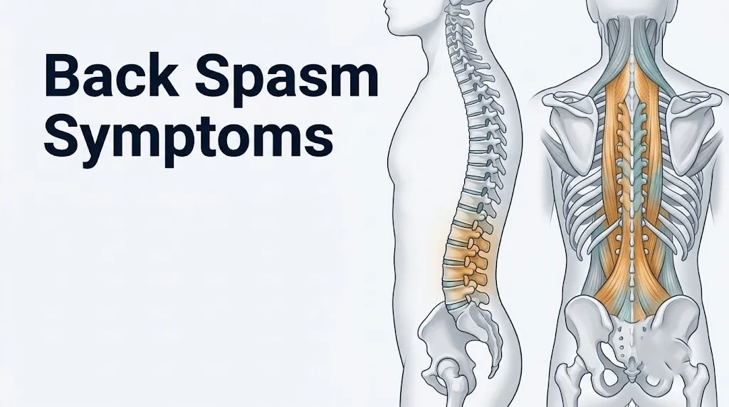
The key muscles involved are the erector spinae group (composed of the iliocostalis, longissimus, and spinalis), the multifidus, and the quadratus lumborum. In the upper back, the rhomboid major, rhomboid minor, trapezius, and thoracic paraspinal muscles are the primary structures affected.
The formation process works like this: mechanical stress, injury, or overuse activates nociceptors (pain-sensing nerve endings) in the muscle tissue. Those nociceptors trigger the release of substance P and prostaglandins into the local tissue environment, initiating a pain and inflammatory signal that travels through the dorsal root ganglia to the spinal cord. The spinal cord’s reflex arc responds by increasing the firing rate of gamma motor neurons, which raises tension in the muscle spindle apparatus. This effectively tells the muscle to contract harder as a protective splinting mechanism against further injury.
Think of it like the muscle locking itself in a protective brace. The body is trying to prevent movement that it has detected as damaging. Unfortunately, the “brace” itself is painful and can become self-sustaining.
The contraction sustains itself through a biochemical problem: calcium ions released from the sarcoplasmic reticulum to drive the muscle contraction cannot be efficiently pumped back in because ATP (the cellular energy currency) becomes depleted during sustained firing. Without ATP, the SERCA pump cannot restore calcium homeostasis. The muscle stays contracted even when the original trigger is gone.
According to the American Academy of Family Physicians (AAFP), most acute muscle spasms in the back are self-limiting when no structural spinal pathology is present. The key qualifier in that statement is “no structural pathology.” When the spasm is secondary to a herniated disc, spinal stenosis, or vertebral fracture, the mechanism sustaining the spasm is not self-resolving.
People with a higher resting level of sympathetic nervous system activation (those under chronic psychological stress, those with anxiety disorders, or those with poorly managed chronic pain) tend to experience more frequent and more severe spasms. Elevated cortisol and sympathetic tone raise baseline muscle tension, lowering the threshold at which a muscle will trigger into protective spasm.
Symptoms of Back Spasms You Will Actually Feel
The symptoms of back spasms a person actually experiences include a spectrum of sensations that range from a brief, sharp grabbing pain during movement to a sustained, incapacitating contraction that makes basic functions like sitting, standing, or taking a full breath acutely difficult.
Here is what people consistently report in clinical settings:
Pain character: Most commonly described as cramping, gripping, seizing, or clenching. Some describe it as a “knot” that locks up. Others compare it to a charley horse, but in the back instead of the calf.
Pain onset: Often sudden. People commonly report the spasm starting during a specific movement (bending to pick something up, twisting to reach for an object, getting up from a chair) or appearing immediately upon waking with no clear trigger.
Postural distortion: The body automatically shifts posture to offload the spasming muscle. This produces a visible lean away from the pain side, forward hunching, or antalgic gait (a limp designed to minimize pain with each step).
Muscle hardness: The spasming muscle becomes visibly or palpably firm, often described as a “rock” under the skin. This is the physically contracted muscle at near-maximal tension.
Symptom pattern during movement vs. rest:
| Situation | Typical Symptom Pattern | Clinical Significance |
|---|---|---|
| Bending forward | Sharp pain increase, movement stops | Anterior flexion loads the posterior muscles |
| Twisting the trunk | Immediate pain spike | Rotational stress on paraspinal and multifidus |
| Lying still | Pain may ease or persist depending on cause | Positional relief suggests mechanical origin |
| Coughing or sneezing | Sudden sharp pain spike | Increased intra-abdominal pressure transmitted to spine |
| Morning on waking | Severe stiffness that eases slightly with movement | Common in inflammatory causes; opposite of mechanical |
Women tend to describe spasm pain as more diffuse, involving a wider area of the back, while men more frequently describe sharp, localized pain at a specific point. Research published in the Journal of Pain has found that biological sex influences both the sensory threshold for musculoskeletal pain and the breadth of the pain referral pattern, likely due to differences in central pain sensitization pathways.
What Does a Back Spasm Feel Like
A back spasm feels like a sudden, involuntary grip or clench deep inside the back muscles that produces intense localized pain, a sensation of something “locking up,” and an immediate inability to move in the direction the body was going.
The most common description: you are bending over, lifting, or twisting, and without warning, the muscle seizes. It feels like someone reached inside your back and grabbed the muscle with a fist. The grip doesn’t let go when you try to straighten up.
Several distinct physical sensations are typically present:
- The initial seizure: A sharp, electric-like pain at the moment of onset. This reflects the rapid firing of nociceptors as the muscle goes into spasm.
- The sustained clench: A deep, aching, gripping pressure that continues after the initial sharp pain. This is the sustained involuntary contraction of the erector spinae or multifidus.
- The guarded breath: Breathing becomes shallow because a full inhalation expands the ribcage and loads the thoracic paraspinal muscles. This causes a secondary anxiety response in many people experiencing their first severe spasm.
- The positional trap: There is often one position (frequently lying flat with knees bent or lying on one side in a fetal position) that reduces pain substantially. Moving out of that position re-triggers the sharp pain.
- The post-spasm ache: After the acute contraction releases, the affected muscle remains sore, tender to touch, and fatigued for 24 to 72 hours, similar to the delayed onset muscle soreness after an intense workout.
Quick Tip:
- If the spasm is at the lumbar level (lower back), pain typically feels deepest and most localized in the region between the bottom of the ribcage and the top of the pelvis.
- If the spasm involves the quadratus lumborum, the sensation often wraps around the flank and can mimic flank or kidney pain.
- If the pain occurs between the shoulder blades, the rhomboids or thoracic erector spinae are likely involved.
Key Takeaway: A back spasm feels like a sudden, involuntary muscle clench that locks movement and is driven by a physiological cascade involving calcium ion trapping and motor neuron hyperexcitability, not simply “tight muscles.”
Muscle Spasm in Back Symptoms by Location
Muscle spasm in back symptoms vary meaningfully by location because different muscle groups at different spinal levels have distinct mechanical roles, different nerve supply patterns, and different referral zones for pain.
Understanding where the spasm is occurring helps clarify the likely cause, the expected symptom pattern, and the appropriate initial response.
| Spasm Location | Primary Muscles Involved | Pain Pattern | Common Trigger |
|---|---|---|---|
| Upper thoracic (T1-T4) | Trapezius, rhomboid major/minor | Between shoulder blades, may refer to neck or shoulders | Poor posture, prolonged desk work, carrying heavy bags |
| Mid-thoracic (T5-T9) | Thoracic erector spinae, iliocostalis thoracis | Band-like tightness across the mid-back | Twisting motion, reaching overhead |
| Lower thoracic/upper lumbar (T10-L2) | Longissimus thoracis, quadratus lumborum (superior fibers) | Diffuse aching from mid-back into the flanks | Bending, lifting, sudden motion |
| Lumbar (L1-L5) | Erector spinae (lumbar), multifidus | Deep, concentrated pain centered in the lower back | Lifting, bending, sustained sitting or standing |
| Lumbosacral junction (L5-S1) | Multifidus, iliocostalis lumborum, quadratus lumborum (inferior fibers) | Lower back pain that may begin to refer into the buttock | Rotational loading, disc-related trigger |
| Sacroiliac region | Gluteus medius, piriformis, sacral erector fibers | Aching pain at the base of the spine, into the upper buttock | Prolonged standing, walking asymmetry, pregnancy |
Location matters for another reason: spasms closer to the L4-L5 and L5-S1 discs are more likely to involve underlying disc pathology, particularly in adults over 35. A spasm at these levels that does not resolve within the expected 48 to 72-hour window warrants clinical investigation.
People with a history of scoliosis experience spasm in asymmetric patterns, with the muscles on the concave side of the curve bearing disproportionate load. This produces a different referral pattern than a spasm in a structurally normal spine.
Symptoms of Muscle Spasm in Upper Back
Symptoms of muscle spasm in the upper back center between the shoulder blades, across the shoulders, or along the upper thoracic spine, and include a burning, squeezing, or knotted sensation that can radiate into the neck, shoulders, or around the ribcage.
The muscles most frequently involved are the rhomboid major, rhomboid minor, trapezius (particularly the middle and lower trapezius fibers), and the thoracic erector spinae at the T1 through T8 vertebral levels.
Upper back spasms have a distinct quality compared to lower back spasms. The pain tends to be:
- More burning or aching than the sharp, clenching quality typical of lumbar spasms
- Aggravated by arm movements, reaching overhead, or turning the head
- Associated with a feeling of tightness “between” the shoulder blades rather than “through” the back
- Sometimes accompanied by a headache at the base of the skull, produced when the upper trapezius or levator scapulae fibers are involved and pull on the suboccipital muscles
According to NIAMS, poor static posture (prolonged sitting with the head forward and the shoulders rounded) is among the most common contributors to upper thoracic paraspinal spasm. In this position, the rhomboids are chronically stretched and weakened while the pectoral muscles shorten, creating a mechanical imbalance that makes the upper back highly susceptible to spasm under load.
Upper back spasms in people who use keyboards or hand-held devices for more than six hours daily tend to involve the trapezius and levator scapulae with greater frequency than other occupational groups, according to clinical observation data published in the European Journal of Pain.
People with pre-existing thoracic kyphosis (an exaggerated forward curve in the upper thoracic spine) experience upper back spasms more frequently and with greater severity, because the mechanical load on the thoracic paraspinal muscles is chronically elevated by the postural abnormality.
Lower Back Spasms Symptoms
Lower back spasms symptoms typically include sudden, intense pain centered between the bottom of the ribcage and the top of the pelvis, with localized muscle hardness, severely restricted bending and lifting capacity, and often a sharp increase in pain with coughing, sneezing, or any sudden trunk movement.
The primary muscles producing lumbar spasm are the erector spinae at the L1-L5 level, the multifidus (which runs along the individual vertebral segments), and the quadratus lumborum, which spans from the twelfth rib to the iliac crest of the pelvis on each side.
Lower back spasms are the most commonly reported type. A 2020 analysis published in The Lancet found that low back pain, for which acute muscle spasm is a primary presenting feature, represents the leading cause of years lived with disability globally, affecting approximately 619 million people worldwide.
Characteristic symptoms of lower back spasms:
- Deep, aching or sharp pain concentrated in the lumbar region
- Inability to fully straighten the torso or stand upright without pain
- Pain that intensifies when transitioning from sitting to standing
- Muscle hardness palpable on both sides of the lumbar spine
- Occasional referral of pain into the upper buttock (gluteal region) without radiating down the leg, produced by quadratus lumborum or gluteus medius involvement
- Antalgic lean: the trunk shifts to one side as the body offloads the spasming muscle
Men and women experience lower back spasm with similar overall prevalence, but women are more likely to report spasm associated with sacroiliac joint dysfunction, particularly during the premenstrual phase of the menstrual cycle. Prostaglandins released during menstruation cause pelvic ligament laxity, increasing sacroiliac joint mobility and reducing lumbopelvic stability, which lowers the threshold for spasm.
Key Takeaway: Lower back spasm most commonly involves the erector spinae and multifidus at the L1-L5 level, and its signature symptom is an inability to transition from sitting to standing without sharp pain, not just general back soreness.
Back Spasm With Radiating Pain and Numbness Symptoms
Back spasm with radiating pain and numbness symptoms indicates that the spasm is either directly compressing or is occurring in response to compression of a spinal nerve root, and this combination of symptoms always warrants prompt clinical evaluation rather than home management alone.
When a spasm occurs in muscles surrounding an already-compromised spinal segment, specifically near a herniated nucleus pulposus at L4-L5 or L5-S1, or near a narrowed spinal canal from lumbar spinal stenosis, the swollen, contracted muscle can press on exiting nerve roots. The result: pain that travels down the buttock, hip, and leg along the path of the compressed nerve.
This is distinct from pure muscular referral. Radiculopathy (nerve root compression) produces:
- Pain that travels in a specific, anatomically predictable band (dermatomal pattern) from the lower back into the leg
- Numbness or tingling in the foot, toes, or calf corresponding to the specific nerve root level (L4: inner knee; L5: top of the foot; S1: outer ankle and heel)
- Weakness in specific muscle groups (L4 compression weakens knee extension; L5 compression weakens big toe extension; S1 compression weakens ankle plantar flexion)
Pure muscle referral from the quadratus lumborum or gluteus medius produces buttock and upper thigh pain but does not travel below the knee and does not produce numbness, tingling, or specific muscle weakness.
According to the American Academy of Orthopaedic Surgeons (AAOS), the combination of back spasm with unilateral leg pain traveling below the knee suggests radiculopathy and should prompt evaluation by a primary care physician or orthopedic spine specialist, particularly if the leg pain is more prominent than the back pain itself.
When both legs are involved simultaneously, or when numbness affects the inner thighs and perineum (saddle area), the clinical picture shifts from routine radiculopathy to cauda equina syndrome, which is a surgical emergency.
How Long Do Back Spasms Last
Back spasms last anywhere from a few seconds to several days depending on the severity of the underlying muscle injury, whether structural spinal pathology is driving the spasm, and how effectively the protective splinting response is managed in the first 24 to 72 hours.
A brief, self-resolving spasm triggered by an awkward movement typically releases within minutes to an hour. Spasms associated with acute lumbar strain typically peak in intensity over the first 24 to 48 hours, then gradually improve over 3 to 7 days with appropriate management.
Here is a general clinical timeline for acute back spasm without underlying structural pathology:
| Time from Onset | Expected Symptom Pattern | What This Typically Means |
|---|---|---|
| 0 to 24 hours | Maximum intensity, severe movement restriction, muscle guarding | Acute inflammatory phase; pain is highest |
| 24 to 72 hours | Intensity begins to ease; postural rigidity persists | Early resolution; protective splinting decreasing |
| 3 to 7 days | Movement progressively less painful; residual aching | Muscle healing phase; gentle activity beneficial |
| 7 to 14 days | Most mechanical spasms substantially improved | Expected resolution for uncomplicated acute strain |
| Beyond 4 to 6 weeks | Persistent or worsening symptoms | Evaluate for structural cause or secondary diagnosis |
The AAFP states that approximately 90% of acute low back pain episodes, including those driven by muscle spasm, improve within 6 weeks without surgical intervention when no serious underlying cause is present.
For older adults, this timeline extends. People over 60 experience slower muscle tissue recovery due to reduced satellite cell activity in skeletal muscle, reduced local blood flow, and more frequent coexisting structural changes in the spine. What resolves in 5 days for a 35-year-old may take 10 to 14 days for a 65-year-old.
Athletes and physically active individuals often experience faster resolution due to higher baseline muscle vascularization and more efficient inflammatory response resolution, though they also tend to re-injure more readily by returning to activity too soon.
Back Spasm Symptoms That Won’t Go Away
Back spasm symptoms that won’t go away beyond the expected 2 to 6-week window signal that the spasm is not a simple acute mechanical event but is instead being sustained by an underlying structural, inflammatory, or systemic cause that requires clinical investigation.
A spasm becomes chronic when the neurophysiological cycle maintaining it has a continuous driver. Several clinical scenarios produce this pattern:
- Ongoing disc pathology: A herniated or degenerated disc at L4-L5 or L5-S1 continuously irritates adjacent nerve roots and paraspinal tissues, keeping the protective splinting response active.
- Spinal stenosis: Narrowing of the spinal canal at the lumbar level compresses nerve roots chronically, triggering persistent reflex muscle guarding.
- Myofascial pain syndrome: Active myofascial trigger points in the erector spinae or quadratus lumborum maintain a cycle of localized hypertonicity and referred pain that does not resolve without targeted treatment.
- Inflammatory arthropathy: Conditions such as ankylosing spondylitis or psoriatic arthritis produce inflammatory back pain that the body responds to with ongoing muscle guarding.
- Vertebral compression fracture: Particularly in postmenopausal women with osteoporosis or older adults with low bone density, a compression fracture at a thoracic or lumbar vertebral body can present as a back spasm that never fully resolves.
According to the American Academy of Orthopaedic Surgeons, any episode of back pain or spasm that persists beyond 6 weeks warrants evaluation with a primary care physician, who should perform a physical examination and determine whether imaging (plain X-ray or lumbar MRI) is appropriate.
Red flags in non-resolving spasm that require prompt evaluation regardless of duration:
- Fever or chills accompanying the back pain (suggests spinal infection or osteomyelitis)
- Unexplained weight loss (raises concern for malignancy)
- Pain that is consistently worse at night and does not ease with position changes (inflammatory or neoplastic origin)
- History of cancer with new-onset back pain (vertebral metastasis must be excluded)
Key Takeaway: Back spasm symptoms that persist beyond 6 weeks are not self-limiting and require clinical evaluation to rule out disc pathology, spinal stenosis, inflammatory arthropathy, vertebral fracture, or in rare cases, spinal infection or malignancy.
Back Spasm Symptoms at Night
Back spasm symptoms at night that wake you from sleep or are consistently worst during nighttime hours carry specific clinical significance because the pattern of nocturnal pain is one of the key distinguishing features between mechanical and non-mechanical spinal pathology.
Mechanical back spasms, the type produced by muscle strain or minor injury, typically improve with rest and lying down. The reduction in axial load on the spine decreases the mechanical stress driving the spasm, and most people find relief in a comfortable sleeping position.
Nocturnal back spasm or pain that is worse lying down, that wakes the person from sleep, or that is not relieved by any position is a clinical red flag. The AAFP and AAOS both flag nocturnal spinal pain as one of the primary “red flag” symptoms requiring investigation for non-mechanical causes. These include:
- Inflammatory spondyloarthropathy: Conditions such as ankylosing spondylitis classically produce back pain and stiffness that is worst in the early morning or during the night, improves with movement, and does not ease with rest.
- Spinal neoplasm: Primary or metastatic tumors of the vertebral column frequently present with constant or nocturnal back pain that is not position-dependent.
- Spinal infection: Vertebral osteomyelitis or discitis produces persistent back pain unrelieved by rest.
- Osteoporotic compression fracture: Acute fractures produce pain at rest that is often at maximum severity in the morning.
People over 50 experiencing back spasm symptoms that reliably wake them from sleep should discuss this specific pattern with a primary care physician at the earliest opportunity. The overnight exacerbation pattern, when clearly present, changes the differential diagnosis from a routine muscle spasm to a clinical picture that warrants imaging.
Nighttime back cramping that occurs during sleep (rather than pain that prevents sleep onset) is more commonly associated with dehydration, electrolyte imbalance (low magnesium or potassium), or circulatory insufficiency in the paraspinal vessels. This is distinct from nocturnal inflammatory pain and is typically brief, cramping in character, and resolves spontaneously within minutes.
Back Spasm Symptoms During Pregnancy
Back spasm symptoms during pregnancy are extremely common, affecting an estimated 50% to 80% of pregnant women at some point during gestation, and they are driven by a specific hormonal and biomechanical mechanism that does not apply to the non-pregnant population.
The primary driver is relaxin, a hormone produced by the corpus luteum and later the placenta during pregnancy. Relaxin causes progressive laxity in the ligaments of the sacroiliac joint, pubic symphysis, and lumbopelvic region. This is biologically necessary to allow the pelvis to widen for delivery, but it comes at a structural cost: the ligamentous support of the lumbar spine and sacroiliac joints is reduced throughout pregnancy.
With less ligamentous stability, the paraspinal muscles (primarily the multifidus and erector spinae) must work harder to maintain lumbar stability. This increased demand raises the risk of spasm, particularly in the second and third trimesters as the uterus enlarges and shifts the body’s center of gravity forward, increasing the lumbar lordosis angle and loading the posterior muscles further.
Pregnant women with back spasm symptoms should be aware of the following:
- Spasm pain localized to the sacroiliac region (at the base of the spine, near the dimples in the lower back) is common and usually relates to sacroiliac joint laxity rather than disc pathology.
- Sudden, severe lumbar spasm in the third trimester that radiates into the pelvis or is accompanied by uterine contractions warrants immediate contact with an obstetrician-gynecologist to rule out preterm labor.
- Sciatica (radiculopathy along the sciatic nerve) occurs in approximately 1% of pregnancies and can produce severe lower back spasm with radiating leg pain.
Management of back spasm in pregnancy is limited by the contraindication of most oral muscle relaxants and many anti-inflammatory medications during gestation. A board-certified obstetrician-gynecologist should be the first contact for managing spasm during pregnancy. Physical therapy with a therapist experienced in prenatal care is frequently recommended as a safe and effective approach.
Key Takeaway: Back spasm symptoms during pregnancy are driven by relaxin-induced lumbopelvic ligament laxity, which forces the paraspinal muscles to compensate for reduced structural support, and management must be coordinated with an obstetrician-gynecologist because most oral muscle relaxants are contraindicated during gestation.
Back Spasm Symptoms in Older Adults
Back spasm symptoms in older adults require a higher index of suspicion for underlying structural or systemic causes because the same symptom pattern that represents a simple mechanical strain in a 30-year-old can, in a person over 65, indicate a vertebral compression fracture, spinal stenosis, inflammatory arthropathy, or in rare cases, vertebral metastasis.
The primary age-related change increasing vulnerability is the progressive reduction in bone mineral density that accelerates after age 50 in women and after age 65 in men. Osteoporosis makes the vertebral bodies susceptible to compression fractures that can occur with ordinary activities (coughing, bending, picking up a light object) and present clinically as sudden, severe back spasm that appears to have a muscular cause.
Key features that distinguish compression fracture-related pain from simple muscle spasm in older adults:
| Feature | Simple Muscle Spasm | Compression Fracture Presentation |
|---|---|---|
| Onset trigger | Lifting, twisting, straining | Minor movement or spontaneous |
| Pain location | Paraspinal muscle bands | Midline spine, sharp and focal |
| Palpation | Muscle tenderness bilateral | Focal midline vertebral point tenderness |
| Position relief | Some positions ease pain | Minimal relief in any position |
| Duration | Improves in 3 to 7 days | Persists and worsens without treatment |
| Associated symptoms | Postural lean, guarding | Height loss, stooped posture over time |
According to NIAMS, vertebral compression fractures affect approximately 700,000 Americans annually, predominantly postmenopausal women and older men with osteoporosis. A significant proportion of these fractures are initially attributed to muscle spasm and treated conservatively, delaying diagnosis.
Older adults also experience a higher rate of spinal stenosis, where the narrowed lumbar spinal canal produces spasm combined with a characteristic symptom called neurogenic claudication: leg pain, weakness, or heaviness that worsens with walking and standing, and relieves with sitting or leaning forward. This pattern distinguishes it from vascular claudication and from standard lumbar spasm.
Any older adult with sudden-onset back spasm following a fall, minor trauma, or high-risk activity should be evaluated by a primary care physician with plain X-ray imaging of the spine before a compression fracture is ruled out.
Back Spasm vs Pulled Muscle Symptoms
Back spasm and a pulled back muscle are related but distinct clinical entities: a spasm is an involuntary muscle contraction, while a pulled muscle (muscle strain) refers to actual tearing of muscle fibers, and the two often occur together but have distinguishing symptom features that help tell them apart.
The confusion is understandable. Both produce back pain, both are triggered by overexertion or sudden movement, and both cause restricted range of motion. The distinction matters because their natural histories and management approaches differ.
How to tell them apart:
- Quality of pain: Spasm pain is gripping, cramping, and feels like something is “clenched.” Pulled muscle pain is more consistently aching, bruised, and tender to direct touch on the affected area.
- Muscle hardness: A spasm produces a visibly or palpably hard, contracted muscle. A pure strain produces soreness and tenderness but the muscle is not necessarily hard or contracted.
- Onset timing: Spasm tends to start immediately at the moment of injury or awkward movement. Strain pain often develops or worsens over several hours (delayed onset soreness pattern) as inflammation builds.
- Response to gentle movement: A spasm typically causes pain that spikes sharply with any movement. A mild to moderate strain often eases slightly with gentle walking or light activity as blood flow increases.
- Duration: Spasm without underlying structural cause often improves within 48 to 72 hours at its peak. A moderate-to-severe muscle strain can remain significantly painful for 1 to 2 weeks as torn fibers repair.
In practice, a back spasm and a muscle strain almost always co-occur. The mechanical disruption of a muscle strain triggers the neurophysiological spasm response, and the spasm in turn limits the tissue’s ability to rest and heal. The distinction that matters clinically: if pain is improving with gentle activity and time, both conditions are likely resolving. If pain is worsening, radiating into the legs, or accompanied by neurological symptoms, a structural cause must be evaluated.
Back Spasm vs Kidney Pain Symptoms
Back spasm and kidney pain are frequently confused because both can produce pain in the posterior flank region, between the bottom of the ribcage and the top of the pelvis, but several clinical features reliably distinguish one from the other.
The kidneys sit in the retroperitoneal space, flanking the spine at approximately the T12 to L3 vertebral level. When a kidney is involved (from infection, stone, or other pathology), pain originates from the renal capsule or ureter, not from muscle tissue.
Key distinguishing features:
| Feature | Back Muscle Spasm | Kidney Pain |
|---|---|---|
| Pain location | Paraspinal muscles (bilateral or one side), responds to palpation of muscles | Costovertebral angle (the angle between the lowest rib and the spine), often exquisitely tender at that specific point |
| Pain character | Cramping, gripping, worse with trunk movement | Aching, constant, or colicky (comes in waves); kidney stone pain is often described as the worst pain ever experienced |
| Movement effect | Movement or bending significantly worsens pain | Position change does not typically relieve kidney pain; kidney stone pain is classic for writhing |
| Associated symptoms | Postural changes, guarding, muscle hardness | Fever, chills, nausea, vomiting, blood in urine, painful urination, frequent urination |
| Palpation | Muscle bands are tender; no costovertebral angle tenderness | Costovertebral angle tenderness is present and often dramatic |
| Urine changes | None | Hematuria (blood in urine), cloudy urine, strong or unusual odor in urinary tract infection |
The Mayo Clinic describes costovertebral angle tenderness (pain when a clinician taps the back at the angle between the lowest rib and the spine) as a primary physical examination finding for kidney involvement. This is absent in a pure muscle spasm.
If flank pain is accompanied by fever above 38.3°C (101°F), chills, painful urination, or blood in the urine, the clinical picture is kidney-related, not musculoskeletal, and requires same-day evaluation by a primary care physician or urgent care provider.
Key Takeaway: Kidney pain differs from back muscle spasm by being located specifically at the costovertebral angle, not relieved by position changes, and accompanied by urinary symptoms, fever, or nausea, while muscle spasm pain is worsened by trunk movement and accompanied by visible muscle hardness and postural guarding.
When Is a Back Spasm Serious
A back spasm becomes a serious or potentially dangerous clinical situation when it is accompanied by neurological symptoms, is not responding to the expected timeline for resolution, occurs in the context of high-risk features (age over 50, history of cancer, osteoporosis, recent trauma, fever), or involves any loss of bowel or bladder function.
The most critical concern is cauda equina syndrome. The cauda equina is the bundle of nerve roots descending from the end of the spinal cord through the lower lumbar canal and sacrum. When a central disc herniation, large epidural hematoma, or spinal tumor compresses these nerve roots at multiple levels simultaneously, the result is a neurological emergency with a narrow window for surgical intervention.
According to the AAOS, cauda equina syndrome requires emergency surgical decompression typically within 24 to 48 hours of onset to prevent permanent neurological deficits. Delay in surgical intervention is directly associated with long-term bowel, bladder, and sexual dysfunction.
Warning signs that a back spasm is a medical emergency:
- New onset of difficulty urinating or inability to urinate with a back spasm
- New onset of bowel incontinence (inability to control bowel movements) with a back spasm
- Numbness in the inner thighs, genitals, or perianal region (“saddle anesthesia”)
- Progressive weakness in both legs simultaneously
- Back pain following significant trauma (fall from height, motor vehicle accident)
- Severe back pain with fever and chills in a person with a compromised immune system or recent infection
Additional serious but non-emergency presentations requiring prompt (same day or next day) medical evaluation:
- Any back spasm associated with bladder urgency or incomplete bladder emptying (not urinary incontinence, which is the emergency)
- Back spasm in a person with known cancer
- Back spasm with significant morning stiffness lasting more than 45 minutes daily (possible inflammatory spondyloarthropathy)
- Back spasm following a fall in a person over 60 with known or suspected osteoporosis
How Doctors Diagnose Back Spasm Symptoms
Doctors diagnose back spasm symptoms through a structured clinical process that begins with a detailed history, proceeds to physical examination, and uses imaging or electrodiagnostic testing only when specific clinical findings suggest an underlying structural or systemic cause beyond simple muscle spasm.
The diagnostic process:
- Medical history: The clinician asks about onset (sudden vs. gradual), specific triggering activity, pain character and location, any radiating pain or neurological symptoms, prior episodes, recent illness, fever, unexplained weight changes, and history of cancer, osteoporosis, or inflammatory arthropathy.
- Physical examination: This includes visual inspection of posture and spinal alignment, palpation of the paraspinal muscles and vertebral spinous processes (midline bony tenderness suggests vertebral rather than muscular pathology), range of motion testing, and neurological testing including muscle strength assessment, deep tendon reflexes, and sensory testing in the lower extremities.
- Straight Leg Raise test (SLR): A key clinical test in which the examiner lifts the patient’s straight leg while the patient lies flat. Reproduction of radiating leg pain at less than 60 degrees of elevation is a positive finding suggesting nerve root compression (L4, L5, or S1 radiculopathy).
- Plain X-ray: Ordered when fracture, spondylolisthesis (forward slippage of a vertebral body), or degenerative changes need to be assessed. X-rays do not show soft tissue structures like discs or muscles.
- Lumbar MRI: The gold standard for visualizing the intervertebral discs, nerve roots, spinal cord, and surrounding soft tissue structures. Ordered when neurological symptoms are present, when spasm does not resolve in the expected timeframe, or when a red flag feature is present.
- Electromyography (EMG) and Nerve Conduction Velocity (NCV) testing: Used when radiculopathy is suspected but the MRI findings are equivocal or do not fully explain the clinical picture. These tests assess the electrical function of specific nerve roots and muscles.
- Blood work: Ordered when inflammatory arthropathy (ESR, CRP, HLA-B27 antigen), spinal infection (CBC, blood cultures), or malignancy (PSA, tumor markers depending on history) are in the differential diagnosis.
For most patients with an acute, uncomplicated back spasm and no red flag features, a primary care physician can establish the diagnosis through history and physical examination alone, without imaging.
Emergency Symptoms: When to Call 911 or Go to the ER
Certain symptoms associated with back spasms require immediate emergency evaluation. Do not wait to see if these resolve on their own.
Call 911 or go to the nearest emergency room immediately if you experience:
- New inability to urinate or sudden urinary retention occurring alongside back pain (possible cauda equina compression)
- New bowel incontinence or loss of control over bowel function with back pain (cauda equina emergency)
- Numbness or loss of sensation in the inner thighs, perineum, genitals, or anal region with back pain (saddle anesthesia indicating cauda equina nerve root compression)
- Progressive weakness in both legs simultaneously occurring with back pain
- Back pain following a high-energy trauma such as a fall from height, motor vehicle collision, or sports collision (possible vertebral fracture with spinal cord involvement)
- Severe back pain with high fever (above 38.9°C / 102°F) and altered mental status (possible spinal epidural abscess or meningitis)
- Sudden onset of severe, tearing back pain radiating into the chest or abdomen in a person with a history of hypertension or aortic disease (possible aortic dissection, which can mimic back spasm)
These presentations can indicate cauda equina syndrome, vertebral fracture with cord compromise, spinal epidural abscess, or aortic dissection, and require emergency medical assessment, not an urgent care appointment or a phone call.
Frequently Asked Questions About Back Spasm Symptoms
What does a back spasm feel like exactly?
A back spasm feels like a sudden, involuntary clenching or gripping sensation deep in the back muscles, often described as something “locking up” or a tight fist grabbing the muscle from inside.
Most people experience sharp or cramping pain at the moment of onset, followed by a sustained aching pressure that limits all trunk movement.
The affected muscle typically becomes visibly or palpably hard, and most positions feel uncomfortable except one specific posture (often lying flat with knees bent) that reduces the load on the contracted muscle.
How do I know if my back pain is a muscle spasm or something more serious?
A simple muscle spasm improves with rest, gentle movement, and heat within 48 to 72 hours and does not produce numbness, tingling, leg weakness, or changes in bowel or bladder function.
Back pain that is accompanied by any neurological symptom, that is consistently worst at night without relief from position changes, that follows significant trauma, or that occurs in the context of fever, unexplained weight loss, or a history of cancer requires clinical evaluation, not watchful waiting.
A primary care physician should be contacted for any back pain episode that does not begin improving within one week or that is accompanied by any red flag feature described above.
Can back spasms cause numbness and tingling in the legs?
Back spasms can cause numbness and tingling in the legs when the spasm occurs in muscles surrounding a compressed or irritated spinal nerve root, typically at the L4-L5 or L5-S1 level.
The numbness and tingling travel along a specific anatomical pattern (dermatomal distribution) corresponding to the compressed nerve root, not diffusely throughout the leg.
This combination of spasm with radiating leg numbness warrants evaluation by a primary care physician to determine whether disc herniation, spinal stenosis, or radiculopathy is driving the symptoms.
How long do back spasms usually last?
An acute back spasm caused by muscle strain without underlying structural pathology typically peaks in severity within the first 24 to 48 hours and substantially improves within 3 to 7 days.
Approximately 90% of acute low back pain episodes, including those involving muscle spasm, improve within 6 weeks according to the American Academy of Family Physicians.
Spasms lasting beyond 6 weeks, or those that are worsening rather than improving, require clinical evaluation for an underlying structural or systemic cause.
Should I use heat or ice for a back spasm?
Ice (cold therapy) is generally more appropriate in the first 24 to 48 hours after a back spasm, when the primary process is acute inflammation, as cold reduces local blood flow, decreases prostaglandin release, and numbs the nociceptors in the affected tissue.
Heat therapy becomes more appropriate after the acute inflammatory phase (after 48 hours), as it increases local blood flow, reduces muscle tension by relaxing contracted fibers, and decreases the pain-signaling threshold in surrounding tissue.
Neither ice nor heat addresses an underlying structural cause; if the spasm is associated with neurological symptoms or does not improve within the expected timeframe, a primary care physician should evaluate the situation.
When should I go to the emergency room for a back spasm?
Go to the emergency room immediately if a back spasm is accompanied by new difficulty urinating, bowel incontinence, numbness in the inner thighs or groin area (saddle anesthesia), or progressive weakness in both legs, as these symptoms suggest cauda equina syndrome, which is a surgical emergency.
Also go immediately if the back pain followed significant trauma, is accompanied by high fever and chills, or involves sudden severe pain radiating into the chest or abdomen in someone with a history of high blood pressure or aortic disease.
These presentations require emergency imaging and evaluation, not urgent care, a telehealth visit, or home management.
What the Evidence Actually Says, and What to Do Next
Back spasm symptoms are real, often severe, and driven by a well-understood neurophysiological process involving paraspinal muscle hyperexcitability, calcium ion trapping, and the body’s protective splinting reflex. The great majority of acute spasms in people without structural spinal pathology improve meaningfully within one week. That statistic is reassuring, but it only applies when the clinical picture matches a simple mechanical event.
Pay attention to the features that don’t fit the expected pattern: pain that is worst at night, numbness or weakness that travels into the legs, any change in bladder or bowel function, or a spasm that isn’t getting better by week two. Those features change the clinical picture entirely, and they are the ones most frequently dismissed or minimized.
The right provider for an uncomplicated spasm that is improving is watchful waiting with appropriate home management. The right provider for anything that doesn’t fit that description is a primary care physician at the earliest opportunity, with immediate emergency evaluation for any neurological symptom involving bowel, bladder, or saddle area sensation.