What Causes an Outie Belly Button: Your Complete 2026 Guide
An outie belly button forms primarily because of two distinct biological processes: excess scar tissue that accumulates above the skin surface after the umbilical cord falls off, or an incompletely closed umbilical ring that allows abdominal contents to push outward, producing a condition known as umbilical hernia. Understanding which of these two mechanisms is responsible for a protruding belly button determines whether the situation is a harmless physical variation or a medical finding that warrants clinical evaluation.
MedlinePlus, published by the U.S. National Library of Medicine, reports that umbilical hernias are among the most common abdominal wall defects in infants, occurring in approximately 10 to 30 percent of all newborns, with considerably higher rates among premature infants and infants of African descent. That frequency means outie belly buttons are not rare developmental outliers. They are a normal part of the spectrum of human anatomy in many cases, and a clinically meaningful finding in others.
This article explains the complete anatomy of how belly buttons form, the specific mechanisms behind each cause of outie belly button, who is at elevated risk, how to tell a harmless cosmetic outie from an umbilical hernia, and the precise clinical thresholds that make evaluation by a primary care physician or general surgeon necessary.
What Causes an Outie Belly Button
What causes an outie belly button comes down to two primary mechanisms: too much scar tissue forming above the skin level after the umbilical cord detaches, or a failure of the umbilical ring in the abdominal wall to close completely, allowing tissue from inside the abdomen to protrude outward.
The cord leaves behind a small wound when it detaches. The body heals that wound through a process called fibroplasia, in which specialized cells called fibroblasts migrate into the area and deposit collagen. This is the same basic process that closes any healing wound. Most of the time, the collagen deposition stays level with or slightly below the skin surface, forming the familiar inward depression. When fibroblasts deposit excess collagen superficially before the umbilical ring narrows and closes, the resulting scar tissue mounds above the surface. That mound is what most people casually describe as an outie.
The second mechanism is anatomically different and clinically more important. If the umbilical ring, the circular opening in the abdominal wall fascia through which the cord passed, does not fully close after birth, a fascial defect remains. The muscular and fascial layers of the abdominal wall generate intra-abdominal pressure with every breath, cough, or strain. That pressure can push abdominal contents, typically omentum (the fatty apron inside the abdomen) or a loop of bowel, through the defect. The result is a soft, reducible bulge at the navel.
Key causes at a glance:
- Excess scar tissue (cosmetic outie, no medical significance in most cases)
- Incomplete umbilical ring closure producing umbilical hernia
- Umbilical granuloma (persistent moist granulation tissue at the stump site)
- Pregnancy-related pressure stretching the abdominal wall outward
- Adult-onset elevated intra-abdominal pressure from obesity, ascites, or chronic straining
What Is an Outie Belly Button and How Common Is It
An outie belly button is a navel that protrudes outward from the abdominal surface rather than forming the recessed hollow that most people have. Medically, the belly button itself is called the umbilicus or navel, and its shape is determined by the interplay of scar tissue formation, umbilical ring closure, and overlying skin tension after the umbilical cord separates.
Population estimates vary based on methodology, but clinical surveys and dermatological literature consistently suggest that approximately 10 percent of the general population has an outie-type belly button as a stable adult physical feature. The majority of these represent benign cosmetic variations rather than pathological findings. A 2018 study in the journal Aesthetic Surgery Journal found that among individuals seeking elective umbilicoplasty (cosmetic belly button surgery), protruding navel shape was the most commonly cited concern, suggesting that cosmetic outies are common enough to drive a specific surgical subspecialty.
Infants show considerably higher rates. Research published in the Journal of Pediatric Surgery found umbilical hernia in approximately 10 to 30 percent of full-term newborns, rising to more than 75 percent in infants born before 28 weeks of gestation. Not every infant with an umbilical hernia has a visibly obvious outie, and not every outie in an infant indicates a hernia. The distinction matters clinically, as discussed in detail in later sections.
Adults can also develop new protrusion at the umbilicus well into midlife. This is almost always caused by a pathological mechanism, typically acquired umbilical hernia, rather than a developmental one.
Innie vs. Outie Belly Button: What Is the Difference
The difference between an innie and an outie belly button is determined by how the umbilical ring closes and how much scar tissue forms above the skin level, not by any aspect of childbirth practice, umbilical cord cutting technique, or the use or non-use of cord clamps.
An innie forms when the linea alba and surrounding fascial structures contract effectively, pulling the edges of the umbilical wound inward as the ring closes. The overlying skin follows that inward contraction, creating the characteristic hollow. An outie forms when either excess scar tissue sits above that plane or the ring does not pull inward enough to retract the skin.
A persistent myth holds that how the umbilical cord is clamped or cut determines belly button shape. This is not supported by any clinical evidence. The cord is cut 1 to 2 centimeters from the abdominal wall, and the stump falls off on its own. The shape of the resulting scar is determined entirely by internal healing biology, not by external clamping decisions.
| Feature | Innie Belly Button | Outie Belly Button |
|---|---|---|
| Umbilical ring closure | Complete | Incomplete (hernia) or complete with scar tissue excess |
| Scar tissue level | Below or at skin surface | Above skin surface |
| Skin retraction | Present | Absent or reversed |
| Clinical significance | None in most cases | Cosmetic (excess scar) or pathological (hernia) |
| Prevalence | Approximately 90% of adults | Approximately 10% of adults |
| Associated condition possible | No | Umbilical hernia in some cases |
For children under age 5 who have a noticeable outie, a pediatrician should determine whether the protrusion represents normal scar tissue variation or an umbilical hernia that should be tracked for spontaneous closure.
How the Umbilical Cord Shapes Your Belly Button
The umbilical cord is the anatomical origin of every belly button. During fetal development, the cord connects the fetus to the placenta and contains two umbilical arteries and one umbilical vein, all embedded in Wharton’s jelly, a gelatinous connective tissue, and encased in a thin layer of amnion (the fetal membrane).
Think of the umbilical cord like a hollow straw passing through a muscular door. The straw delivers everything the fetus needs through the door. When birth happens and the straw is cut, the door has to close. How completely and quickly that door closes determines a great deal about belly button shape.
After the cord is clamped and cut, several processes begin simultaneously. The umbilical vessels thrombose (clot) within hours. Wharton’s jelly, which had maintained the cord’s shape, desiccates rapidly because it is no longer perfused. The stump dries, shrinks, and turns from yellowish-green to black over 5 to 15 days, then separates from the abdominal wall. What remains is a raw wound at the base of the umbilical ring.
According to the American Academy of Pediatrics, the average cord stump falls off between 1 and 3 weeks after birth. During this period, the umbilical ring simultaneously contracts through fibrosis of the surrounding fascial tissue. The degree of fibroblast activity, collagen deposition pattern, and fascial contraction speed collectively determine whether the healed belly button sits below, level with, or above the surrounding abdominal skin surface.
For infants born prematurely, the abdominal wall fascial structures are less mature. The umbilical ring is larger relative to body size and closes more slowly, which is why premature infants have substantially higher rates of umbilical hernia and more prominent outie belly button appearance.
Umbilical Ring Anatomy and Closure
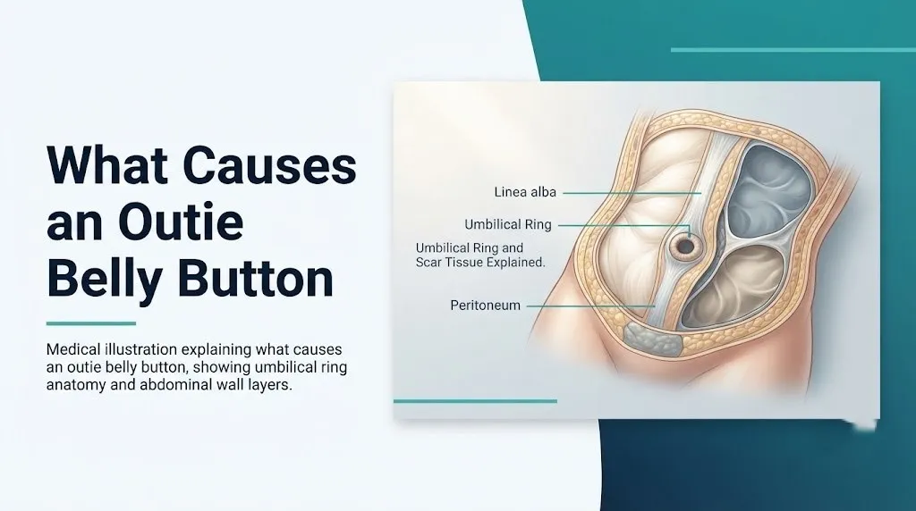
The umbilical ring is a circular defect in the linea alba, the fibrous midline structure of the abdominal wall formed by the interlocking aponeuroses of the three flat abdominal muscles: the external oblique, internal oblique, and transversus abdominis. During fetal life, this opening is the passage through which the umbilical cord traverses the abdominal wall.
Beneath the linea alba lies the transversalis fascia, a continuous layer of connective tissue that lines the inner surface of the abdominal wall. Beneath that is the peritoneum, the serous membrane encasing the abdominal cavity and its organs. These three layers (linea alba, transversalis fascia, peritoneum) form the anatomical barrier that, when intact, keeps abdominal contents inside the abdomen.
During normal postnatal development, the umbilical ring narrows progressively as fibroblasts deposit collagen around its edges. The National Institutes of Health notes that in most infants, this closure is complete by 12 to 18 months of age, though smaller defects may close as late as 4 to 5 years. Ring size at birth predicts closure likelihood: defects smaller than 1 centimeter in diameter close spontaneously in more than 90 percent of cases. Defects larger than 2 centimeters are far less likely to close without surgical intervention.
When the ring fails to close fully, a fascial defect persists into childhood or adulthood. The peritoneum, under cyclic intra-abdominal pressure generated by respiration, physical activity, and bowel function, can bulge through this defect. That bulge, covered only by skin, is the hernia sac that produces the protruding appearance. Adults with residual small defects that were clinically silent throughout childhood can develop symptomatic hernias later in life when intra-abdominal pressure increases due to weight gain, pregnancy, or abdominal muscle weakening with age.
Excess Scar Tissue and Outie Belly Button Formation
Excess scar tissue is the most common cause of a cosmetic outie belly button in people who have no underlying hernia. This type of outie forms when the wound healing process at the umbilical stump site produces more collagen than needed, and that collagen accumulates above the level of the surrounding skin.
The cellular mechanism involves fibroblasts, the primary connective tissue cells responsible for wound repair. After the cord stump separates, fibroblasts migrate into the wound under the direction of signaling molecules including transforming growth factor-beta (TGF-beta) and platelet-derived growth factor (PDGF). These cells synthesize type I and type III collagen to close the wound. In individuals who develop outie belly buttons through this mechanism, fibroblast activity produces a thicker collagen scaffold than the surrounding tissue retracts. The result is a raised scar that sits proud of the skin plane, similar in biological principle to a hypertrophic scar on any other area of the body.
This type of outie is entirely benign. It does not contain abdominal contents. It does not pose any health risk. It does not change in size with coughing, straining, or standing versus lying down. That last feature (the absence of positional change) is a clinically practical way to distinguish scar-tissue outies from hernia-based outies at home.
According to Mayo Clinic, cosmetic outie belly buttons caused by excess scar tissue require no medical treatment. The only reasons to address them are aesthetic preference, in which case elective umbilicoplasty is an option, or if the tissue becomes irritated by clothing, in which case a primary care physician can assess whether any minor intervention is indicated.
People with a personal or family history of keloid scarring or hypertrophic scar formation may be more likely to develop prominent outie belly buttons through this mechanism. Keloid-prone skin, more common in individuals of African, Asian, and Hispanic descent, generates exaggerated fibroblast responses to wound healing. This does not make the outie medically concerning. It does mean that any surgical correction carries a higher risk of visible scarring as a procedural complication.
Key Takeaway: An outie belly button caused by excess scar tissue is a benign anatomical variant that does not change in size with positional changes, coughing, or straining. An outie that grows larger when you stand or strain and shrinks when you lie down is behaving like a hernia and warrants evaluation by a primary care physician.
Umbilical Hernia as a Cause of Outie Belly Button
An umbilical hernia is the most clinically important cause of an outie belly button. It occurs when a defect in the umbilical ring allows abdominal contents, most commonly the fatty omentum or, less frequently, a loop of small intestine, to protrude through the fascial opening and push against the overlying skin.
The herniated tissue sits inside a hernia sac, a thin pouch of peritoneum that has bulged through the defect. From the outside, this appears as a soft, rounded protrusion at the navel. Unlike the scar-tissue outie, a hernia-based protrusion typically reduces (returns into the abdomen) when the person lies flat or gently presses on it. It re-emerges with standing, physical activity, coughing, or any other action that raises intra-abdominal pressure.
The National Institute of Diabetes and Digestive and Kidney Diseases (NIDDK) describes umbilical hernia as one of the most common types of hernia in both children and adults, accounting for approximately 10 percent of all abdominal hernias diagnosed and repaired annually in the United States.
| Feature | Scar Tissue Outie | Umbilical Hernia Outie |
|---|---|---|
| Changes size with position | No | Yes, larger when upright or straining |
| Reducible by gentle pressure | No | Often yes |
| Tender to touch | No | Can be if omentum is inside |
| Soft and compressible | No (firm scar) | Yes (soft tissue inside) |
| Increases with coughing | No | Yes |
| Requires medical evaluation | No | Yes |
| Potential for complications | None | Incarceration, strangulation |
Any adult who develops a new outie-type belly button protrusion that was not present previously should be evaluated by a primary care physician to determine whether umbilical hernia is the underlying cause. Adults with confirmed umbilical hernias are typically referred to a general surgeon for repair planning.
Umbilical Granuloma and Belly Button Appearance
An umbilical granuloma is a small, moist, pinkish-red growth of granulation tissue that forms at the base of the belly button after the umbilical cord stump falls off. It is one of the less commonly recognized causes of a protruding or abnormal-appearing belly button, particularly in newborns and young infants.
Normally, after the stump separates, the wound epithelializes (covers itself with skin cells) completely within a few days. In some infants, the moist environment created by residual moisture, delayed healing, or minor infection prevents full epithelialization. Instead, the wound heals with granulation tissue: a mixture of new blood vessels (driven by vascular endothelial growth factor, VEGF), macrophages, and newly forming collagen. This tissue remains soft, moist, and pink, and it persists beyond normal healing time, sometimes for weeks.
According to Mayo Clinic, umbilical granuloma is one of the most common umbilical cord complications in newborns, occurring in approximately 1 in 500 births, though the actual incidence may be higher because many cases resolve without formal medical diagnosis.
The granuloma does not cause an outie in the same anatomical sense as scar tissue or hernia. What it produces is a persistent pink tissue tag or moist lump at the navel base that may look like an outie or may be confused with one. It can bleed minimally when rubbed, and it may produce a small amount of clear or slightly yellowish discharge.
Granulomas do not resolve on their own as reliably as umbilical hernias do. The standard treatment is cauterization using silver nitrate applied by a pediatrician, which destroys the granulation tissue and allows normal skin to form. The procedure is performed in the office and is not painful for the infant. Parents who notice a persistent moist, pink tissue mass at their newborn’s belly button site after the stump has fallen off should bring this to their pediatrician’s attention within the week rather than waiting for the next scheduled visit.
Key Takeaway: Umbilical granuloma looks like a persistent moist pink lump at a newborn’s navel after the cord falls off. It is not a hernia, but it does need assessment and a quick office treatment by a pediatrician rather than watchful waiting at home.
Outie Belly Button in Newborns and Infants
Outie belly buttons are significantly more common in newborns and infants than in older children or adults, primarily because the umbilical ring is still in the process of closing during the first months of life. A prominent outie in a newborn is often the visible sign of an umbilical hernia, not excess scar tissue, because scar tissue formation takes longer to accumulate.
MedlinePlus states that umbilical hernias are present in approximately 10 to 30 percent of full-term infants at birth. Among premature infants born before 28 weeks of gestation, rates exceed 75 percent. Infants of African descent have roughly 3 times the prevalence of umbilical hernia compared to infants of European or Asian descent, according to epidemiological data reviewed in the Journal of Pediatric Surgery. The reason for this racial disparity is not fully characterized but is thought to involve differences in umbilical ring closure timing and fascial development rates.
The practical assessment a parent can do at home:
- Gently lay the infant flat on their back on a firm surface.
- Observe whether the belly button protrusion reduces (goes back in) when the baby is relaxed and lying flat.
- Check whether the protrusion enlarges visibly when the baby cries, strains, or is held upright.
- Note whether the skin over the protrusion looks normal in color (skin-toned) versus red, purple, or darkened.
- Feel whether the protrusion is soft (hernia with reducible contents) or firm (scar tissue).
- Report findings to the pediatrician at the next visit, or sooner if the baby shows pain, vomiting, or color change at the site.
Most umbilical hernias in infants under 1.5 centimeters in diameter close without treatment by age 2 to 4. Defects larger than 2 centimeters, hernias that have not closed by age 4 to 5, or hernias that become symptomatic require evaluation by a board-certified pediatric surgeon to discuss surgical repair options.
Causes of Outie Belly Button in Adults
An outie belly button that develops in adulthood almost always has a pathological cause. Adults do not spontaneously develop new scar tissue protrusions at the navel. A new or enlarging protrusion in an adult is a hernia until proven otherwise by clinical examination.
Adult umbilical hernias form through the same basic mechanism as in infants (a defect in the linea alba at the umbilical ring) but arise from different triggers. Elevated intra-abdominal pressure over time weakens the fascial edges of even a fully closed umbilical ring and can cause re-opening of the defect. Common triggers in adults include:
- Obesity, which increases chronic resting intra-abdominal pressure throughout the abdominal wall
- Multiple pregnancies, which stretch and weaken the linea alba repeatedly
- Chronic heavy lifting or strenuous physical labor
- Chronic constipation requiring repeated forceful straining
- Persistent ascites from conditions including liver cirrhosis and portal hypertension
- Chronic obstructive pulmonary disease (COPD), in which chronic forceful coughing generates repeated pressure spikes against the abdominal wall
- Previous abdominal surgery, which can create focal areas of fascial weakness
According to research published in the Journal of the American College of Surgeons, umbilical hernias account for approximately 175,000 surgical repairs annually in the United States. The peak incidence in adults occurs between ages 35 and 60, with women who have had multiple pregnancies representing the largest demographic subgroup.
Adults with connective tissue disorders including Ehlers-Danlos syndrome and Marfan syndrome have inherently weaker fascial tissue throughout the body. For these individuals, umbilical hernia can develop at a younger age and with smaller mechanical triggers than in the general population. A board-certified general surgeon familiar with connective tissue disorder complications should manage hernia repair planning in these patients.
Key Takeaway: A new outie belly button in an adult is a hernia until a clinical examination says otherwise. It needs evaluation by a primary care physician, not a wait-and-see approach, because adult umbilical hernias carry a higher complication risk than infant hernias.
Outie Belly Button During and After Pregnancy
Pregnancy produces a temporary outie belly button through a mechanism completely different from the developmental mechanism in newborns. During pregnancy, the growing uterus pushes the abdominal contents upward and forward, dramatically increasing intraperitoneal pressure throughout the third trimester.
This increased pressure pushes on the inner surface of the umbilical ring from inside the abdominal cavity. Even a fully closed umbilical ring that has been stable for decades can be pushed outward as the uterus enlarges, causing the navel to flatten and then protrude. This is a pressure-driven mechanical phenomenon, not a hernia, and it resolves in the majority of women within weeks to months after delivery as the uterus returns to normal size and abdominal wall pressure normalizes.
However, pregnancy also increases the risk of developing a true umbilical hernia for the first time. The combination of elevated intra-abdominal pressure, hormonal effects on connective tissue laxity (specifically the action of relaxin, a hormone that loosens ligaments and fascial structures to prepare for labor), and the physical stretching of the linea alba creates conditions where the umbilical ring can partially re-open. Postpartum women who find that their outie belly button has not resolved within 3 to 4 months after delivery, or whose protrusion is painful or changes size with physical activity, should discuss this with their obstetrician-gynecologist or primary care physician.
Women who develop diastasis recti (separation of the two sides of the rectus abdominis muscles along the midline) during pregnancy are at particularly elevated risk for persistent post-pregnancy umbilical protrusion. Diastasis recti weakens the entire linea alba, reducing the tension that would otherwise keep the umbilical ring from re-opening under pressure. A physical therapist specializing in pelvic floor and abdominal wall rehabilitation can assess diastasis recti and guide appropriate abdominal wall strengthening exercises. These exercises are safe and specific; they differ from standard core exercises, which can worsen diastasis in some presentations.
Risk Factors for Umbilical Hernia in Adults
Certain anatomical, physiological, and lifestyle factors significantly increase the likelihood that an adult will develop an umbilical hernia producing an outie belly button. These factors work by either increasing intra-abdominal pressure chronically, weakening the fascial structures of the abdominal wall, or both simultaneously.
Risk factors supported by clinical evidence:
- Obesity: Body mass index (BMI) above 30 is associated with chronically elevated intra-abdominal pressure due to the mechanical load of intra-abdominal and retroperitoneal fat. This places constant outward force on all abdominal wall fascial structures.
- Multiple pregnancies: Each pregnancy stretches the linea alba and umbilical ring. Women who have had three or more pregnancies have substantially higher umbilical hernia rates than nulliparous women, according to data from the Annals of Surgery.
- Liver cirrhosis with ascites: Ascites (accumulation of fluid in the peritoneal cavity) can generate enormous intra-abdominal pressure. Patients with portal hypertension-related ascites develop umbilical hernias at rates between 20 and 40 percent, far exceeding the general adult population.
- Chronic cough: Conditions producing persistent forceful coughing, including COPD, asthma, and chronic bronchitis, generate repeated high-pressure episodes within the abdominal cavity that stress all fascial structures.
- Previous abdominal surgery: Surgical incisions disrupt fascial integrity. Incisions near the umbilicus can leave areas of relative fascial weakness that evolve into hernias over years.
- Connective tissue disorders: Ehlers-Danlos syndrome and related conditions produce structurally abnormal collagen throughout the body. Fascial tissue in these individuals has reduced tensile strength, making hernia formation more likely with less provocation.
| Risk Factor | Mechanism | Relative Risk vs. General Population |
|---|---|---|
| BMI over 30 | Elevated resting intra-abdominal pressure | 2 to 3 times higher |
| Multiple pregnancies | Linea alba stretching, relaxin-mediated laxity | 3 to 4 times higher in women with 3+ pregnancies |
| Liver cirrhosis with ascites | Extreme intra-abdominal fluid pressure | 5 to 10 times higher |
| Ehlers-Danlos syndrome | Structurally abnormal collagen in fascial tissue | Significantly elevated; exact ratio not well-quantified |
| Chronic COPD with cough | Repeated coughing-generated pressure spikes | Elevated; varies with cough severity |
Strangulated Umbilical Hernia: Symptoms and Emergency Signs
A strangulated umbilical hernia is a surgical emergency. It occurs when abdominal contents that have protruded through the umbilical ring defect become trapped and their blood supply is cut off. The pressure of the fascial edges of the ring can compress the blood vessels supplying the herniated tissue, leading to ischemia and, if untreated within hours, tissue necrosis (death).
This complication is more common in adults than in infants. The American Hernia Society identifies adult umbilical hernias as carrying a higher strangulation risk than inguinal (groin) hernias because the umbilical ring tends to have narrower, less yielding fascial edges. Research published in JAMA Surgery found that approximately 5 to 10 percent of unrepaired adult umbilical hernias will eventually require emergency surgery for incarceration or strangulation.
Emergency Symptoms: When to Call 911 or Go to the ER
Certain symptoms associated with an umbilical hernia require immediate emergency evaluation. Do not wait to see if these resolve on their own.
Call 911 or go to the nearest emergency room immediately if you experience:
- Sudden, severe pain at the belly button protrusion that did not resolve within minutes and is getting worse
- A hernia protrusion that was previously soft and reducible but is now firm, hard, and cannot be pushed back in
- Skin over the belly button protrusion that has turned red, purple, dark blue, or black
- Nausea or vomiting accompanying belly button pain and an irreducible hernia
- Fever combined with a painful, firm belly button protrusion
- A hernia protrusion that has been growing rapidly over hours rather than days or weeks
These presentations indicate strangulated or incarcerated umbilical hernia, conditions that involve ischemic tissue injury and potential bowel obstruction, and require emergency surgical evaluation and intervention. Urgent care centers are not equipped for this. This is an emergency room presentation.
Can an Innie Belly Button Become an Outie
An innie belly button can become an outie later in life under specific anatomical circumstances. This transition is not a cosmetic quirk. It is almost always a sign that the umbilical ring has been forced partially open by chronic intra-abdominal pressure, producing a new umbilical hernia in a previously normal fascial structure.
The most common circumstances producing this change in adults include significant weight gain, the advanced stages of pregnancy, and the development of ascites from liver disease. In these situations, the pressure rise inside the abdominal cavity exceeds the structural capacity of the closed umbilical ring. The fascial ring, which was adequately strong for normal intra-abdominal pressure, fails under the new mechanical load.
Innie-to-outie transitions can also occur after abdominal surgeries that disrupt the integrity of tissues near the umbilicus, or as a very gradual change with age-related loss of fascial tensile strength in people with other risk factors.
What a transition change does not mean: it does not mean the belly button is “choosing” to look different. It does not mean a cosmetic concern has developed. It means a structural change has occurred in the abdominal wall. Any adult who notices their previously flat or innie belly button beginning to protrude should report this to their primary care physician. A clinical examination (physical examination, sometimes followed by abdominal ultrasound) will determine whether a hernia defect is present and how large it is.
According to Mayo Clinic, a new umbilical protrusion in an adult that was not previously present should be evaluated within a few weeks rather than monitored at home indefinitely. This is because adult umbilical hernias do not close spontaneously the way infant hernias often do, and early evaluation allows the surgeon to plan elective repair under better conditions than emergency repair.
Key Takeaway: When an adult’s belly button changes from an innie to an outie, it is not cosmetic. It is a new finding that needs physical examination by a primary care physician or general surgeon within a few weeks, not months.
Outie Belly Button Treatment and Correction Options
The appropriate treatment for an outie belly button depends entirely on the underlying cause. Benign cosmetic outies caused by excess scar tissue require no medical treatment and pose no health risk. Umbilical hernias, umbilical granulomas, and new adult-onset protrusions each have specific clinical management pathways.
For cosmetic scar tissue outies:
The only interventions are elective. A board-certified plastic surgeon can perform umbilicoplasty, a surgical procedure that reshapes the belly button by removing excess scar tissue and repositioning the skin to create a more recessed appearance. This is done as an outpatient procedure under local anesthesia in most cases. Results are permanent for the cosmetic variant. Patients with a history of keloid or hypertrophic scarring should discuss this risk explicitly with their surgeon before proceeding.
For umbilical hernias in infants:
Clinical guidelines from the American Pediatric Surgical Association support watchful waiting for infant umbilical hernias that are asymptomatic and under 1.5 centimeters in diameter, as these close spontaneously in the majority of cases by age 4 to 5. Surgical repair (herniorrhaphy) is indicated for defects larger than 2 centimeters, hernias that have not closed by age 5, or any hernia that becomes symptomatic (painful, irreducible, or associated with incarceration). A board-certified pediatric surgeon performs the repair under general anesthesia as a short outpatient procedure.
For umbilical hernias in adults:
Adult umbilical hernias do not close spontaneously. The standard of care is elective surgical repair, performed as either open herniorrhaphy (small incision at the navel, direct fascial closure with or without mesh) or laparoscopic repair (minimally invasive approach through small ports). Research in the Annals of Surgery demonstrates lower recurrence rates with mesh-reinforced repair compared to primary fascial closure alone, particularly for defects larger than 1 centimeter. A general surgeon will discuss the appropriate approach based on hernia size, patient health status, and surgical history.
For umbilical granuloma:
Silver nitrate cauterization applied by a pediatrician over one to three office visits is the standard first-line treatment. Salt cauterization (using table salt applied in small amounts to the granuloma) is sometimes mentioned in parenting resources but lacks the clinical evidence base to replace the established medical approach. Parents should not attempt home treatment without physician guidance.
When to See a Doctor About an Outie Belly Button
Most outie belly buttons do not require medical evaluation. A stable cosmetic outie in a healthy child or adult that has been present since infancy, does not change in size with posture or activity, is not painful, and has no overlying skin changes is a benign anatomical variation. It warrants attention if it changes.
See a primary care physician if:
- A belly button protrusion is new in an adult (was not present previously)
- The protrusion changes size with standing, lying down, coughing, or straining
- The protrusion is tender or painful to touch
- The skin over the protrusion appears different from surrounding skin in color or texture
- A newborn’s belly button has not fully healed within 3 weeks of cord stump separation
- A pink, moist, persistent tissue mass is visible at a newborn’s navel after stump separation (possible umbilical granuloma)
- An infant’s umbilical hernia has not closed by age 4 to 5
- A belly button protrusion in any age group enlarges rapidly over days or weeks rather than remaining stable
See a general surgeon or board-certified pediatric surgeon if:
- A primary care physician has confirmed the presence of an umbilical hernia
- An adult hernia is growing in size or has become uncomfortable
- An infant hernia has not closed by age 4 to 5 years
Go to the emergency room immediately or call 911 if any belly button protrusion becomes suddenly more painful, cannot be pushed back in when it previously could, changes color over the overlying skin, or is accompanied by nausea, vomiting, or fever. These are the signs of strangulated or incarcerated umbilical hernia, a surgical emergency.
People with chronic conditions including liver cirrhosis, ascites, COPD, or connective tissue disorders should mention their abdominal wall history to their managing specialist at routine visits. These populations are at elevated risk for umbilical hernia and benefit from earlier surveillance rather than waiting for a symptomatic presentation.
Frequently Asked Questions About Outie Belly Buttons
What causes an outie belly button?
An outie belly button is caused by either excess scar tissue forming above the skin surface after the umbilical cord stump separates, or by an incompletely closed umbilical ring in the abdominal wall that allows abdominal contents to protrude outward, which is called an umbilical hernia.
The scar tissue type is a benign cosmetic variation with no medical significance.
The hernia type requires evaluation by a primary care physician or general surgeon to determine the appropriate management.
Is an outie belly button a sign of a hernia?
An outie belly button is not always a hernia. Many outie belly buttons are simply cosmetic variations caused by excess scar tissue at the umbilical cord healing site.
A hernia-related outie typically changes size with posture (larger when standing or straining, smaller when lying flat) and feels soft and compressible, while a scar tissue outie stays the same size regardless of position and feels firm.
Any adult with a new outie belly button that was not present previously should be evaluated by a primary care physician, as adult-onset umbilical protrusions are more likely to represent hernia than cosmetic variation.
Can an outie belly button go away on its own?
In infants with umbilical hernia, the outie belly button often resolves without treatment as the umbilical ring closes spontaneously, typically by age 2 to 4 for defects under 1.5 centimeters.
In adults, hernia-related outies do not resolve without surgical treatment because the fascial defect in adults does not close on its own the way it does in developing infants.
Cosmetic outie belly buttons caused by scar tissue do not go away on their own, but they require no treatment because they pose no health risk.
Is an outie belly button dangerous?
A cosmetic outie belly button caused by excess scar tissue is not dangerous and requires no medical management.
An umbilical hernia-related outie can become dangerous if the herniated tissue becomes trapped and loses its blood supply, a complication called strangulation that requires emergency surgery.
The American Hernia Society estimates that approximately 5 to 10 percent of unrepaired adult umbilical hernias will eventually require emergency intervention, which is one reason elective surgical repair is recommended for most adults with diagnosed umbilical hernias.
What causes an outie belly button in adults when they never had one before?
A new outie belly button in an adult who did not have one previously is almost always caused by an acquired umbilical hernia, where the umbilical ring in the abdominal wall has been partially forced open by elevated intra-abdominal pressure.
Common triggers include significant weight gain, multiple pregnancies, liver disease with fluid accumulation (ascites), or chronic heavy coughing from conditions like COPD.
A primary care physician should evaluate any new adult-onset belly button protrusion within a few weeks of first noticing it, because adult umbilical hernias do not close spontaneously and carry a strangulation risk.
When should I be worried about an outie belly button?
Be concerned and seek same-day evaluation at an emergency room if any belly button protrusion becomes suddenly more painful, cannot be pushed back in when it previously could, has overlying skin that looks red, purple, or darkened, or is accompanied by nausea, vomiting, or fever.
These signs indicate a possible strangulated or incarcerated umbilical hernia, which requires emergency surgical evaluation and cannot be safely managed at home or at urgent care.
For non-emergency situations, see a primary care physician for any outie belly button that is new, painful, changes size with activity, or occurs in a newborn whose umbilical wound has not healed within 3 weeks.
Understanding what causes an outie belly button starts with recognizing that not all outies are the same thing. A stable cosmetic outie that has been present since birth is not the same clinical finding as a new protrusion in a 45-year-old, and neither of those is the same as a soft reducible bulge in a 3-month-old infant that retracts when the baby lies flat. The mechanism matters.
For most people reading this, the practical next step is simple: note whether the outie is new or long-standing, whether it changes size with posture or activity, and whether it is painful. If the answer to any of those last two questions is yes, a conversation with a primary care physician is the right move. Early evaluation of an umbilical hernia allows for planned elective surgical repair under far better conditions than emergency repair after a strangulation event.
Your anatomy is specific to you. The best assessment of a belly button that concerns you comes from a clinical examination by a physician who can feel the defect, estimate the ring size, and give you a plan grounded in your actual anatomy rather than a generic description.