What Causes Your Water to Break: Full Clinical Guide
What causes your water to break is a biochemical and mechanical process in which the two-layered amniotic sac surrounding your baby is weakened by a coordinated cascade of inflammatory enzymes, hormones, and physical pressure until it ruptures, releasing amniotic fluid. At term, this process is driven by your body’s normal labor cascade. When it happens too early, it reflects a disruption of that process by infection, cervical weakness, uterine overdistension, or structural factors that accelerate membrane breakdown prematurely.
According to ACOG Practice Bulletin No. 188, premature rupture of membranes complicates approximately 8% of all pregnancies and is the leading identifiable cause of preterm birth in the United States. The National Institute of Child Health and Human Development (NICHD) reports that preterm birth affects approximately 1 in 10 births, and membrane rupture before 37 weeks plays a role in a substantial proportion of those cases.
This article covers the specific biochemical mechanism behind term membrane rupture, the distinct causes of early water breaking including infection, cervical insufficiency, and polyhydramnios, how to tell whether fluid loss is amniotic fluid or urine, what to do in the first minutes after rupture, and which scenarios require calling 911 without waiting to call your obstetrician.
What Causes Your Water to Break: The Direct Answer
Your water breaks when inflammatory enzymes called matrix metalloproteinases (MMPs), activated by a combination of fetal hormones, prostaglandins, and immune cells in the uterine lining, degrade the structural collagen of the amniotic membranes until the mechanical pressure of the uterus causes them to rupture.
This is not a passive tearing of tissue. It is an active, biochemically orchestrated process that involves your baby’s hormonal signaling, your immune system, and the physical forces of uterine contractions all converging on membranes that have been progressively weakened over the final weeks of pregnancy.
The process differs depending on the timing:
| Type of Rupture | Timing | Primary Driver | Clinical Response |
|---|---|---|---|
| Spontaneous term SROM | At or after 37 weeks, with labor | Normal labor cascade; MMP activation at term | Proceed to labor and delivery |
| PROM at term | At or after 37 weeks, before labor | MMP cascade without contractions yet | Hospital evaluation; often labor induction within 24 hours |
| PPROM | Before 37 weeks | Infection, inflammation, cervical weakness, or overdistension | Immediate evaluation; management depends on gestational age |
| Pathological rupture | Any gestational age | Chorioamnionitis, cervical insufficiency, polyhydramnios, trauma | Urgent obstetric evaluation |
| Amniotomy | Any gestational age | Provider-performed artificial rupture with an amniohook | Standard labor induction or augmentation procedure |
Women who have experienced a prior preterm birth have an approximately 17 to 32% chance of recurrent PPROM in subsequent pregnancies, according to data cited in the American Journal of Obstetrics and Gynecology, making prior preterm birth the strongest single modifiable risk factor for surveillance.
What Is the Amniotic Sac and What Does Breaking Mean?
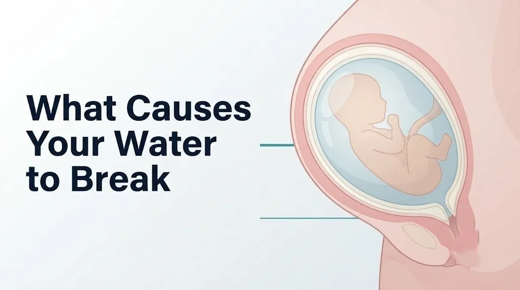
The amniotic sac is the fluid-filled double-layered membrane that completely surrounds and protects the fetus throughout pregnancy, composed of an inner layer called the amnion and an outer layer called the chorion, and “breaking” refers to the rupture of this sac at any point in pregnancy, which allows amniotic fluid to escape through the vagina.
The amnion is the layer that directly contacts the amniotic fluid. It is a thin, avascular (bloodless) membrane composed of a layer of columnar epithelial cells sitting on a basement membrane made primarily of type IV collagen, fibronectin, and laminin. These proteins form the structural scaffolding that gives the amnion its tensile strength. The chorion lies outside the amnion and is in contact with the maternal decidua. It is slightly thicker and has its own collagen framework.
Think of the amniotic sac like a double-walled water balloon. The inner balloon (amnion) holds the water directly. The outer balloon (chorion) provides an additional structural layer and interfaces with the uterine lining. What “breaks” is specifically this double membrane, and where it breaks first matters: rupture at the cervical os releases a rush or trickle of fluid, while a high rupture can produce only a slow intermittent leak that is much harder to recognize.
The amount of fluid released depends on where the tear occurs and whether it seals partially after rupture. At term, total amniotic fluid volume ranges from approximately 800 to 1,000 milliliters. A complete rupture at the lowest point releases a dramatic gush. A small tear higher in the sac releases a slow trickle that many pregnant women mistake for urinary leakage.
For women carrying twins or higher-order multiples, the anatomy is different: each fetus typically has its own amniotic sac (in dichorionic-diamniotic twins), meaning the “water breaking” refers to rupture of one or both sacs, and the clinical implications differ from singleton pregnancies.
How Do the Amniotic Membranes Rupture at Term?
The amniotic membranes rupture at term through a well-characterized biochemical cascade initiated by the fetal hypothalamic-pituitary-adrenal (HPA) axis, which produces increasing amounts of corticotropin-releasing hormone (CRH) from the fetal hypothalamus and placenta as the pregnancy approaches its natural endpoint, triggering a sequence of hormonal and inflammatory events that progressively weaken the membrane’s collagen framework until it can no longer withstand the physical forces of labor.
As gestational age increases, fetal CRH production rises exponentially. This drives increased fetal cortisol production from the fetal adrenal cortex, which is converted in the placenta to estrogens. Rising estrogen relative to progesterone shifts the uterus from a quiescent state to a pro-labor state. Decidual macrophages (immune cells resident in the decidua basalis and decidua parietalis) are activated, releasing interleukin-1 beta (IL-1beta) and tumor necrosis factor-alpha (TNF-alpha).
These cytokines, combined with prostaglandin E2 (produced when phospholipase A2 cleaves arachidonic acid from membrane phospholipids and cyclooxygenase-2 (COX-2) oxidizes it), upregulate MMP production in the amniotic membrane’s own stromal cells and the macrophages infiltrating the membrane. MMP-1 (collagenase-1) cleaves fibrillar collagen. MMP-3 (stromelysin-1) cleaves multiple matrix components and activates other MMPs. MMP-9 (gelatinase B) degrades the type IV collagen that forms the basement membrane structure of the amnion.
The result is progressive structural weakening at what researchers call the “zone of altered morphology,” a region of the membrane closest to the cervical os that shows the most MMP activity and the earliest collagen degradation. When uterine contractions apply cyclical pressure to this already-weakened zone, rupture occurs. At term, this is the designed outcome of normal parturition biology.
According to research published in the American Journal of Obstetrics and Gynecology, the balance between MMPs and their natural inhibitors (tissue inhibitors of metalloproteinases, or TIMPs) is the key regulatory mechanism. When MMP activity exceeds TIMP inhibitory capacity, the membrane becomes vulnerable to rupture.
Key Takeaway: Your water breaking at term is not a random event. It is the endpoint of a weeks-long biochemical process in which fetal hormones, maternal immune cells, and enzymatic collagen degradation collaborate to prepare the membrane for rupture at exactly the right moment.
What Causes Your Water to Break Early?
Your water breaks early (before 37 weeks of gestation) when the normal term membrane rupture cascade is triggered prematurely by factors including intrauterine infection, cervical weakness, uterine overdistension from excess amniotic fluid, structural abnormalities, or prior uterine surgery that disrupts the lower uterine segment’s structural integrity.
Early water breaking, specifically preterm premature rupture of membranes (PPROM), is clinically distinct from water breaking at term because the fetal organs, particularly the lungs, may not be mature enough to function outside the uterus. The gestational age at rupture determines the clinical urgency and management approach.
The causes of early water breaking range in frequency:
- Intrauterine infection (chorioamnionitis): the most common identifiable cause, found in approximately 25 to 40% of PPROM cases
- Cervical insufficiency: structural weakness of the cervix produces early dilation and exposes membranes to vaginal bacteria and mechanical stretching
- Polyhydramnios: excess amniotic fluid increases intrauterine pressure beyond what the membranes can sustain
- Multiple gestation: twins and higher-order multiples produce significantly greater uterine distension and higher intrauterine pressure than singletons
- Prior uterine surgery: cesarean section or myomectomy scars alter the lower uterine segment architecture and may create areas of reduced membrane support
- Tobacco smoking: components of cigarette smoke reduce TIMP levels in amniotic membranes, shifting the MMP-to-TIMP balance toward premature collagen degradation
- Placenta previa: low placental implantation over the internal os alters local membrane perfusion and immune environment
- Fetal anomalies: certain structural fetal anomalies reduce fetal swallowing, producing polyhydramnios and consequent membrane over-tension
Black pregnant women experience PPROM at higher rates than white pregnant women, a disparity documented in research published in Obstetrics and Gynecology that reflects higher rates of intrauterine infection from sexually transmitted infections and bacterial vaginosis, along with broader structural social determinants of health rather than inherent biological differences.
What Causes PPROM (Preterm Premature Rupture of Membranes)?
PPROM (preterm premature rupture of membranes) is rupture of the amniotic membranes before 37 weeks of gestation, caused in most cases by a combination of intrauterine infection, progressive cervical changes, and the same MMP-mediated collagen degradation that drives term rupture, but occurring prematurely because one or more amplifying factors have accelerated the biochemical cascade beyond what the membranes can withstand.
The most mechanistically specific pathway in PPROM involves bacterial colonization of the amniotic space or decidua. Certain bacteria, including Ureaplasma urealyticum, Mycoplasma hominis, and Gardnerella vaginalis (the organism most associated with bacterial vaginosis), produce two types of products that directly attack the amniotic membranes. First, they produce phospholipases that cleave arachidonic acid from membrane phospholipids, feeding directly into the prostaglandin E2 synthesis pathway and triggering uterine contractions and membrane inflammation. Second, they produce bacterial proteases and collagenases that directly degrade the type IV collagen and fibronectin of the amnion’s basement membrane, bypassing the need for the normal MMP cascade entirely.
The American College of Obstetricians and Gynecologists, in ACOG Practice Bulletin No. 188, identifies the following as established independent risk factors for PPROM: prior preterm birth, current intrauterine infection, cervical cerclage in place, maternal smoking, low maternal body mass index, prior surgical procedures on the cervix, second or third trimester bleeding, uterine distension from multiple gestation or polyhydramnios, and prior preterm delivery specifically from PPROM.
For women with a history of prior PPROM, the recurrence risk varies by the gestational age of the prior event. PPROM before 28 weeks in a prior pregnancy carries a recurrence risk of approximately 32%, making close surveillance with serial transvaginal cervical length measurement and fetal fibronectin testing a standard component of prenatal care in subsequent pregnancies.
How Chorioamnionitis Causes Water to Break
Chorioamnionitis (intrauterine infection, also called intra-amniotic infection) causes membrane rupture through direct bacterial enzymatic attack on the amniotic membrane collagen and through intense local immune activation that dramatically amplifies the MMP-driven membrane degradation far beyond what occurs in normal term labor.
The anatomical pathway of ascending infection begins in the vagina and cervix. Bacteria ascend through the cervical canal, penetrate the chorion-decidual interface, colonize the space between the chorion and amnion (called the extra-amniotic space), and eventually can reach the amniotic fluid itself and the fetal surface of the membranes. At each step, both the bacteria and the host immune response contribute to membrane damage.
Bacterially-produced phospholipases activate the prostaglandin E2 synthesis pathway. The resulting uterine contractions increase mechanical pressure on membranes that are simultaneously being chemically attacked. Activated decidual neutrophils and macrophages responding to the bacterial invasion release even higher concentrations of IL-1beta, TNF-alpha, and IL-8 than occur in normal labor, producing a far more aggressive MMP upregulation. The infected membrane essentially degrades itself much faster than a healthy membrane under normal labor conditions.
Clinical signs that infection may be driving or has accompanied membrane rupture:
- Maternal fever above 100.4 degrees Fahrenheit (38 degrees Celsius)
- Uterine tenderness on palpation
- Foul-smelling or cloudy amniotic fluid rather than the normal clear or slightly yellow-tinted fluid
- Fetal tachycardia above 160 beats per minute on cardiotocography
- Elevated maternal white blood cell count above 15,000 cells per microliter
Pregnant women with bacterial vaginosis are two to six times more likely to develop PPROM compared to those without it, based on data from multiple prospective studies cited by the Society for Maternal-Fetal Medicine. Screening and treatment of bacterial vaginosis in early pregnancy is an area of active clinical investigation.
Key Takeaway: Infection is not just a risk factor for PPROM. It is the most common direct mechanical cause, because bacteria physically destroy the amniotic membrane’s structural proteins through their own enzymes, independently of the normal labor cascade.
How Cervical Insufficiency Causes Early Membrane Rupture
Cervical insufficiency (historically called incompetent cervix) causes early membrane rupture through a mechanical mechanism in which the cervix dilates silently and progressively before the normal onset of labor, gradually exposing the lower pole of the amniotic membranes to the vaginal environment, where they are subjected to physical stretching beyond their structural tolerance and to the bacterial colonization and inflammatory conditions of the vaginal microbiome.
In a normally functioning cervix, the internal os remains closed throughout pregnancy, supported by the cervical stroma’s collagen-rich connective tissue matrix and a mucous plug that seals the cervical canal from ascending bacteria. Cervical insufficiency represents a structural failure of this supportive mechanism. The internal os dilates silently, without labor contractions, typically during the second trimester, funneling the amniotic membranes downward into the upper vagina. This “funneling” is visible on transvaginal ultrasound as a U-shaped or V-shaped opening at the top of the cervical canal.
Once the membranes funnel through the dilated internal os, they are exposed to:
- Direct mechanical over-stretching as they bulge into the cervical canal and potentially beyond the external os
- Contact with vaginal bacteria that would normally be excluded by the closed cervix and mucous plug
- A new mechanical loading pattern that concentrates stress at the zone of altered morphology rather than distributing it across the full membrane surface
Cervical length below 25 millimeters measured by transvaginal ultrasound between 16 and 24 weeks of gestation is a well-validated predictor of PPROM and preterm birth, established by multiple large prospective studies. The American College of Obstetricians and Gynecologists recommends routine cervical length screening between 18 and 24 weeks of gestation for women with a prior preterm birth.
Women who have had prior cervical procedures including loop electrosurgical excision procedure (LEEP) or cone biopsy for abnormal cervical cells have anatomically shorter cervices and higher rates of cervical insufficiency, making them a group requiring specific prenatal surveillance.
Can Polyhydramnios Cause Your Water to Break?
Polyhydramnios (excess amniotic fluid, defined as an amniotic fluid index above 24 to 25 cm or a single deepest pocket above 8 cm by ultrasound) can cause the water to break by increasing the intrauterine pressure against the amniotic membranes to a level that exceeds their structural capacity, particularly in membranes that have already been biochemically weakened by the normal gestational aging process.
The mechanism is straightforwardly mechanical. The amniotic membranes have a finite tensile strength determined by their collagen and fibronectin content. As polyhydramnios increases the volume and pressure within the amniotic sac, the membranes experience greater circumferential wall tension (described by Laplace’s law: tension equals pressure times radius). Increased volume means increased radius, and increased pressure compounds this, producing dramatically higher membrane wall tension at any given pressure compared to a normal-fluid pregnancy.
Polyhydramnios causes and their PPROM implications:
- Gestational diabetes mellitus: poorly controlled blood glucose produces fetal hyperglycemia, which drives fetal osmotic diuresis (excess urine production) and polyhydramnios. Pregnant women with uncontrolled gestational diabetes have measurably higher rates of polyhydramnios and associated PPROM.
- Fetal structural anomalies: conditions affecting fetal swallowing (esophageal atresia, central nervous system anomalies that impair the swallowing reflex) prevent fetal reabsorption of amniotic fluid, producing progressive polyhydramnios.
- Twin-to-twin transfusion syndrome (TTTS): in monochorionic twin pregnancies, vascular connections between the twins produce fluid imbalance, with the recipient twin developing polyhydramnios in its sac.
- Idiopathic polyhydramnios: in some cases, no underlying cause is identified; mild idiopathic polyhydramnios carries a lower PPROM risk than polyhydramnios from fetal anomaly.
Women diagnosed with polyhydramnios should discuss the specific underlying cause with their obstetrician-gynecologist and may be referred to a maternal-fetal medicine specialist for additional evaluation, particularly if the fluid index is severely elevated or rising rapidly on serial ultrasound examinations.
Can Sex or Physical Activity Cause Your Water to Break?
Sexual intercourse and normal physical activity do not cause your water to break in a healthy pregnancy at normal gestational age, because the amniotic membranes at term require biochemical weakening by the labor cascade before they become susceptible to mechanical rupture, and the physical forces involved in intercourse or typical exercise are far below the level required to rupture structurally intact membranes.
This question is one of the most frequently searched by pregnant women and is worth addressing specifically because the anxiety around it can affect quality of life unnecessarily. The type IV collagen framework of the healthy amnion has substantial tensile strength that normal physical forces cannot overcome. Medical organizations including the American College of Obstetricians and Gynecologists explicitly state that sexual intercourse is safe throughout a normal pregnancy and does not increase the risk of membrane rupture or preterm birth in low-risk pregnancies.
There are specific exceptions where sexual intercourse may be clinically restricted:
- Placenta previa, where the placenta overlies the cervical os, because intercourse can cause cervical contact with the placenta and bleeding
- Vasa previa, where fetal blood vessels overlie the cervical os
- Active preterm labor or PPROM in a current pregnancy
- Cervical cerclage in place, where the provider may restrict penetrative intercourse
Semen contains prostaglandins that could theoretically affect cervical ripening, and this has prompted some clinicians to recommend abstaining from intercourse in the final weeks of pregnancy in high-risk situations. However, the ACOG does not routinely recommend abstinence from intercourse at term in low-risk pregnancies based on current evidence.
Strenuous physical exertion, heavy lifting, or sudden trauma (such as a car accident or a direct blow to the abdomen) can theoretically increase intrauterine pressure acutely, but in a biochemically intact membrane, the forces involved in normal physical activity are insufficient to produce rupture. Any trauma to the pregnant abdomen should be evaluated by an obstetrician regardless of whether membrane rupture occurs.
Key Takeaway: Normal sex and exercise do not cause your water to break in a healthy pregnancy. Membrane rupture requires prior biochemical weakening of the collagen framework, not just mechanical pressure. The exceptions are specific high-risk conditions where your OB-GYN will explicitly restrict intercourse.
How to Tell If Your Water Broke or If You Peed
Distinguishing amniotic fluid leakage from urinary incontinence or increased vaginal discharge is one of the most practically important skills for a pregnant woman to have, because the two presentations are often confused and the clinical implications are completely different.
The key distinguishing characteristics are controllability, odor, color, and pH. Urine can be partially controlled by pelvic floor muscle contraction; amniotic fluid cannot be stopped by any voluntary muscle action because it comes from above the pelvic floor. Amniotic fluid has a neutral to slightly alkaline pH (7.1 to 7.3) compared to the normally acidic vaginal secretions (pH 3.8 to 4.5) and typically acidic urine (pH 4.6 to 8.0). Amniotic fluid is typically clear to slightly yellow-tinged, odorless, or has a very faint sweetish odor different from the ammonia scent of urine.
A practical self-assessment approach if you are unsure:
- Empty your bladder completely, then place a clean dark-colored pad or liner in your underwear.
- Wait 15 to 30 minutes lying down or sitting quietly.
- After 15 to 30 minutes, stand up or change position, which may cause pooled fluid to flow out.
- Examine the pad. If you see continued fluid accumulation that you cannot voluntarily stop, or if the fluid appears clear rather than yellow and has no urine odor, call your obstetrician-gynecologist immediately.
- Do not use an internal tampon or douche to assess the fluid, as this can introduce bacteria.
- Do not wait more than one hour before calling if you genuinely cannot determine whether your water has broken.
The American College of Obstetricians and Gynecologists recommends that all suspected membrane rupture be confirmed by a healthcare provider, because self-assessment is unreliable and the clinical consequences of missed rupture (undetected infection, cord prolapse risk) are too serious.
Women who have urinary incontinence throughout pregnancy, which is very common due to the pressure of the uterus on the bladder, may find it particularly difficult to distinguish. In any case of genuine uncertainty, calling the labor and delivery unit is the appropriate action.
What to Do Immediately When Your Water Breaks
When you believe your water has broken, call your obstetrician-gynecologist or the labor and delivery unit of your planned delivery hospital immediately, note the time the fluid started, its color and odor, whether you feel contractions, and how much fluid you have seen, and proceed to the hospital as instructed by your clinical team.
The immediate actions after suspected membrane rupture depend on your gestational age and whether you are experiencing contractions, but the common thread is that no confirmed amniotic membrane rupture is a “wait and see at home” situation.
To describe the situation accurately to your provider:
- Note the exact time you first noticed the fluid loss and how it began (sudden gush or slow trickle).
- Observe the color: clear or slightly yellow is typical of normal amniotic fluid; green or brown fluid suggests the presence of meconium (fetal bowel movement), which changes the management plan; foul-smelling or cloudy fluid may indicate infection.
- Note whether you feel regular contractions (tightening of the abdomen at regular intervals, typically at least 6 per hour) or whether you feel no contractions at all.
- Check whether you feel the baby moving. Fetal movement should be present and normal. If you have not felt the baby move in the past 2 hours, tell your provider immediately.
- Put on a clean pad (not a tampon) to track fluid loss volume during transport.
- Do not have sexual intercourse, use an internal tampon, or take a bath after rupture, as these can introduce bacteria.
At 37 weeks or later, most guidelines including ACOG Practice Bulletin No. 107 support proceeding to hospital admission for assessment and, in most cases, labor induction if contractions do not begin spontaneously within 12 to 24 hours. Before 37 weeks, the management is more complex and depends on the specific gestational age, as described in the section on PPROM.
Women who are Group B Streptococcus (GBS) positive on prior vaginal-rectal culture (typically performed between 35 and 37 weeks) should inform their provider at the time of rupture, as GBS positive status affects the timing and approach to antibiotic administration during labor.
What Causes Water to Break Without Contractions Starting?
Water breaks without contractions starting in approximately 8 to 10% of term pregnancies (PROM at term) when the MMP-driven biochemical membrane weakening reaches its rupture threshold before the oxytocin-driven uterine contraction pattern of active labor has fully established, meaning the collagen degradation process and the myometrial activation process simply reach their respective thresholds at slightly different times.
This is not a clinical emergency if you are at 37 weeks or beyond. The membranes and the uterus are following the same biological program, but not with perfect synchrony. The uterus typically begins contracting within 12 hours of membrane rupture at term in the majority of women, often sooner. When contractions do not begin spontaneously, most obstetric providers begin labor induction to reduce the risk of intrauterine infection that rises with the time interval between rupture and delivery.
The specific question of why membranes can rupture before contractions clinically confirms what the biochemistry shows: the MMP cascade is the primary driver of rupture, and it proceeds on a timeline that is biochemically distinct from the oxytocin-uterotonic contraction cascade. These two processes normally overlap, but they are not perfectly coupled.
Preterm water breaking without contractions (PPROM without active preterm labor) is a distinct and clinically more complex situation. In PPROM, the rupture may be caused by infection or structural factors that do not trigger uterine contractions, meaning the woman may leak fluid for days to weeks before labor begins. Management in this scenario, detailed in the section on PPROM, depends entirely on gestational age.
According to the American College of Obstetricians and Gynecologists, women at term whose water breaks without contractions should proceed to their delivery hospital for evaluation and should not wait at home for contractions to begin before calling, because the window for infection risk begins at the moment of rupture.
Risk Factors That Make Your Water More Likely to Break Early
The risk factors that most reliably predict early membrane rupture (PPROM before 37 weeks) can be grouped into four categories: infectious, structural, mechanical, and behavioral, each with a distinct physiological mechanism by which it accelerates the normal membrane weakening process.
| Risk Factor | Category | Mechanism | Magnitude of Risk Increase |
|---|---|---|---|
| Prior preterm birth (any cause) | Historical | Complex; higher baseline inflammation, possible anatomical | 17 to 32% recurrence risk |
| Prior PPROM specifically | Historical | Recurrent structural or infectious vulnerability | Up to 32% recurrence risk |
| Bacterial vaginosis | Infectious | Bacterial phospholipases and proteases attack membranes | 2 to 6 times higher risk |
| Group B Streptococcus colonization | Infectious | Local inflammatory activation | Moderately increased |
| Cervical length below 25 mm | Structural | Membrane exposure and over-stretching | Multiple-fold increased risk |
| Multiple gestation | Mechanical | Uterine overdistension, increased intrauterine pressure | Substantially increased |
| Polyhydramnios | Mechanical | Increased membrane wall tension by Laplace’s law | Substantially increased |
| Active tobacco smoking | Behavioral | Reduced TIMP levels, imbalanced MMP activity | Approximately 2 times higher risk |
| Low BMI or poor nutrition | Nutritional | Reduced collagen precursor availability for membrane synthesis | Moderately increased |
| Prior cesarean or uterine surgery | Structural | Altered lower uterine segment architecture | Moderately increased |
| Placenta previa | Placental | Altered local immune environment at internal os | Moderately increased |
Adolescent pregnant women under 18 have higher baseline rates of sexually transmitted infections that increase intrauterine colonization risk, placing them at elevated PPROM risk relative to adult pregnant women.
According to data published in Obstetrics and Gynecology, smoking cessation before and during pregnancy is one of the few modifiable behavioral interventions with evidence of reducing PPROM risk, making tobacco cessation counseling a standard component of prenatal care discussions for at-risk patients.
How Doctors Confirm Your Water Has Broken
Doctors confirm that your water has broken through a combination of clinical examination and laboratory tests that identify the biochemical characteristics of amniotic fluid, distinguishing it from urine and vaginal secretions with a high degree of accuracy.
The standard diagnostic approach unfolds as:
- Sterile speculum examination: the provider visually inspects the cervix and vaginal vault for pooling of clear fluid in the posterior fornix (the rear of the vagina behind the cervix). Visible pooling of fluid that increases when the patient coughs (Valsalva maneuver) is a positive pooling test.
- Nitrazine paper pH test: a strip of pH-sensitive paper is placed in contact with the pooled vaginal fluid. Amniotic fluid has a pH of 7.1 to 7.3. Vaginal secretions are normally acidic (pH 3.8 to 4.5). A positive test (the paper turns blue-black) indicates alkaline fluid consistent with amniotic fluid. False positives can occur with blood, semen, or bacterial vaginosis, all of which can raise vaginal pH.
- Ferning (arborization) test: a sample of vaginal fluid is dried on a microscope slide. When dried amniotic fluid crystallizes, it forms a distinctive fern-like branching pattern visible under a microscope. This occurs because amniotic fluid contains sodium chloride, proteins, and other crystalloids that produce arborization on drying. Cervical mucus can also fern, so the test requires careful interpretation.
- PAMG-1 immunoassay (AmniSure): a rapid bedside immunoassay that detects placental alpha microglobulin-1 (PAMG-1), a protein present in amniotic fluid at concentrations 100 to 1,000 times higher than in normal vaginal secretions. This test has a high sensitivity and specificity for membrane rupture and is less affected by blood contamination than the nitrazine test.
- Amniocentesis with indigo carmine dye: the gold standard for ambiguous cases. Indigo carmine dye is injected into the amniotic sac via amniocentesis. If a blue-tinged fluid subsequently appears on a vaginal pad, membrane rupture is confirmed definitively.
The fetal fibronectin (fFN) test is used differently: it predicts the risk of preterm rupture before it occurs rather than confirming rupture that has already happened, and is most useful in evaluating women with preterm labor symptoms.
Key Takeaway: A single clinical test cannot always definitively confirm membrane rupture. When the standard bedside tests (pooling, nitrazine, ferning) are inconclusive, the PAMG-1 immunoassay or amniocentesis provides definitive confirmation, and your OB team will use the combination of findings to make the clinical decision.
What Happens If Water Breaks Before 37 Weeks?
If water breaks before 37 weeks of gestation (PPROM), the management depends critically on the specific gestational age at rupture and the presence or absence of signs of infection, fetal distress, or umbilical cord prolapse, with the primary tension being between the risks of prematurity if the pregnancy continues and the risks of intrauterine infection if expectant management is prolonged.
The American College of Obstetricians and Gynecologists summarizes the gestational age-stratified approach from ACOG Practice Bulletin No. 188:
| Gestational Age at PPROM | General Management Approach | Key Clinical Considerations |
|---|---|---|
| Less than 24 weeks (periviable) | Individualized counseling; may attempt expectant management or delivery based on parental preference and clinical status | Extremely high neonatal morbidity and mortality; maternal infection risk substantial |
| 24 to 33 weeks 6 days | Expectant management; antenatal corticosteroids; antibiotics to prolong latency; GBS prophylaxis if GBS positive | Antibiotics shown to reduce neonatal infection; steroids accelerate fetal lung maturity |
| 34 to 36 weeks 6 days | Delivery generally recommended; steroids if not given within prior 7 days | Neonatal outcomes improve substantially at 34+ weeks; infection risk of expectant management outweighs prematurity risk |
| 37 weeks or later (PROM at term) | Proceed to delivery (induction or spontaneous labor); antibiotics per GBS status | Standard labor management; infection risk rises with latency period |
Antenatal corticosteroids (a course of betamethasone) are administered to accelerate fetal lung maturity between 24 and approximately 34 weeks when PPROM is diagnosed and delivery within 7 days is anticipated. This is one of the most evidence-supported interventions in obstetrics, with the NICHD supporting its use based on multiple controlled trials showing reduced rates of neonatal respiratory distress syndrome.
Women whose water breaks before 24 weeks are in the periviable period, where the clinical and ethical complexity of the situation requires counseling by a maternal-fetal medicine specialist and potentially a neonatologist to discuss realistic expectations for neonatal outcomes at the specific gestational age.
When Water Breaking Is a Medical Emergency
Water breaking becomes a medical emergency when it is accompanied by visible umbilical cord prolapse, signs of placental abruption, evidence of fetal distress, signs of maternal sepsis, or preterm rupture at an extremely early gestational age that requires immediate specialist evaluation for periviability counseling.
The most time-critical emergency associated with membrane rupture is umbilical cord prolapse, in which the umbilical cord falls through the open cervix into the vagina after the membranes rupture, and the presenting fetal part (head or buttocks) then compresses the cord against the cervix, blocking blood flow from the placenta to the fetus. Without immediate intervention, cord compression produces fetal hypoxia and brain damage within minutes.
Emergency Symptoms: When to Call 911 or Go to the ER
Certain situations associated with membrane rupture require immediate emergency evaluation. Do not wait to see if these resolve on their own.
Call 911 or go to the nearest emergency room immediately if you experience:
- Visible umbilical cord in the vagina or at the vaginal opening after your water breaks: this is a cord prolapse emergency. If you can see or feel the cord, get onto your hands and knees (to reduce pressure on the cord) and call 911 immediately. Do not drive yourself. Do not push the cord back.
- Heavy bright red vaginal bleeding after your water breaks: this can indicate placental abruption (premature separation of the placenta from the uterine wall), which can cause rapid fetal and maternal compromise
- Severe, sudden, or constant abdominal pain after your water breaks, with a rigid abdomen that does not soften: this is a pattern consistent with placental abruption rather than normal labor contractions, which come and go
- Fever above 101 degrees Fahrenheit (38.3 degrees Celsius), shaking chills, and rapid heart rate after your water has broken: these are signs of maternal sepsis from intrauterine infection and require immediate emergency evaluation
- No fetal movement for 2 hours or more after your water breaks: fetal movement should continue normally after rupture; absence of movement warrants immediate emergency evaluation
- Your water has broken before 24 weeks of gestation: periviable rupture requires immediate evaluation at a facility with a level III or IV neonatal intensive care unit and maternal-fetal medicine specialist availability
These presentations can indicate umbilical cord prolapse, placental abruption, or maternal sepsis, all of which are immediately life-threatening to the mother, the baby, or both, and require emergency medical assessment, not a call to your obstetrician’s office or an urgent care visit.
Frequently Asked Questions About What Causes Your Water to Break
What causes your water to break naturally at the end of pregnancy?
Your water breaks naturally at the end of pregnancy when a coordinated biochemical cascade driven by fetal corticotropin-releasing hormone (CRH), decidual inflammatory cytokines (IL-1beta, TNF-alpha), and matrix metalloproteinases (MMP-1, MMP-3, MMP-9) degrades the type IV collagen of the amniotic membrane until the physical forces of labor contractions produce rupture.
This process is designed to occur at or near the point when the baby’s lungs and organ systems are mature enough for independent function, typically at 38 to 40 weeks, making the timing of membrane rupture part of the normal parturition program rather than a random structural failure.
According to the American College of Obstetricians and Gynecologists, spontaneous membrane rupture before the onset of active labor occurs in approximately 8 to 10% of term pregnancies and is managed by proceeding to the hospital for evaluation and, if labor does not begin spontaneously within 12 to 24 hours, by labor induction.
Can your water break slowly without you noticing?
Your water can break slowly with only a trickle of fluid rather than a dramatic gush, particularly when the rupture occurs high in the amniotic sac rather than at the lowest point near the cervix.
A high rupture produces a slow intermittent leak of amniotic fluid that many pregnant women mistake for increased vaginal discharge or stress urinary incontinence, which is extremely common in the third trimester.
Any persistent or recurrent trickle of clear, uncontrollable fluid from the vagina warrants calling your obstetrician-gynecologist or the labor and delivery unit rather than assuming it is urine or discharge, because undetected membrane rupture carries infection and cord prolapse risk.
What does it feel like when your water breaks?
When your water breaks as a complete rupture at the cervical os, most women describe a sudden gush of warm fluid that is impossible to control with pelvic floor muscle contraction, often without pain or warning.
A slow, high rupture feels more like a continuous warm, wet sensation that recurs with position changes and cannot be controlled, often described as feeling like wet underwear that gets wetter whenever you stand from a seated position.
Unlike urine, amniotic fluid cannot be voluntarily stopped, has no ammonia odor, and continues to leak because the baby’s kidneys continuously produce urine that replenishes the amniotic fluid.
Can stress or emotional upset cause your water to break early?
Psychological stress does not directly cause your water to break through a defined, well-established clinical mechanism, though research has found an association between chronic psychological stress and preterm birth in epidemiological studies.
The proposed biological links involve stress-driven HPA axis activation increasing corticotropin-releasing hormone levels, which could theoretically accelerate the parturition cascade, but this relationship is complex, bidirectional, and modified by social support, access to prenatal care, and other factors.
Women experiencing high levels of stress during pregnancy should raise this with their obstetrician-gynecologist, not because stress is a confirmed direct cause of PPROM, but because addressing stress is part of overall prenatal wellness and may intersect with other PPROM risk factors like infection susceptibility.
How long after your water breaks does labor usually start?
At term (37 weeks or later), labor begins spontaneously within 12 hours of membrane rupture in approximately 70% of women, and within 24 hours in approximately 90% of women, according to clinical data cited by the American College of Obstetricians and Gynecologists.
When labor does not begin spontaneously within 12 to 24 hours, most obstetric protocols initiate labor induction to reduce the risk of intrauterine infection, which rises progressively with the time interval between rupture and delivery.
In PPROM (before 37 weeks), the latency period between rupture and delivery can last days to weeks if expectant management is chosen, and this is managed in a hospital or with very close outpatient surveillance depending on the gestational age and clinical circumstances.
Is it safe to drive yourself to the hospital after your water breaks?
Driving yourself to the hospital after your water breaks is generally safe at term in the absence of emergency signs (no heavy bleeding, no cord visible, contractions are manageable, baby is moving normally), but it is preferable to have someone else drive so you can focus on monitoring symptoms.
If you experience any of the emergency signs listed in this article, including visible cord, heavy bleeding, no fetal movement, or high fever with chills, call 911 immediately rather than driving yourself, because these situations require emergency intervention that cannot wait for transport time.
The decision should be based on your gestational age, whether you have symptoms beyond the water breaking itself, and how quickly you can safely reach your planned delivery facility.
Acting on What You Now Know
Most women experience their water breaking as an uncomplicated event near the end of a healthy term pregnancy, and the path from that moment to a safely delivered baby is well-established and well-supported by clinical medicine. What this article has given you is the clinical knowledge to recognize when your specific situation fits that reassuring picture and when it does not.
The two facts most worth carrying from this article: membrane rupture before 37 weeks always requires immediate contact with your obstetrician-gynecologist or labor and delivery unit, regardless of whether you feel contractions; and any rupture accompanied by visible cord, heavy bleeding, severe constant pain, or maternal fever is a 911 emergency, not an office call.
The moment you are uncertain whether your water has broken, call your obstetric provider. Every hour of confirmed membrane rupture without professional evaluation is an hour of unnecessary infection risk.