What Causes Moles to Suddenly Appear: 2026 Causes Guide
Moles suddenly appear when melanocytes, the pigment-producing cells in the deepest layer of the skin, multiply and cluster together rather than spreading evenly, producing a raised or flat pigmented spot on the surface. The most common triggers include cumulative and acute UV radiation exposure, genetic predisposition, hormonal shifts, and, in some cases, medication use or immune system changes.
According to the American Academy of Dermatology (AAD), most adults have between 10 and 40 moles, and new moles can develop throughout adulthood, though most acquired nevi emerge before age 40. New moles appearing after age 50, or any mole that changes rapidly, carry a higher risk of representing a dysplastic or malignant lesion and warrant evaluation.
This article covers every clinically established reason why moles suddenly appear, including the molecular mechanisms no other source explains, the specific populations most at risk, why moles appear on the neck specifically, and how to recognize a new mole that requires prompt dermatology evaluation.
What Causes Moles to Suddenly Appear
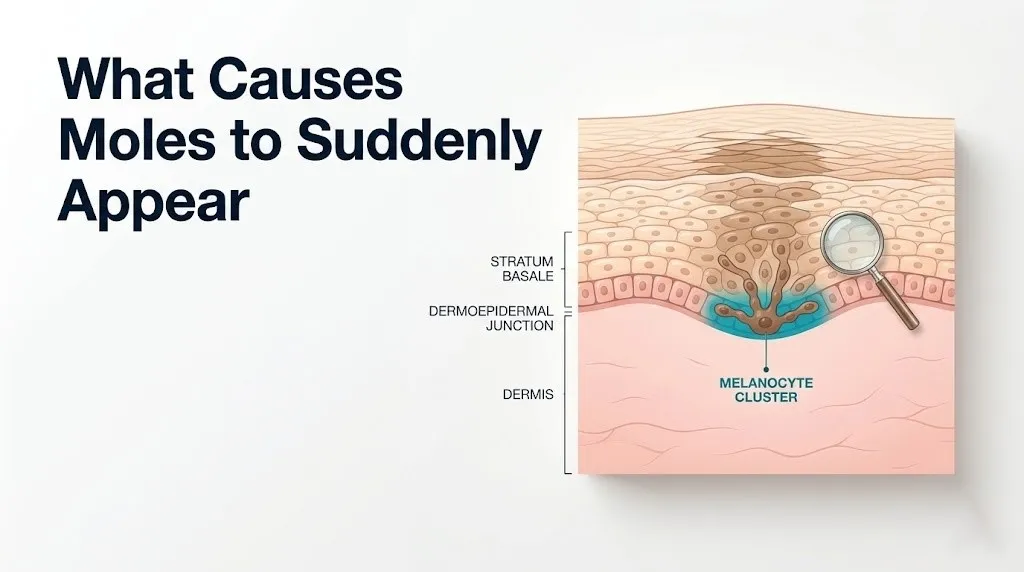
Moles suddenly appear when melanocytes in the stratum basale of the epidermis undergo localized proliferation and cluster into nests, driven by some combination of UV radiation exposure, genetic predisposition, hormonal signaling, or impaired immune surveillance.
The process is not random. A single melanocyte acquires a somatic (non-inherited) mutation that gives it a mild growth advantage over surrounding cells. That cell divides. Its daughter cells divide again. Over weeks to months, the cluster becomes visible at the surface as a pigmented lesion. From the outside, it looks like something that appeared suddenly. From the inside, the process has usually been underway for longer than it seems.
Most new moles in people under 40 are benign acquired nevi. The National Cancer Institute reports that the average person develops the majority of their lifetime mole count before age 30, with new moles becoming less frequent and more clinically notable after age 40. That does not mean new moles after 40 are automatically dangerous, but they do warrant closer scrutiny.
| Cause of New Mole | Underlying Mechanism | Who Is Most Affected |
|---|---|---|
| UV radiation exposure | BRAF mutation, MAPK pathway activation | Fair-skinned adults with cumulative sun exposure |
| Genetic predisposition | CDKN2A variants, family history of atypical nevi | Individuals with FAMM syndrome or multiple first-degree relatives with melanoma |
| Hormonal changes | Estrogen/progesterone receptor activation on melanocytes | Pregnant women, adolescents, oral contraceptive users |
| Medication or immunosuppression | Impaired T-cell surveillance of melanocyte clusters | Organ transplant recipients, people on chronic immunosuppressants |
| Aging and cumulative sun damage | Accumulated UV-induced DNA mutations over decades | Adults over 40, outdoor workers, people with history of sunburns |
| Puberty | MSH and sex hormone elevation during rapid development | Adolescents aged 12 to 20 |
The neck is a specific site of concern for new mole development because of its chronic UV exposure and the clinical importance of lesions near the dense cervical lymphatic network. That site-specific context is covered in its own section below.
What Is a Mole and How Does It Form
A mole (medical term: melanocytic nevus, ICD-10: D22) is a benign cluster of melanocytes that have accumulated in a localized area of skin rather than distributing individually throughout the epidermis the way they normally do.
Melanocytes originate from neural crest cells during fetal development. They migrate from the neural crest to the epidermis during the seventh to twelfth weeks of gestation, settling in the stratum basale at a density of roughly one melanocyte per ten to twenty surrounding keratinocytes. Think of them like individual ink cartridges scattered evenly across the base of the skin. When those cartridges stay separated, they produce the even, background pigmentation of normal skin tone. When a cluster of cartridges fuses together, the concentrated pigment becomes visible as a mole.
Three structural types of melanocytic nevi exist based on where in the skin the melanocyte clusters reside:
- Junctional nevus: Melanocyte clusters located at the dermoepidermal junction. Typically flat and dark brown. The earliest structural form of a developing acquired nevus.
- Compound nevus: Clusters at both the dermoepidermal junction and within the dermis. Slightly raised, often lighter in the center. The intermediate structural form.
- Intradermal nevus: Clusters entirely within the dermis, with no junctional component. Usually dome-shaped, flesh-colored or light brown. The most common type in adults.
The Mayo Clinic notes that most moles a person acquires during childhood and early adulthood begin as junctional nevi and gradually migrate into the dermis over years, which is why older adults tend to have more raised, flesh-colored moles while children typically have flat, dark ones. A mole that appears flat and dark in an adult, rather than following this established pattern, warrants closer examination.
Melanocyte Proliferation and the MAPK Pathway
The molecular reason most moles form is a somatic activating mutation in the BRAF gene, specifically the BRAF V600E substitution, which constitutively activates the MAPK (mitogen-activated protein kinase) signaling pathway in affected melanocytes.
Under normal conditions, the MAPK pathway functions like a controlled assembly line. An external growth signal (such as a growth factor binding to a surface receptor) triggers a chain reaction: RAS activates RAF (BRAF), which activates MEK, which activates ERK. ERK then enters the cell nucleus and promotes genes that drive cell division. The key word is “controlled.” When the signal is present, the line runs. When it stops, the line stops.
The BRAF V600E mutation breaks the off switch. The mutated BRAF kinase remains permanently active regardless of upstream signaling. ERK is constitutively activated. The melanocyte divides when it should not. Research published in the Journal of Investigative Dermatology has confirmed that the BRAF V600E mutation is detectable in approximately 70 to 82% of common acquired benign melanocytic nevi in adults, making it the most frequent molecular event in benign mole formation.
Why does this mutation cause a benign mole rather than immediate cancer in most cases? The answer is oncogene-induced senescence. Once BRAF V600E drives a certain number of cell divisions, the cell activates a permanent growth arrest program mediated partly by the p16/INK4a protein (encoded by the CDKN2A gene). The cluster stops growing. The result is a stable benign nevus. In people with inherited CDKN2A mutations, this braking system is impaired, which is why they develop numerous atypical nevi and carry elevated melanoma risk.
Individuals with a personal or family history of familial atypical mole and melanoma syndrome (FAMM syndrome) should discuss genetic counseling with their primary care physician and receive annual full-body skin examinations by a board-certified dermatologist, regardless of age.
Key Takeaway: Most moles form because a single melanocyte acquires a BRAF V600E mutation that activates uncontrolled cell division through the MAPK pathway. The cell cluster is then stabilized by oncogene-induced senescence via the CDKN2A/p16 system. When that braking system is inherited as defective, moles become numerous and atypical.
UV Radiation as a Cause of New Moles
UV radiation is the most well-established environmental cause of new mole development. Both UV-B radiation (wavelengths of 280 to 315 nm) and UV-A radiation (wavelengths of 315 to 400 nm) damage melanocyte DNA, but through different mechanisms that are each clinically relevant.
UV-B radiation causes direct DNA damage in the form of pyrimidine dimers, cyclobutane pyrimidine dimers, and (6-4) photoproducts at adjacent thymine bases. These lesions, if not repaired by the nucleotide excision repair (NER) pathway before the cell divides, become permanent mutations. The BRAF V600E mutation is a C-to-T transition that bears the molecular signature of UV-B-induced pyrimidine dimer damage. UV-A radiation generates reactive oxygen species (ROS) that damage DNA indirectly through oxidative base modification, particularly at guanine residues.
The National Cancer Institute reports that the association between UV exposure and melanocytic nevus count is well-established across epidemiological cohort studies: children who receive intermittent intense UV exposure (beach vacations, outdoor sports) develop more new moles per year than matched children with lower UV exposure, regardless of baseline skin type. The pattern for adults is similar: a summer with high cumulative UV exposure typically produces new nevi that become visible in the months following.
It is not only beach days that matter. The neck and upper chest are chronically UV-exposed in most people who spend time outdoors without high-necked clothing, even during incidental daily exposure like walking to a car or eating outside. People who work outdoors (construction, landscaping, agriculture, outdoor instruction) accumulate decades of UV exposure to the neck that is rarely matched by the level of sunscreen protection applied there.
Quick Tip:
- Broad-spectrum SPF 30 or higher sunscreen applied to the neck every morning reduces UV-B and UV-A exposure to melanocytes in that region year-round, not just in summer.
- People with Fitzpatrick skin type I (always burns, never tans) and type II (usually burns, sometimes tans) have lower baseline epidermal melanin density, meaning UV-induced melanocyte DNA damage is less buffered and new nevi are more likely to develop at lower UV doses.
- Tanning bed users are not protected from UV-A radiation by broad-spectrum outdoor sunscreen if they are simultaneously using indoor tanning equipment. Tanning beds emit primarily UV-A at intensities up to 10 to 15 times that of natural sunlight, per NCI data.
Genetics and Hereditary Mole Development
Genetic factors determine both how many moles a person develops over their lifetime and how likely those moles are to become atypical or malignant. The two most clinically established genetic contributors are MC1R gene variants and CDKN2A mutations.
The MC1R gene encodes the melanocortin-1 receptor on the surface of melanocytes. This receptor determines which type of melanin a melanocyte produces. When MC1R is fully functional, UV stimulation drives production of eumelanin (brown-black pigment that absorbs UV and protects DNA). Common MC1R variants, associated with red hair, fair skin, and freckles, shift production toward pheomelanin (red-yellow pigment that is less effective at UV absorption and may generate ROS upon UV activation). Individuals with MC1R variants develop more nevi per unit of UV exposure than individuals with fully functional MC1R. Research published in JAMA Dermatology has found that MC1R variant carriers are at approximately 1.4 to 2.0 times the baseline risk of developing melanoma compared to non-carriers with equivalent UV exposure histories.
CDKN2A is the tumor suppressor gene that encodes p16/INK4a and p14/ARF. As described in the MAPK pathway section, these proteins are the brakes that halt BRAF V600E-driven melanocyte proliferation. Inherited loss-of-function variants in CDKN2A remove those brakes, allowing melanocyte clusters to grow larger, become atypical, and fail to achieve stable senescence. This is the molecular basis of familial atypical mole and melanoma syndrome (FAMM syndrome), characterized by more than 50 atypical nevi, a personal or family history of melanoma, and autosomal dominant inheritance.
Family history matters independently of identified gene mutations. The American Academy of Dermatology states that individuals with a first-degree relative (parent, sibling, or child) diagnosed with melanoma have at least a twofold increased risk of developing melanoma themselves, supporting annual total-body skin examinations by a board-certified dermatologist from age 30 onward in this group.
Hormonal Causes of New Moles
Hormones are an underappreciated but clinically well-observed cause of new mole development, operating through specific receptor-mediated mechanisms on melanocytes rather than through a vague systemic effect.
Melanocytes express estrogen receptor alpha (ERα) and progesterone receptors on their surface. When circulating estrogen and progesterone rise significantly, as they do during puberty, pregnancy, and with exogenous hormone use (combined oral contraceptives, hormone replacement therapy), receptor activation on melanocytes upregulates melanogenesis through the MSH-MC1R signaling axis. Elevated estrogen also appears to increase melanocyte sensitivity to UV stimulation, meaning the same UV dose produces a greater melanocyte response in a hormonally active individual compared to a hormonal baseline.
The result, observed clinically across reproductive medicine and dermatology, is a tendency for existing moles to darken during hormonal peaks and for new melanocytic lesions to appear. Clinical observation consistently shows that adolescents develop the highest rate of new nevus formation during puberty, coinciding with rapid elevation of sex hormones. The majority of a person’s lifetime acquired nevus count is established between puberty and age 30.
Melanocyte-stimulating hormone (MSH), produced by the anterior pituitary gland, directly stimulates the MC1R receptor on melanocytes, driving melanin synthesis and, at elevated levels, melanocyte activity. MSH shares a portion of its molecular sequence with adrenocorticotropic hormone (ACTH), which is why conditions that chronically elevate ACTH (such as Addison’s disease or Cushing’s disease depending on the subtype) can produce increased skin pigmentation.
Women using combined oral contraceptives or receiving hormone replacement therapy (HRT) with estrogen and progestin should discuss their mole history with their prescribing obstetrician-gynecologist and, if new atypical-appearing moles develop during hormonal therapy, should be referred promptly to a board-certified dermatologist for evaluation.
Key Takeaway: Hormones cause new moles because melanocytes carry estrogen and progesterone receptors that, when stimulated by elevated circulating hormones during puberty, pregnancy, or hormonal medication use, increase melanocyte activity and can trigger localized cell clustering. This is not a vague “hormonal effect”; it is a specific receptor-mediated mechanism.
Medication and Immunosuppression Causing New Moles
Certain medications and immune-altering therapies are established causes of new mole formation and deserve specific attention because this category is almost entirely absent from general health content about moles.
Immunosuppressive drugs work by suppressing T lymphocyte and natural killer cell activity to prevent organ rejection or control autoimmune disease. Among the normal functions of these immune cells is surveillance of skin-resident melanocytes: abnormal or excessively proliferating melanocyte clusters are identified and eliminated before they become visible. When this surveillance is pharmacologically suppressed, BRAF-mutated melanocyte clusters that would normally be cleared can develop into visible nevi.
Specific drug classes and their mechanisms:
- Cyclosporine (calcineurin inhibitor): Suppresses T lymphocyte activation by blocking interleukin-2 production. Organ transplant recipients on long-term cyclosporine have documented elevated rates of new melanocytic lesion formation and melanoma. Per National Cancer Institute data, organ transplant recipients have approximately a 2.4-fold increased risk of melanoma compared to the age-matched general population.
- Azathioprine (purine synthesis inhibitor): Generates DNA-reactive thiopurine metabolites that, when incorporated into melanocyte DNA under UV exposure, produce a characteristic pattern of UV-independent DNA mutations. Research published in the British Journal of Dermatology found that azathioprine combined with UV exposure causes a unique mutational signature in skin cells distinct from standard UV-induced mutations.
- PUVA therapy (psoralen plus UV-A): Used for psoriasis and other inflammatory skin conditions. Psoralen is a photosensitizing compound that, when activated by UV-A, forms DNA crosslinks. Long-term PUVA therapy is associated with new nevus formation and elevated melanoma risk.
- BRAF inhibitor targeted therapy drugs (used in metastatic melanoma treatment): Paradoxically can trigger new benign nevi formation by activating the MAPK pathway in wild-type BRAF melanocytes. This is a known and monitored side effect.
People receiving any of these therapies should ask their treating physician (oncologist, rheumatologist, transplant medicine specialist, or dermatologist) about their need for increased skin surveillance frequency, specifically annual or semi-annual full-body skin examinations by a board-certified dermatologist.
What Causes Moles to Suddenly Appear on the Neck
Moles appear on the neck more visibly than on many other body areas because the neck receives chronic UV radiation exposure, has relatively thin overlying skin, and is an anatomical site where new pigmented lesions carry specific clinical significance due to proximity to the cervical lymphatic system.
The neck’s skin differs from the torso in two important ways for mole formation. First, the skin overlying the sternocleidomastoid muscle and platysma muscle is thinner and more mobile, which means melanocytic nevi in this region may develop a different tactile character (softer, more compressible) than those on the thicker skin of the back or thighs. Second, the posterior and lateral neck is chronically UV-exposed in people with short hair or those who wear open collars, yet sunscreen is applied to the neck far less consistently than to the face. A 2021 survey-based study found that fewer than 30% of adults regularly apply sunscreen to the posterior neck during summer outdoor activities.
New moles appearing specifically on the neck warrant attention for a second anatomical reason. The cervical lymph nodes (both the deep and superficial cervical chains running along the internal jugular vein and the external jugular vein respectively) are the first drainage station for lymphatic fluid from the scalp, face, and upper neck. Melanoma arising in a neck mole can metastasize through these regional lymph nodes before distant spread. This regional drainage pattern is why a board-certified dermatologist evaluating a suspicious neck mole should also perform palpation of the cervical lymph node chains as part of the examination.
To describe a new neck mole accurately to a dermatologist at your appointment:
- Note whether the mole is on the anterior (front), posterior (back), or lateral (side) neck, and how far it is from the jawline or clavicle.
- Record when you first noticed it and whether anything changed between that date and your appointment.
- Photograph it in consistent lighting with a ruler or coin for scale reference.
- Note any associated symptoms: itching, bleeding, crusting, or tenderness.
- Tell the dermatologist about any medications you take, your sun exposure history at this site, and any family history of melanoma or atypical nevi.
Women in perimenopause and postmenopause are a specific group who may notice new neck moles coinciding with hormonal shifts, particularly when transitioning off combined oral contraceptives or starting hormone replacement therapy. Any new neck mole in this context should be evaluated by a board-certified dermatologist, not attributed solely to hormonal changes without professional examination.
Key Takeaway: The neck is a high-risk site for new moles because it combines chronic underprotected UV exposure with thin skin and proximity to the cervical lymph nodes, which are the first drainage pathway for any malignancy arising in a neck mole.
New Moles in Adults Over 40
New moles in adults over 40 are less common than in younger adults and are statistically more likely to represent a concerning lesion than an equivalent new mole in a 25-year-old.
The biological reason is cumulative. By age 40, most people have already acquired their primary nevus burden (the collection of moles established during childhood, adolescence, and early adulthood). The mechanisms that drive benign nevus formation, primarily hormonal stimulation and direct BRAF mutation induction by UV, are operating in a skin that has accumulated decades of UV-induced DNA damage. New melanocyte clusters forming in this context are forming in a microenvironment that is more genomically unstable than younger skin.
According to the American Academy of Dermatology, melanoma incidence increases with age, with the median age at diagnosis being approximately 65 years as of current U.S. epidemiological data. Men over 50 have a disproportionately higher melanoma mortality rate than women of the same age, partly because men are less likely to perform skin self-examinations and less likely to report new or changing moles to a physician. Men in this age group should specifically examine the back of the neck and the posterior scalp, areas that receive significant UV exposure and are difficult to self-examine.
A useful clinical rule of thumb, supported by the USPSTF’s evidence review on skin cancer screening, is that any new pigmented lesion appearing in an adult over 50 who has no recent history of new benign nevi should be evaluated by a board-certified dermatologist within 4 to 6 weeks of being noticed, regardless of whether the lesion appears clinically benign to the patient. The USPSTF acknowledges that the evidence base for routine population-wide skin cancer screening is still developing, but expert clinical consensus from the AAD supports annual full-body skin examinations in adults over 40 with risk factors.
| Age Group | Likelihood That a New Mole Is Benign | Recommended Action |
|---|---|---|
| Under 30 | High, especially with sun exposure history | Self-monitor; dermatology if ABCDE features present |
| 30 to 40 | Moderate to high | Dermatology evaluation within 4 to 8 weeks |
| 40 to 50 | Moderate; warrant evaluation | Dermatology evaluation within 4 to 6 weeks |
| Over 50 | Lower than younger adults | Dermatology evaluation within 2 to 4 weeks; do not delay |
| Any age with rapid change | Concerning regardless of age | Urgent dermatology evaluation within 1 to 2 weeks |
New Moles During Pregnancy
New moles during pregnancy are driven primarily by the dramatic hormonal shifts of gestation, specifically the surge in human chorionic gonadotropin (hCG), estrogen, and progesterone that characterize the first and second trimesters.
Human chorionic gonadotropin is produced by the placental syncytiotrophoblast cells and peaks in the first trimester. It shares structural similarity with thyroid-stimulating hormone (TSH) and can cross-react with certain G-protein-coupled receptors, including receptors on melanocytes. Elevated hCG combined with rising estrogen and progesterone creates a hormonal environment that markedly upregulates melanocyte activity. The most visible result is linea nigra (the midline abdominal pigmented line), melasma (facial hyperpigmentation), and, in some pregnancies, darkening of existing moles or appearance of new ones.
Clinicians generally observe that most pregnancy-associated mole changes are benign responses to the hormonal environment. However, pregnancy does not protect against melanoma. Per research summarized by the National Cancer Institute, melanoma is one of the most common malignancies diagnosed during pregnancy, and the hormonal environment of pregnancy may potentially accelerate the growth of a pre-existing early melanoma. The difficulty is that some normal pregnancy-related mole darkening can superficially resemble the color variation of a dysplastic or malignant lesion.
The clinical standard, supported by the American College of Obstetricians and Gynecologists (ACOG), is that any new or changing mole noticed during pregnancy should be evaluated by a board-certified dermatologist, not deferred until after delivery. Dermoscopy is safe in pregnancy. If a biopsy is needed, local anesthesia with lidocaine is considered safe during all trimesters, and biopsy should not be delayed out of concern about the pregnancy.
Women who are pregnant and notice a new mole that meets any ABCDE criterion, bleeds, itches persistently, or has appeared and changed within weeks should request same-week evaluation by a board-certified dermatologist and mention the pregnancy status explicitly when booking the appointment.
Dysplastic Nevus Sudden Appearance
A dysplastic nevus (also called an atypical nevus or Clark’s nevus) is a melanocytic nevus with histologically abnormal cellular features that place it between a common benign mole and early melanoma on the biological spectrum.
Dysplastic nevi can appear suddenly or develop through rapid evolution of a pre-existing flat mole. They are distinguished from common nevi by several macroscopic and microscopic features. At the molecular level, they typically carry not only the BRAF V600E mutation of benign nevi but additional second-hit mutations in tumor suppressor genes (CDKN2A, PTEN) that destabilize the oncogene-induced senescence response, allowing continued, albeit slow, growth.
The American Academy of Dermatology estimates that up to 10% of the U.S. population has at least one clinically atypical mole. Individuals with more than five clinically atypical moles have approximately a tenfold increased risk of developing melanoma compared to the general population, per published epidemiological data.
Distinguishing features of a dysplastic nevus:
- Size: Typically larger than 5 to 6 millimeters in diameter (larger than a pencil eraser).
- Color variation: Two or more shades of brown, tan, pink, or red within the same lesion.
- Border: Irregular, ill-defined, or fading gradually into surrounding skin rather than a sharp edge.
- Surface: Often has a “fried egg” appearance: darker raised center with a lighter, flatter surrounding component.
- Location: Dysplastic nevi are more common on the trunk but do appear on the neck, scalp, and extremities.
A dysplastic nevus that appears and then grows or changes within weeks to a few months is not behaving like a stable benign nevus. This growth pattern, called rapidly evolving nevus, warrants evaluation by a board-certified dermatologist with dermoscopy within 1 to 2 weeks, not at the next available routine appointment.
Key Takeaway: A dysplastic nevus is molecularly unstable compared to a common benign mole: it has acquired additional mutations that impair the oncogene-induced senescence braking system, which is why it can continue to grow and evolve in ways a common benign mole will not.
New Mole Risk Factors and Who Is Most Affected
The risk factors for developing new moles in adulthood are well-defined, and understanding them helps distinguish someone who is experiencing a biologically expected new mole from someone whose new mole history places them at elevated melanoma risk.
Primary risk factors for new mole development:
- Fitzpatrick skin type I or II: People who consistently burn rather than tan have lower baseline melanin density in the epidermis and generate less UV-protective eumelanin per UV stimulus. Their melanocytes accumulate UV-induced DNA damage more rapidly per unit of sun exposure.
- Cumulative UV exposure: Outdoor workers, athletes, and people who live at high altitude or closer to the equator have higher annual UV dose exposure than matched individuals with indoor occupational patterns.
- History of blistering sunburns: A single severe blistering sunburn in childhood has been associated with approximately double the lifetime melanoma risk compared to no severe burns, per NCI epidemiological data. Each blistering sunburn represents an episode of acute, high-intensity UV-induced melanocyte DNA damage.
- Greater than 50 moles already present: The total mole count itself is an independent risk factor. Individuals with 50 or more common nevi have approximately a 5-fold increased melanoma risk compared to those with fewer than 10.
- Family history of melanoma or atypical nevi: First-degree relative with melanoma confers at least a 2-fold increased risk.
- Personal history of melanoma: Previous melanoma diagnosis increases the risk of a second primary melanoma.
- Immunosuppression: As described in the medication section. Includes people on immunosuppressive drugs for organ transplant, autoimmune disease, or inflammatory conditions.
- Xeroderma pigmentosum: A rare inherited DNA repair disorder in which the nucleotide excision repair pathway is defective. Affected individuals accumulate UV-induced DNA mutations at a rate orders of magnitude higher than the general population and develop numerous atypical nevi and skin cancers in childhood.
Individuals who have three or more of the risk factors listed above should discuss an individualized skin surveillance plan with a board-certified dermatologist, including the appropriate frequency of total-body skin examinations and the use of standardized mole photography (total-body photography) for change tracking.
ABCDE Warning Signs of a Dangerous New Mole
The ABCDE criteria are the clinical framework developed by dermatologists and endorsed by the American Academy of Dermatology to help patients and clinicians identify melanocytic lesions that warrant biopsy and histological examination.
Each letter corresponds to a specific measurable or observable feature:
| ABCDE Criterion | What It Means | Clinical Threshold That Warrants Evaluation |
|---|---|---|
| A: Asymmetry | One half of the mole does not match the other half when divided by any central axis | Any asymmetry in a new mole |
| B: Border | The edge is irregular, scalloped, notched, or poorly defined | Any border that is not smooth and sharply demarcated |
| C: Color | Multiple shades within one lesion: varying browns, black, red, white, or blue | Two or more distinct colors within one mole |
| D: Diameter | Size greater than 6 millimeters (approximately the diameter of a pencil eraser) | Any lesion larger than 6 mm in its widest dimension |
| E: Evolving | The mole is changing in any way: size, shape, color, surface, or symptom | Any change noticed over days to weeks |
The “E” for Evolving is the criterion most dermatologists consider most clinically urgent. A stable, asymmetric, 7-millimeter dark mole that has looked the same for five years is statistically less concerning than a symmetric, 4-millimeter light brown mole that appeared two months ago and has already changed color.
A sixth criterion has been proposed in the dermatology literature: “EFG” for Elevated, Firm, and Growing, which better captures nodular melanoma, the subtype least likely to be caught by ABCDE because it may be symmetric, have a regular border, and be uniformly dark. Research published in the British Journal of Dermatology found that nodular melanoma accounts for a disproportionate share of melanoma deaths because it is often undetected by patient self-examination until advanced.
Adolescents may develop rapidly growing Spitz nevi (a benign variant that mimics melanoma in some dermoscopic and histological features) that trigger ABCDE concerns. Any concerning mole in a person under 18 should be evaluated by a board-certified pediatric dermatologist or a general board-certified dermatologist with experience in pediatric lesions, given the diagnostic complexity of Spitz nevi in this age group.
New Mole vs Seborrheic Keratosis
A seborrheic keratosis is a benign epidermal growth that is frequently mistaken for a new mole, particularly in adults over 40, and understanding the distinction is practically important because the two lesions have different implications and management approaches.
Seborrheic keratoses are not melanocytic in origin. They arise from keratinocytes (the structural cells of the epidermis) rather than melanocytes, making them a completely different cell type from melanocytic nevi. They commonly appear from the fourth decade onward and become more numerous with age, particularly on the trunk, face, and neck. They tend to look “pasted on,” as though someone placed a waxy, slightly raised, scaly growth on top of the skin surface. Their color ranges from light tan to very dark brown, and their surface has a characteristic stuck-on, warty, or verrucous texture.
| Feature | New Melanocytic Mole (Nevus) | Seborrheic Keratosis |
|---|---|---|
| Cell of origin | Melanocyte | Keratinocyte |
| Surface texture | Smooth or slightly raised, not warty | Warty, verrucous, “stuck-on” appearance |
| Color | Brown shades, may have irregular distribution | Tan to very dark, often uniform within lesion |
| Age of typical onset | Any age; peaks in adolescence and young adulthood | Most common after age 40 |
| Associated concern | Melanoma risk if atypical features present | Benign; no malignant potential as a lesion type |
| Dermoscopy appearance | Pigment network, globules, specific nevus patterns | Milia-like cysts, comedo-like openings, fissures and ridges |
| Action if uncertain | Evaluation by board-certified dermatologist | Evaluation by board-certified dermatologist to confirm |
The practical problem is that a dark, irregularly colored seborrheic keratosis can closely mimic a pigmented melanoma to the naked eye, and a pigmented melanoma can be mistaken for a seborrheic keratosis. The Cleveland Clinic notes that this diagnostic overlap is one reason that dermoscopy, the handheld magnified polarized-light examination tool used by trained dermatologists, is now considered standard practice rather than optional in the evaluation of new pigmented lesions. The pattern of a lesion under dermoscopy resolves most cases of clinical ambiguity. If it does not, a biopsy with histopathological examination by a dermatopathologist is the only definitive way to distinguish the two.
When to See a Dermatologist for a New Mole
Any new mole that meets at least one ABCDE criterion, appeared rapidly in an adult over 40, or has any associated symptom (bleeding, itching, pain, or crusting) warrants evaluation by a board-certified dermatologist rather than a wait-and-see approach.
The urgency of that evaluation depends on the specific characteristics of the lesion:
- Within 1 to 2 weeks (urgent evaluation): A new mole that appeared and changed within 4 weeks; any mole that has bled spontaneously; a lesion that is firm, raised, rapidly growing, and darkly pigmented (possible nodular melanoma); any new neck mole with simultaneous palpable cervical lymphadenopathy (enlarged neck lymph nodes).
- Within 2 to 4 weeks (prompt evaluation): A new mole in an adult over 50 with no recent history of new benign nevi; a new mole with two or more ABCDE features; a new mole during pregnancy that meets any ABCDE criterion.
- Within 4 to 8 weeks (routine but scheduled evaluation): A new mole in an adult over 40 with one mild ABCDE feature; a new mole in a person with multiple known risk factors; any new mole on the neck in someone with a family history of melanoma.
- At the next annual skin examination: A new mole in a person under 40, with no ABCDE features, that has been stable since it appeared and is smaller than 5 millimeters.
To prepare for a dermatology appointment about a new mole:
- Photograph the mole in consistent, natural daylight next to a ruler or coin for scale.
- Write down exactly when you first noticed it and whether it has changed.
- List all medications you currently take, including oral contraceptives, immunosuppressants, and photosensitizing agents.
- Note your sun exposure history: occupation, typical sun protective behaviors, history of tanning bed use, and any history of blistering sunburns.
- Bring a list of first-degree relatives diagnosed with melanoma or multiple atypical nevi.
- Ask the dermatologist specifically whether dermoscopy will be performed and what the next step would be if the lesion cannot be definitively characterized by dermoscopy alone.
According to the American Academy of Dermatology, adults with any combination of fair skin, family history of melanoma, personal history of sunburns, and more than 50 total moles should receive a full-body skin examination by a board-certified dermatologist at least annually, not only when a new mole prompts concern.
When to See a Dermatologist Urgently: Key Warning Signs
Certain presentations associated with new mole appearance require prompt professional evaluation. Do not wait for a routine appointment if you notice any of the following.
Book an urgent appointment with a board-certified dermatologist within 1 to 2 weeks if you notice:
- A new mole that appeared and changed in size, shape, or color within 4 weeks of first appearing
- A mole that bleeds without being scratched or injured
- A rapidly growing, firm, raised, darkly pigmented lesion anywhere on the body, including the neck
- A new pigmented lesion accompanied by a palpable lump (swollen lymph node) in the neck, armpit, or groin
- A mole that is painful, tender to the touch, or itches persistently for more than 2 weeks
- Any new lesion in an immunosuppressed person (organ transplant recipient, person on chronic immunosuppressive therapy) that has any ABCDE feature
These presentations can indicate dysplastic nevus, nodular melanoma, or metastatic melanoma spread to regional lymph nodes, and require in-person dermoscopic evaluation and potential biopsy, not a telephone assessment or wait-and-watch approach.
If you cannot access a board-certified dermatologist within 2 weeks, present to your primary care physician who can perform an initial evaluation, photograph the lesion for documentation, and arrange an expedited referral.
Frequently Asked Questions About What Causes Moles to Suddenly Appear
Can a mole appear suddenly overnight?
A mole cannot develop entirely from scratch overnight, because the underlying melanocyte clustering process requires weeks to months of cell division to produce a visible lesion.
What people notice “overnight” is typically a mole that has been developing slowly and becomes visible once it reaches a size or pigmentation level that catches their attention, or a different lesion entirely such as a blood blister, pigmented insect bite reaction, or rapidly appearing seborrheic keratosis.
A lesion that appears truly within 24 to 48 hours and has any ABCDE features should be evaluated by a board-certified dermatologist within 1 to 2 weeks rather than assumed to be a benign nevus.
Why am I getting new moles at 40 or 50 years old?
New moles after age 40 are less common than in younger adults and are driven primarily by cumulative UV-induced DNA mutations in skin melanocytes that have accumulated over decades of sun exposure.
The American Academy of Dermatology notes that adults with fair skin, significant cumulative sun exposure, or a history of sunburns are the most likely to develop new nevi in middle adulthood.
Any new mole in an adult over 40 should be evaluated by a board-certified dermatologist within 4 to 6 weeks because the statistical likelihood of a concerning lesion is higher in this age group than in younger adults.
What does it mean when a mole appears on your neck suddenly?
A new mole on the neck most commonly reflects accumulated UV radiation exposure to an area that is frequently sun-exposed but often inadequately protected by sunscreen.
The neck is anatomically significant for new mole evaluation because it is close to the cervical lymph nodes, which are the first drainage points for any melanoma metastasizing from a neck-area primary lesion.
A new neck mole with any ABCDE feature, or any associated palpable neck lump, should be evaluated by a board-certified dermatologist within 1 to 2 weeks.
Are new moles always a sign of cancer?
New moles are not always or even usually a sign of cancer. The vast majority of new acquired nevi in adults are benign melanocytic lesions driven by UV exposure, hormonal changes, or genetic factors.
The American Academy of Dermatology estimates that the lifetime risk of melanoma in the United States is approximately 1 in 38 for white Americans, meaning that most people who develop new moles throughout their lives will not develop melanoma.
The concern is not that every new mole is dangerous, but that some new moles in specific contexts, particularly in adults over 40, rapidly changing lesions, or moles with ABCDE features, warrant professional evaluation to identify the small proportion that are not benign.
Can stress cause new moles to appear?
There is no established direct causal mechanism by which psychological stress produces new mole formation in the way UV radiation or hormones do.
The available preliminary evidence suggests an association between chronic psychological stress and impaired immune surveillance, through elevated cortisol-mediated suppression of natural killer cell activity, which could theoretically reduce the immune clearance of aberrant melanocyte clusters, but this pathway has not been confirmed in controlled human clinical research as a cause of new nevus formation.
If you are noticing new moles during a period of high stress, UV exposure, hormonal changes, or medication changes occurring concurrently are far more likely mechanistic contributors, and a board-certified dermatologist can evaluate the lesions regardless of what triggered them.
How quickly should I see a doctor about a new mole?
The answer depends on the features of the specific mole. A new mole in an adult over 50 with no ABCDE features warrants a dermatology appointment within 4 to 6 weeks. A new mole with one or more ABCDE features, or one that has bled or changed rapidly, warrants evaluation within 1 to 2 weeks.
A board-certified dermatologist is the appropriate provider for this evaluation because they can perform dermoscopy, a handheld polarized-light magnification examination that resolves most cases of clinical ambiguity that a naked-eye assessment cannot.
Do not defer a new mole evaluation until your next annual physical with a primary care physician if the lesion has any concerning features: book directly with a dermatologist and specify that the appointment is for evaluation of a new pigmented lesion.
Moving From Awareness to Action
Understanding why moles suddenly appear is genuinely useful information, but it is most useful when it points you toward a specific next step. A new mole appearing on the neck or anywhere else on the body in an adult over 40 deserves a scheduled dermatology evaluation, not months of self-monitoring.
If the mole you noticed has any ABCDE feature, has changed since you first saw it, or appeared and grew within a few weeks, book an appointment with a board-certified dermatologist this week. Bring a dated photograph and a list of your current medications and sun exposure history.
The molecular biology covered in this article makes one practical point clear: most new moles are the result of a BRAF-mutated melanocyte cluster that has successfully entered benign senescence. The ones that have not are identifiable by trained eyes with dermoscopy. Getting those eyes on a concerning lesion early is the single most effective thing you can do with this information.