Light Sensitivity Causes: All Causes Fully Explained 2026
Light sensitivity causes range from benign, self-resolving conditions like digital eye strain and mild dehydration to neurological emergencies like bacterial meningitis and subarachnoid hemorrhage, with the specific physiological mechanism distinguishing each cause determining both the urgency and the appropriate clinical response. Most cases of photophobia (the clinical term for light sensitivity) arise from local eye inflammation, migraine, or medication effects, but the subset that reflects serious neurological disease requires recognition and emergency evaluation without hesitation.
According to the American Migraine Foundation, approximately 80% of migraine attacks involve photophobia, making migraine the single most common neurological cause of recurrent light sensitivity in adults. Light sensitivity also affects a large proportion of people with dry eye disease, uveitis, and corneal conditions, making it one of the most frequently reported visual complaints in both ophthalmology and primary care settings according to the National Eye Institute (NEI).
This article explains every recognized cause of light sensitivity with its specific anatomical and neurophysiological mechanism, distinguishes ocular from central causes, identifies the populations at highest risk for each cause type, and gives you clear clinical thresholds for when emergency evaluation is necessary versus when monitoring is appropriate.
Light Sensitivity Causes: A Clinical Overview
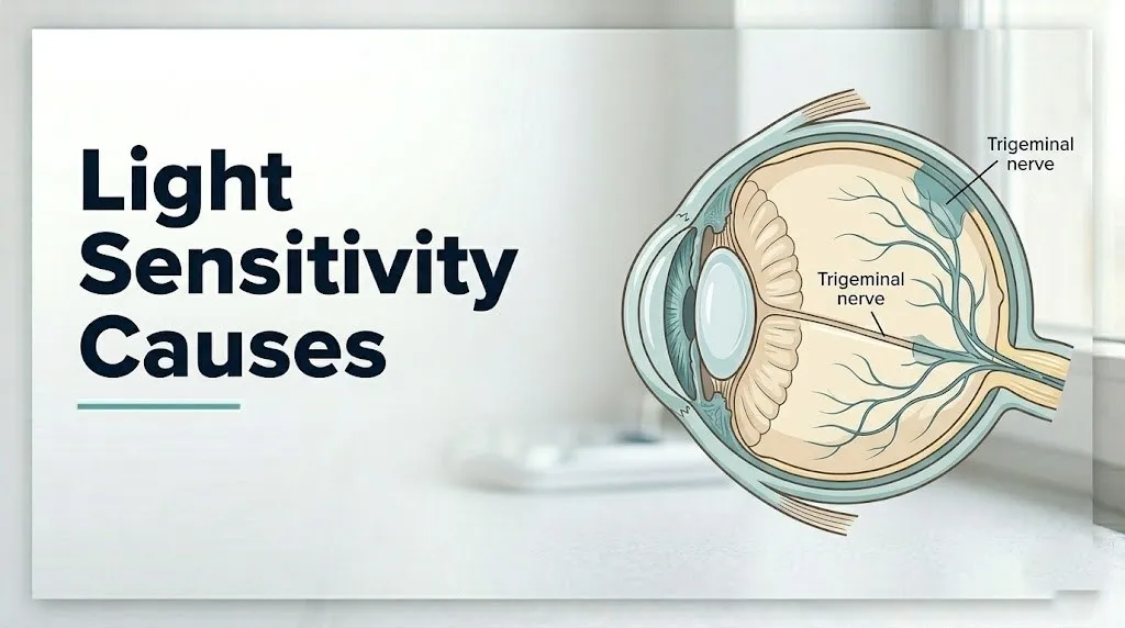
Light sensitivity causes include a spectrum of ocular, neurological, systemic, and pharmacological conditions, each producing the discomfort of photophobia through distinct physiological pathways involving the eye, the trigeminal nerve, or the brain’s pain processing centers.
The clinical term photophobia (from the Greek, meaning “fear of light”) does not reflect an actual fear. It refers to an abnormal painful or uncomfortable response to light that would not affect a person without the underlying condition. Two fundamentally different pathways produce photophobia, and understanding which pathway is active in a given person points directly toward the cause.
| Photophobia Type | Mechanism | Typical Cause Examples | Laterality |
|---|---|---|---|
| Ocular photophobia | Local trigeminal nerve activation at cornea or uveal tract | Corneal abrasion, iritis, glaucoma | Usually unilateral |
| Central photophobia | Thalamic light-pain convergence via ipRGC pathway | Migraine, meningitis, TBI | Usually bilateral |
| Mixed photophobia | Both pathways involved | Severe uveitis, post-TBI with dry eye | Variable |
The frequency hierarchy of causes matters clinically. Migraine and dry eye disease account for the largest proportion of photophobia cases in adults in primary care and ophthalmology settings, according to clinical observational data from the National Eye Institute. Serious neurological causes (meningitis, subarachnoid hemorrhage) account for a smaller proportion but carry life-threatening consequences if missed.
People with lighter eye color (blue, green, or hazel irides) have less melanin in the uveal tract to attenuate incoming light, and consistently report lower thresholds for light discomfort than those with dark irides. This population-level variation in photophobia threshold is supported by observational data and is clinically relevant when assessing whether a patient’s light sensitivity represents a new pathological change or reflects their baseline sensitivity.
What Is Photophobia and How Does It Work?
Photophobia is a symptom defined as an abnormal sensitivity or painful intolerance to light that occurs in the context of an underlying condition affecting either the eye itself or the brain’s pain processing pathways, rather than representing simple discomfort from bright light that any person might experience.
The distinction between normal squinting in bright sunlight and true photophobia is clinically important. Normal pupillary constriction in response to bright light is mediated by the pupillary light reflex, which runs from the retina through the optic tract to the olivary pretectal nucleus in the midbrain, then to the Edinger-Westphal nucleus, and finally through the oculomotor nerve (cranial nerve III) to the iris sphincter muscle, constricting the pupil to reduce light entry. This reflex is preserved and functioning normally in photophobia. The pain of photophobia comes from a separate pathway entirely.
The painful component of photophobia requires a sensitized pain system, either at the level of the eye (inflamed cornea or uveal tract) or at the level of the brain (sensitized trigeminal neurons and thalamic pain centers). This is why a person with normal eyes sitting in the same bright room does not experience pain while a migraine patient finds that same room agonizing.
People who are albino (lacking melanin in the iris and choroid) have no uveal pigment to filter incoming light, meaning their photoreceptors and retinal ganglion cells receive substantially more light energy per exposure than pigmented individuals. This structural difference produces a pronounced, permanent form of photophobia that is present from birth and is not pathological in the disease sense but reflects an anatomical variant with lifelong functional implications.
The Neurophysiology of Light Sensitivity and Pain
The neurophysiology of light sensitivity involves a specialized population of retinal cells called melanopsin-containing intrinsically photosensitive retinal ganglion cells (ipRGCs) that detect light independently of the rods and cones, project to the olivary pretectal nucleus, and send collateral signals to the posterior thalamus, where light-responsive neurons receive convergent input from sensitized trigeminal nociceptors, converting light into pain.
A landmark 2010 study published in Nature Neuroscience by Noseda and colleagues directly demonstrated this pathway in animal models and provided the mechanistic explanation for why migraine patients experience photophobia even in dim light and even when blind in one eye. The ipRGCs respond maximally to blue-spectrum light around 480 nanometers, which explains why blue-wavelength light from LED and fluorescent sources is especially intolerable during migraine attacks.
Think of the posterior thalamus in a sensitized migraine patient like a smoke detector whose sensitivity has been turned up to maximum. When the trigeminal fire alarm system is hyperactive from CGRP-driven meningeal inflammation, even a faint signal (dim light reaching the ipRGCs) is enough to trigger a full alarm response in the thalamus. The pain you feel is real and neurologically produced, not imagined.
The calcitonin gene-related peptide (CGRP), released from trigeminal nerve terminals surrounding meningeal blood vessels during a migraine attack, is the primary molecular driver of trigeminovascular sensitization that makes the thalamus hypersensitive to ipRGC light input. This is confirmed by clinical research: CGRP-blocking medications (gepants and anti-CGRP monoclonal antibodies) reduce not only migraine headache but also migraine photophobia, according to research published in Cephalalgia.
Older adults with age-related neurological changes, including those with early dementia or Parkinson’s disease, can develop abnormal light sensitivity through altered thalamic and cortical processing of sensory stimuli, even without primary eye pathology. A neurologist should evaluate any older adult with new-onset unexplained photophobia alongside other new neurological symptoms.
Key Takeaway: Photophobia is not just “too much light entering the eye.” It is a pain signal generated in the posterior thalamus where light signals from melanopsin-containing retinal cells converge with sensitized trigeminal pain fibers, which is why migraine photophobia is bilateral, persists even with eyes closed, and is worst for blue-spectrum light.
Migraine as a Leading Cause of Light Sensitivity
Migraine causes light sensitivity through trigeminovascular sensitization, in which calcitonin gene-related peptide (CGRP) released from trigeminal nerve terminals surrounding meningeal blood vessels produces neurogenic inflammation that sensitizes the posterior thalamus, making the brain’s light-pain convergence pathway exquisitely reactive during an attack.
Photophobia is one of the most consistent features of migraine, present in approximately 80% of migraine attacks according to the American Migraine Foundation. It can precede the headache phase (appearing during the prodrome or aura), intensify during the peak pain phase, and persist into the postdrome for hours after the headache resolves. This persistence into the postdrome reflects ongoing thalamic sensitization even after the acute CGRP surge has passed.
The temporal pattern of migraine photophobia is clinically distinctive. It is characteristically bilateral, worsened by bright or flickering light (including fluorescent lighting and screen glare), maximally triggered by blue-spectrum wavelengths around 480 nm, and accompanied by phonophobia (sound sensitivity) in the majority of attacks. This bilateral, diffuse quality distinguishes migraine photophobia from the typically unilateral photophobia of local eye conditions like corneal abrasion or iritis.
Women experience migraine at approximately three times the rate of men, according to epidemiological data from the National Institute of Neurological Disorders and Stroke (NINDS). Hormonal fluctuations, particularly the estrogen withdrawal that occurs premenstrually, are a primary trigger for migraine and associated photophobia in women of reproductive age. Women in this population should specifically discuss the relationship between their menstrual cycle and photophobia episodes with their primary care physician or neurologist, as hormonal management may reduce attack frequency.
Eye Conditions That Cause Light Sensitivity
Eye conditions cause light sensitivity through direct activation of the dense network of trigeminal nerve fibers (ophthalmic branch, V1) in the cornea and uveal tract, which interpret ocular inflammation, elevated intraocular pressure, or corneal surface disruption as nociceptive pain signals that are amplified by light-driven pupillary movement and iris muscle contraction.
The eye is one of the most densely innervated tissues in the body. The cornea alone contains approximately 7,000 nerve terminals per square millimeter, all carrying nociceptive input via the nasociliary branch of the ophthalmic division of the trigeminal nerve. When these nerve terminals are inflamed, abraded, infected, or exposed to abnormal intraocular pressure, any additional stimulus including light-driven pupillary constriction (which involves the ciliary body and iris sphincter muscle) amplifies the pain signal.
Key ocular conditions producing photophobia through this mechanism include:
- Acute angle-closure glaucoma: Sudden elevation of intraocular pressure compresses corneal and uveal trigeminal nerve terminals, producing severe unilateral eye pain, photophobia, blurred vision, and a hazy cornea. This is an ophthalmic emergency.
- Episcleritis and scleritis: Inflammation of the episcleral or scleral layers activates adjacent trigeminal fibers, producing eye redness, tenderness, and photophobia. Scleritis is more severe and associated with autoimmune disease.
- Conjunctivitis (viral or bacterial): Conjunctival inflammation with discharge produces mild to moderate photophobia from conjunctival trigeminal nerve activation.
- Keratitis (corneal inflammation from infection): Herpes simplex virus keratitis and bacterial keratitis produce intense photophobia from direct corneal nerve inflammation. Contact lens wearers who develop painful red eye with photophobia need same-day ophthalmology evaluation to rule out infectious keratitis.
Contact lens wearers have an elevated risk for photophobia-producing eye conditions, particularly microbial keratitis from Acanthamoeba or Pseudomonas aeruginosa organisms. Any contact lens wearer developing new eye pain with photophobia and redness should remove lenses immediately and seek same-day evaluation by an ophthalmologist.
Corneal Abrasion and Surface Eye Injuries Causing Photophobia
Corneal abrasion causes photophobia by directly damaging the epithelial surface of the cornea and exposing the underlying stroma and its dense concentration of trigeminal nerve terminal endings, producing immediate, intense photophobia because light-driven pupillary constriction moves the iris against the sensitized cornea and because any light energy reaching the raw nerve terminals intensifies pain.
A corneal abrasion can result from a fingernail scratch, a foreign body (sand, metal shaving, contact lens edge), or chemical splash. The photophobia is typically unilateral, severe, and appears immediately after the injury alongside a foreign body sensation, tearing, and blepharospasm (involuntary eye closing to avoid light). The pain is worse when blinking because the eyelid moves across the exposed nerve terminals.
The clinical significance of corneal abrasion photophobia lies in the risk of secondary infection. An abraded corneal surface is vulnerable to bacterial and viral infection. According to the American Academy of Ophthalmology (AAO), any traumatic eye injury with photophobia, reduced vision, or foreign body that cannot be easily flushed away with sterile saline should be evaluated by an ophthalmologist or emergency medicine physician the same day.
Quick Tip:
- Do not rub a painful, photophobic eye after a suspected corneal injury, as rubbing can extend the abrasion and introduce bacteria
- Patching a painful eye after corneal abrasion is no longer recommended by the AAO because patching increases risk of anaerobic bacterial growth without reducing pain
- Contact lens wearers with corneal abrasion symptoms should not replace their lenses until fully cleared by an ophthalmologist, as lens wear over a healing abrasion dramatically increases infection risk
Occupational exposure is a major risk factor. Workers in manufacturing, construction, metalworking, or woodworking who experience photophobia after any workplace eye exposure should treat the episode as a potential corneal injury until ophthalmological examination confirms otherwise.
Uveitis and Iritis as Causes of Eye Light Sensitivity
Uveitis and iritis (anterior uveitis) cause photophobia through two concurrent mechanisms: direct inflammatory activation of trigeminal nociceptive fibers in the ciliary body and iris stroma, and a reflex ciliary muscle spasm triggered by the iris sphincter’s response to light, which produces a cramping pain in the eye that intensifies with every pupillary constriction.
The uveal tract (iris, ciliary body, and choroid) is richly supplied with trigeminal nociceptive fibers and autonomic nerve terminals. When the uveal tissue is inflamed by infection, autoimmune disease, or trauma, these nerve terminals become sensitized. Each time light triggers the pupillary constriction reflex (causing the iris sphincter to contract), the inflamed ciliary muscle contracts against the inflamed uveal tissue, producing a pain spike that is directly caused by the light stimulus.
According to the National Eye Institute, anterior uveitis (iritis) is the most frequent form of uveitis and is associated with HLA-B27-positive seronegative spondyloarthropathies (ankylosing spondylitis, reactive arthritis, psoriatic arthritis) in approximately 50% of recurrent cases. Adults with a known diagnosis of one of these inflammatory conditions who develop unilateral eye pain and photophobia should contact their ophthalmologist or rheumatologist promptly rather than waiting, as untreated uveitis can cause structural damage to the eye including posterior synechiae (adhesions between the iris and lens) and secondary glaucoma.
Clinical features that distinguish uveitis/iritis photophobia from other ocular causes:
- Unilateral eye pain and photophobia with circumcorneal redness (perilimbal injection)
- Pain worsened specifically by light, particularly through the affected eye
- No significant discharge (which distinguishes it from conjunctivitis)
- Often associated with inflammatory joint symptoms or skin findings in HLA-B27-positive patients
- Diagnosed by slit-lamp examination showing anterior chamber cells and flare
Key Takeaway: Uveitis and iritis produce photophobia through a reflex ciliary spasm mechanism: every time light triggers pupillary constriction, the inflamed iris and ciliary body contract against sensitized nerve terminals, producing a pain spike that is directly caused by the light stimulus. This is why covering one eye during iritis photophobia provides partial relief.
Dry Eye Disease and Light Sensitivity
Dry eye disease causes light sensitivity by producing chronic low-grade inflammation and epithelial damage on the corneal surface, which sensitizes the dense network of trigeminal nociceptive nerve terminals in the corneal epithelium and stroma, lowering the threshold at which normal light exposure triggers discomfort.
The corneal surface is protected by a stable tear film composed of three layers: a mucin layer secreted by goblet cells in the conjunctiva, an aqueous layer from the lacrimal glands, and a lipid layer from the meibomian glands. In dry eye disease, dysfunction of any of these layers produces an unstable tear film that exposes the corneal epithelium to air and environmental irritants between blinks. The exposed epithelial cells and the underlying nerve terminals become chronically sensitized, a process similar to the allodynia (heightened pain sensitivity) seen in chronic pain conditions.
According to the American Academy of Ophthalmology, dry eye disease affects an estimated 16 million Americans, making it one of the most prevalent eye conditions and one of the most frequent causes of chronic photophobia in adults. The photophobia of dry eye is typically bilateral, worse in low-humidity environments (airplane cabins, heated or air-conditioned rooms), and worsened by prolonged screen use, which reduces blink rate and accelerates tear evaporation.
Postmenopausal women are at substantially elevated risk for dry eye disease because estrogen supports lacrimal gland aqueous secretion and goblet cell density. The hormonal changes of menopause reduce both, making postmenopausal women the demographic most consistently affected by dry eye disease and its associated photophobia. Women in this population experiencing new or worsening light sensitivity should specifically discuss dry eye evaluation with their ophthalmologist, asking about tear osmolarity testing and meibomian gland imaging as part of the diagnostic workup.
Meningitis and Light Sensitivity as a Neurological Emergency
Meningitis causes photophobia through inflammatory irritation of the trigeminal nerve fibers in the dura mater, the outermost meningeal layer, which is densely innervated by meningeal branches of the trigeminal nerve. When the meninges are inflamed by bacterial or viral infection, these nerve fibers become sensitized, and any additional sensory stimulus including light (via the ipRGC-to-thalamus pathway) amplifies the pain response in the now-hyperexcitable trigeminal system.
Photophobia in meningitis is not an eye condition. The eyes themselves are normal. The light sensitivity arises because the trigeminovascular system that mediates light-pain processing in the posterior thalamus is severely sensitized by the meningeal inflammatory process. This is the same physiological reason that meningitis photophobia, like migraine photophobia, is typically bilateral.
The clinical context of meningitis photophobia is what makes it an emergency. According to the Centers for Disease Control and Prevention (CDC), bacterial meningitis is a life-threatening infection with a mortality rate of approximately 10 to 15% even with treatment, and neurological complications occur in approximately 11 to 20% of survivors. Photophobia in the context of bacterial meningitis virtually always occurs alongside fever, severe headache, and neck stiffness (meningismus), forming the classic triad.
Call 911 or go to the nearest emergency room immediately if photophobia is accompanied by:
- Fever (temperature above 100.4°F / 38°C)
- Neck stiffness or inability to touch the chin to the chest
- Sudden onset severe headache described as the worst of your life
- Confusion, altered consciousness, or unusual behavior
- Skin rash, particularly a non-blanching petechial or purpuric rash (a sign of meningococcal septicemia)
Children and older adults may present with atypical meningitis symptoms. Infants may show bulging fontanelle, high-pitched cry, and irritability with photophobia rather than the classic adult triad. Older adults may have confusion and photophobia without prominent fever or neck stiffness. These atypical presentations are recognized by the American Academy of Pediatrics and NINDS as requiring the same emergency evaluation.
Concussion and Traumatic Brain Injury Causing Light Sensitivity
Concussion and traumatic brain injury (TBI) cause photophobia through multiple concurrent neurophysiological disruptions: mechanical damage to axons in the visual processing pathways, abnormal thalamic excitability from diffuse axonal injury, and dysfunction of the normal cortical inhibitory mechanisms that filter sensory input under normal conditions.
The posterior thalamus, which mediates the convergence of light signals and trigeminal pain input in the photophobia pathway, is particularly vulnerable to diffuse axonal injury in TBI because of the white matter tracts connecting it to the visual cortex and brainstem. When these connections are disrupted, the normal inhibitory gating that prevents sensory overload is reduced, and ordinary light levels become overwhelming.
According to the National Institute of Neurological Disorders and Stroke (NINDS), photophobia is among the most frequently reported symptoms following concussion, affecting an estimated 50 to 65% of concussion patients in the acute phase. In a subset of patients with postconcussion syndrome, photophobia persists for months to years after the injury, representing a form of chronic central sensitization rather than acute neural injury.
The photophobia of TBI responds differently to light wavelengths than migraine photophobia. Post-TBI patients often report that all light wavelengths are equally intolerable (not specifically blue-spectrum), suggesting that the mechanism involves broader thalamic excitability rather than the specific melanopsin-driven pathway sensitized in migraine. This distinction has implications for management, as tinted lenses designed to filter blue light may provide less benefit for TBI-related photophobia than for migraine-related photophobia.
Athletes with a history of repetitive concussions (common in contact sports including football, hockey, and boxing) face elevated risk for persistent photophobia as part of chronic traumatic encephalopathy (CTE) spectrum symptoms. Any athlete with persistent light sensitivity following head impact should be evaluated by a sports medicine physician or neurologist before returning to play, per the American Academy of Neurology concussion return-to-play guidelines.
Key Takeaway: Concussion photophobia reflects disrupted thalamic inhibitory gating from diffuse axonal injury, not a local eye problem, which is why it responds poorly to eye drops or sunglasses and requires neurological evaluation and management rather than ophthalmological treatment alone.
Medication-Induced Light Sensitivity
Medication-induced light sensitivity occurs through several distinct pharmacological mechanisms depending on the drug class involved, ranging from direct iris sphincter muscle blockade (producing fixed mydriasis that allows excess light entry) to corneal drug deposition and retinal toxicity that sensitize the visual and pain processing pathways.
Specific drug classes and their photophobia mechanisms:
| Drug Class | Example Medications | Mechanism of Photophobia | Clinical Note |
|---|---|---|---|
| Anticholinergics / belladonna alkaloids | Atropine, scopolamine, cyclopentolate | Block iris sphincter (parasympathetic), causing fixed pupil dilation that prevents normal light attenuation | Resolves when medication wears off; mydriasis can last days with atropine |
| Antimalarials | Hydroxychloroquine, chloroquine | Corneal drug deposition producing keratopathy; later, retinal toxicity | Corneal deposits are often asymptomatic early; retinal toxicity requires monitoring |
| Tetracyclines | Doxycycline, minocycline | Photosensitizing effect on the skin and ocular surface; can trigger drug-induced pseudotumor cerebri with associated papilledema and photophobia | Photophobia with headache and doxycycline use warrants evaluation for pseudotumor cerebri |
| Antiepileptics | Zonisamide, topiramate | Central serotonergic and dopaminergic effects; also linked to secondary angle-closure glaucoma from ciliary body changes | Acute vision change with photophobia on topiramate requires same-day ophthalmology evaluation |
| Certain antihypertensives | Amlodipine (rarely) | Possible effects on iris vascular tone and ciliary body blood flow | Uncommon; should be reported to prescribing physician |
According to the American Academy of Ophthalmology, any patient beginning a new medication who develops new photophobia should inform their prescribing physician and an ophthalmologist, as drug-induced eye conditions are often reversible when identified early but can progress if the medication is continued.
People taking doxycycline for acne or Lyme disease treatment who develop photophobia alongside headache and visual changes should seek evaluation by a primary care physician or neurologist for pseudotumor cerebri (idiopathic intracranial hypertension), which is a recognized but underappreciated complication of tetracycline-class antibiotic use, particularly in overweight women of reproductive age.
Systemic Conditions That Cause Light Sensitivity
Multiple systemic conditions cause light sensitivity through shared mechanisms of either central nervous system sensitization, uveal inflammation driven by immune dysregulation, or meningeal involvement that activates the trigeminal pain processing system.
Systemic lupus erythematosus (SLE) produces photophobia through lupus-associated uveitis, lupus retinitis, and lupus cerebritis affecting thalamic and cortical light processing. The antinuclear antibody (ANA) test and anti-double-stranded DNA antibody testing can confirm SLE as a cause in patients with photophobia alongside other systemic features (malar rash, joint pain, fatigue).
Fibromyalgia produces central sensitization affecting multiple sensory modalities simultaneously, including light. The allodynia and hyperalgesia of fibromyalgia involve dysregulation of descending pain inhibitory pathways from the periaqueductal gray matter and rostral ventromedial medulla, meaning sensory inputs including light are processed as painful even when the stimulus is objectively normal. Research published in Pain has documented that photophobia affects a clinically meaningful proportion of fibromyalgia patients independent of comorbid migraine.
Lyme disease (caused by Borrelia burgdorferi) can produce photophobia through Lyme meningitis, cranial neuropathy affecting cranial nerves II or VII, or uveitis. Photophobia occurring in the context of tick exposure, facial palsy, joint swelling, or the characteristic expanding rash (erythema migrans) warrants evaluation by a primary care physician with Lyme serology (enzyme-linked immunosorbent assay followed by Western blot).
People with autoimmune conditions including rheumatoid arthritis, Sjögren’s syndrome, and inflammatory bowel disease have elevated rates of uveitis and secondary dry eye disease, making them at higher baseline risk for photophobia from multiple overlapping causes. Their rheumatologist should be informed of any new light sensitivity so that ocular involvement of the underlying autoimmune condition can be assessed alongside any medication effects.
Light Sensitivity in Pregnancy and Hormonal Changes
Light sensitivity during pregnancy is caused primarily by the substantial increase in migraine frequency that occurs in the first trimester due to rapidly fluctuating estrogen levels, which sensitize the trigeminal pain system and lower the migraine threshold, producing photophobia as part of migraine attacks at a time when many migraine-specific medications have restricted use.
Estrogen has a complex, dose-dependent relationship with migraine physiology. Rising estrogen levels in the first trimester can trigger migraine in women who were previously well-controlled, while the sustained high estrogen of the second and third trimesters typically reduces migraine frequency in women with hormonally driven migraine patterns. The postpartum drop in estrogen after delivery is another high-risk period for migraine and photophobia recurrence.
Beyond migraine, pregnancy-associated hyperemesis gravidarum (severe morning sickness) produces electrolyte imbalances and dehydration that can temporarily sensitize the nervous system, contributing to light sensitivity. The preeclampsia spectrum, including severe preeclampsia and eclampsia, can produce photophobia as part of the cerebral manifestations of hypertensive brain dysfunction, alongside headache, visual disturbances, and altered consciousness.
Photophobia in a pregnant woman in the second or third trimester with headache and elevated blood pressure requires immediate evaluation by an obstetrician-gynecologist on the same day, to rule out preeclampsia or eclampsia. This is a clinical emergency that cannot wait for a scheduled prenatal appointment.
For pregnant women with known migraine, management of photophobia requires coordination between their obstetrician-gynecologist and neurologist to identify safe treatment options that avoid medications with teratogenic potential, particularly specific vasoconstrictive triptans and ergot derivatives whose safety profiles in pregnancy are restricted.
Light Sensitivity in Children and Specific Populations
Light sensitivity in children has a distinct set of causes and presentations compared to adults, with meningitis, migraine, and congenital glaucoma representing the three most clinically urgent causes that a parent or pediatrician must recognize.
Infants with congenital glaucoma present with a triad of photophobia, tearing (epiphora), and blepharospasm (forceful eye closing) in the first months of life. The elevated intraocular pressure from blocked aqueous humor drainage activates corneal trigeminal nerve terminals continuously, producing a child who squints in normal lighting, buries their face to avoid light, and may have enlarged, hazy corneas. This presentation requires urgent evaluation by a pediatric ophthalmologist because congenital glaucoma is treatable but causes permanent vision loss without early surgical intervention.
Children with pediatric migraine experience photophobia during attacks at a high rate, though their migraine attacks are often shorter than adult attacks and may lack the classic unilateral headache of adult migraine. According to the American Academy of Pediatrics, children with migraine may present primarily with bilateral headache, nausea, and a strong preference for dark, quiet rooms. Photophobia in a child alongside recurrent headache lasting 30 minutes to 72 hours warrants evaluation by a pediatric neurologist.
Photophobia in children with fever and neck stiffness follows the same emergency protocol as in adults. Call 911 or go to the emergency room immediately for any child with photophobia plus fever plus altered behavior or neck stiffness, because bacterial meningitis has a rapidly progressive course in children and the window for antibiotic effectiveness is narrow.
People with albinism experience photophobia from birth due to absent melanin in the uveal tract, retinal pigment epithelium, and choroid. Without uveal pigment to filter and absorb incoming light, light passes through the iris and scatters within the eye rather than being absorbed, producing glare and photophobia at ambient light levels that pigmented individuals tolerate without difficulty. Tinted lenses and optical aids are standard management for this population, coordinated by a low-vision specialist or ophthalmologist.
Key Takeaway: In children, photophobia with fever and neck stiffness is a medical emergency requiring 911 or immediate emergency department evaluation regardless of age, because bacterial meningitis progresses rapidly and the diagnostic and treatment window is narrow. Do not wait to see if it resolves.
How Light Sensitivity Is Diagnosed
Light sensitivity is diagnosed through a systematic clinical evaluation that combines detailed symptom history, neurological examination, and targeted ophthalmological and laboratory testing to identify whether the cause is ocular, neurological, systemic, or pharmacological.
The diagnostic pathway begins with history. The laterality of photophobia (one eye vs. both eyes), the timing (acute onset vs. gradual chronic), the associated symptoms (headache, neck stiffness, eye redness, vision change), and the medication history provide the clinical framework that directs the examination.
The standard ophthalmological examination for photophobia includes:
- Visual acuity testing: to assess whether the photophobia is accompanied by vision loss (which raises urgency)
- Slit-lamp examination: to identify corneal abrasion, keratitis, anterior chamber cells and flare (uveitis/iritis), or corneal drug deposits
- Tonometry (intraocular pressure measurement): to identify acute angle-closure glaucoma, where pressure above 21 mmHg with pain and photophobia requires same-day treatment
- Pupillary reflex testing: to assess whether the pupillary light reflex is preserved and symmetric, or whether there is an afferent pupillary defect (APD) suggesting optic nerve disease
- Fundoscopy (ophthalmoscopy): to assess the optic disc, retina, and choroid for papilledema, chorioretinitis, or retinal lesions
For neurological evaluation of photophobia, a brain MRI with and without contrast is ordered when TBI, MS plaques, meningeal disease, or intracranial pathology is suspected. Lumbar puncture (spinal tap for cerebrospinal fluid analysis) is the definitive diagnostic test for meningitis, with a sample sent for cell count, glucose, protein, bacterial culture, and viral PCR.
According to the American Academy of Ophthalmology, a primary care physician should be the first point of contact for photophobia without red eye, vision change, or neurological symptoms, while an ophthalmologist should be the first contact for photophobia with red eye, eye pain, or vision change.
When to See a Doctor and Emergency Warning Signs for Light Sensitivity
See a primary care physician for light sensitivity that has persisted for more than 48 hours without an obvious, resolving cause such as a brief screen-heavy day or mild dehydration, and see an ophthalmologist the same day for any light sensitivity accompanied by eye redness, eye pain, vision change, or contact lens use.
For non-emergency physician referral, the following symptoms warrant an appointment within 24 to 72 hours:
- New photophobia lasting more than two days without an identified cause
- Photophobia that is progressively worsening over days to weeks
- Photophobia in a person with a known autoimmune condition (SLE, ankylosing spondylitis, IBD, psoriasis)
- Photophobia after starting a new medication
- Photophobia in a person with a history of migraine that is behaving differently from prior attacks
- Photophobia in a pregnant woman in any trimester alongside headache
Things to tell your doctor or bring to the appointment:
- When the photophobia started and whether it is getting better or worse
- Whether it affects one eye or both
- Whether it is associated with headache, neck stiffness, fever, or vision change
- A complete medication list including vitamins, herbal supplements, and eye drops
- Whether you wear contact lenses and the type and age of your current lenses
- Any recent head injury, even a mild one from weeks ago
Emergency Symptoms: When to Call 911 or Go to the ER
Certain presentations of light sensitivity require immediate emergency evaluation. Do not wait to see if these resolve on their own.
Call 911 or go to the nearest emergency room immediately if light sensitivity is accompanied by:
- Fever above 100.4°F (38°C) with neck stiffness or inability to flex the neck (possible bacterial meningitis, a life-threatening infection)
- Sudden onset severe “thunderclap” headache described as the worst headache of your life (possible subarachnoid hemorrhage from a ruptured brain aneurysm)
- Confusion, altered consciousness, difficulty speaking, or arm or leg weakness alongside light sensitivity (possible encephalitis, stroke, or severe meningitis)
- Sudden loss of vision in one or both eyes alongside light sensitivity (possible acute angle-closure glaucoma emergency or retinal vascular event)
- A non-blanching purplish or red skin rash alongside fever and light sensitivity (possible meningococcal septicemia, which progresses within hours)
- Photophobia in an infant with bulging fontanelle, high-pitched cry, or extreme irritability (possible infant meningitis)
These presentations can indicate bacterial meningitis, subarachnoid hemorrhage, acute angle-closure glaucoma, or encephalitis, all of which require emergency medical assessment in a hospital with appropriate diagnostic and treatment capabilities, not an urgent care appointment or a telephone consultation.
Frequently Asked Questions About Light Sensitivity Causes
What is the most common cause of light sensitivity?
Migraine is the most common neurological cause of recurrent light sensitivity, affecting approximately 80% of migraine attacks according to the American Migraine Foundation.
Dry eye disease is the most common chronic ocular cause of photophobia in adults, affecting an estimated 16 million Americans according to the American Academy of Ophthalmology.
In any individual patient, the cause is identified by evaluating whether the photophobia is unilateral or bilateral, whether it accompanies red eye or vision change, and whether it occurs in the context of headache or systemic symptoms.
Can dry eyes cause light sensitivity?
Yes, dry eye disease causes light sensitivity by producing chronic inflammation and surface damage on the cornea that sensitizes the trigeminal nerve terminals densely embedded in the corneal epithelium.
This corneal nerve sensitization lowers the threshold at which normal light exposure triggers discomfort, producing bilateral photophobia that is typically worse in dry environments, during prolonged screen use, and in air-conditioned spaces.
Postmenopausal women are at the highest risk for dry eye-related photophobia because estrogen decline after menopause reduces lacrimal gland secretion and goblet cell density in the conjunctiva.
Is sudden light sensitivity a sign of something serious?
Sudden light sensitivity can indicate a serious or life-threatening condition when it is accompanied by fever, neck stiffness, severe headache, confusion, or vision loss.
Meningitis and subarachnoid hemorrhage both present with sudden photophobia alongside these accompanying symptoms and require immediate emergency room evaluation.
Sudden unilateral photophobia with eye pain, redness, and vision change can indicate acute angle-closure glaucoma, which also requires same-day ophthalmological emergency evaluation to prevent permanent vision loss.
What does light sensitivity in migraine feel like?
Migraine photophobia is a bilateral, painful intolerance to light that is characteristically worst for bright or flickering light and for blue-spectrum wavelengths from LED and fluorescent sources.
It is caused by thalamic sensitization through the trigeminovascular pathway, not by a problem with the eyes themselves, which is why it persists even when the eyes are closed and continues into the hours after the headache phase resolves.
People describe migraine photophobia as an immediate worsening of head pain upon light exposure, a deep aching behind the eyes, and an overwhelming need to seek a dark room regardless of how bright the environment actually is.
Can medications cause sensitivity to light?
Yes, several medication classes cause light sensitivity through distinct mechanisms, including anticholinergics that block iris sphincter function to produce fixed pupil dilation, tetracyclines that can trigger pseudotumor cerebri, and antimalarials that deposit in the cornea or cause retinal toxicity.
Anyone who develops new light sensitivity after starting a medication should report this to their prescribing physician and an ophthalmologist promptly.
Doxycycline-associated photophobia with headache and visual changes is a specific combination that requires same-day evaluation for pseudotumor cerebri (idiopathic intracranial hypertension), particularly in overweight women of reproductive age.
Why do some people have permanently sensitive eyes to light?
Some people have permanently reduced thresholds for light discomfort due to structural factors, including albinism (absent uveal melanin that normally absorbs light within the eye), aniridia (absence of the iris that normally controls light entry), and blue or light-colored irises (less melanin to filter light).
Chronic conditions including fibromyalgia, postconcussion syndrome, and long-standing dry eye disease can produce persistent central or peripheral sensitization that sustains photophobia beyond the acute phase of the underlying illness.
People with permanent photophobia should be evaluated by an ophthalmologist to confirm the structural or functional basis and to discuss appropriate optical aids, including tinted lenses that filter high-energy wavelengths, as part of an individualized management plan.
Closing
The most practical takeaway from this article is the laterality test. Unilateral photophobia with a red eye almost always points to a local ocular cause requiring same-day ophthalmological evaluation. Bilateral photophobia without a red eye almost always points to a neurological or systemic cause that requires primary care or neurology evaluation. This single clinical observation can help you direct yourself to the right type of provider without delay.
The symptom combination that changes everything is photophobia with fever and neck stiffness. That combination requires a 911 call or an immediate emergency room visit, not an urgent care appointment or a phone consultation. Bacterial meningitis does not wait.
If your light sensitivity is chronic and unexplained, ask your primary care physician for a referral to both an ophthalmologist and a neurologist. You want both pathways evaluated, because in conditions like fibromyalgia, postconcussion syndrome, and autoimmune disease, ocular and central mechanisms often coexist, and addressing only one rarely resolves the full picture.