C5-C6 and C6-C7 Disc Disease in Neck: Symptoms Explained
Degenerative disc disease in the neck at C5-C6 and C6-C7 produces two distinct symptom patterns: cervical radiculopathy, where compressed nerve roots cause pain, numbness, and weakness radiating into the arm and hand in a specific anatomical territory, and cervical myelopathy, where spinal cord compression produces bilateral hand dysfunction, gait changes, and bladder symptoms that require urgent neurosurgical evaluation. Knowing which pattern your symptoms match determines both the urgency and the appropriate clinical pathway.
The National Institute of Neurological Disorders and Stroke (NINDS) identifies cervical spondylosis and disc degeneration as among the most prevalent musculoskeletal conditions affecting adults over 40, with C5-C6 and C6-C7 the two most frequently degenerated levels in the entire cervical spine. The North American Spine Society (NASS) reports that cervical radiculopathy, the nerve root compression syndrome that frequently results from disc degeneration at these levels, has an incidence of approximately 83 per 100,000 adults annually in the United States.
This article maps every major symptom of cervical DDD at C5-C6 and C6-C7 to the specific nerve root or spinal cord structure producing it, distinguishes the C6 nerve root symptom pattern from the C7 pattern in anatomical detail, identifies the myelopathy signs that change the clinical urgency completely, and explains exactly when and which provider to contact based on what you are experiencing.
Degenerative Disc Disease in Neck C5-6 and C6-7 Symptoms: Overview
Degenerative disc disease in the neck at C5-C6 and C6-C7 produces symptoms through two mechanisms: compression of the C6 or C7 nerve roots in their neuroforaminal exit canals, producing unilateral arm and hand symptoms, and compression of the cervical spinal cord itself by central disc material or osteophytic ridging, producing bilateral neurological dysfunction.
The symptom picture that a person with C5-C6 and C6-C7 disc disease experiences depends entirely on where the degenerative changes are compressing tissue. A posterolateral disc herniation or uncovertebral osteophyte compressing the C6 nerve root produces a completely different clinical picture from a central disc bulge pressing against the anterior spinal cord. Both can coexist in the same patient, which makes the clinical assessment more complex than single-level or single-mechanism conditions.
The most practically useful framework for understanding these symptoms is to start with the anatomy. The C6 nerve root exits the spinal canal through the C5-C6 neuroforamen (the bony channel between the C5 and C6 vertebrae). The C7 nerve root exits through the C6-C7 neuroforamen. Each nerve root has its own specific territory: muscles it powers, skin it senses, and reflex it mediates. When that root is compressed, every function in its territory is affected.
Observable symptoms across both levels include:
- Neck pain with or without stiffness that worsens with specific head positions
- Shoulder or periscapular aching that is difficult to localize precisely
- Pain radiating down one arm in a pattern specific to the affected nerve root
- Numbness, tingling, or altered sensation in specific finger distributions
- Weakness in specific arm or hand muscles
- Changes in deep tendon reflexes at the elbow or triceps (detectable by a clinician)
- In advanced cases: bilateral arm or leg symptoms, gait changes, or bladder dysfunction
Why C5-C6 and C6-C7 Are the Most Commonly Affected Cervical Levels
C5-C6 and C6-C7 are the most frequently degenerated levels in the cervical spine because they experience the highest cumulative mechanical stress in the lower cervical region, where the greatest arc of flexion and extension motion occurs and where axial compressive loads from the weight of the head are concentrated.
The cervical spine functions like a crane arm, with the head as the load at the end of the arm. The lower cervical segments, particularly C5-C6 and C6-C7, are the pivot points that absorb the greatest mechanical energy with each movement of the neck. Over years of repetitive flexion, extension, and rotation, the intervertebral discs at these levels undergo more cumulative compressive and shear loading than the upper cervical segments.
The biomechanical stress concentrates at C5-C6 and C6-C7 for a specific anatomical reason. The C5-C6 and C6-C7 segments lie at the junction between the relatively mobile lower cervical spine and the less mobile upper thoracic spine. Motion above and below this junction converges at these two levels. Research published in the Spine journal has documented that the C5-C6 disc is the single most frequently herniated and degenerated level in the cervical spine across multiple large imaging cohort studies, followed closely by C6-C7.
The uncovertebral joints of Luschka, the small joints on the lateral margins of the cervical vertebral bodies that are unique to the cervical spine and absent in the thoracic and lumbar spine, are particularly prone to developing osteophytes (bone spurs) at C5-C6 and C6-C7 as disc degeneration and height loss progress. These osteophytes directly border the neuroforamen, making foraminal narrowing and nerve root compression a predictable consequence of degeneration at these specific levels.
Older adults over age 60 show radiographic evidence of disc degeneration at C5-C6 in the majority of imaging studies, even when they are asymptomatic, according to observational imaging data referenced in Mayo Clinic clinical resources, underscoring how prevalent these changes are at these specific levels.
How Cervical Disc Degeneration Produces Symptoms
Cervical disc degeneration at C5-C6 and C6-C7 produces symptoms through the biochemical degradation of the nucleus pulposus proteoglycan matrix, subsequent disc height loss, reactive osteophyte formation at adjacent joint surfaces, and the resulting mechanical compression and neurochemical irritation of the C6 or C7 nerve root or the cervical spinal cord.
The process begins inside the disc itself, long before any structural compression is visible. The nucleus pulposus, the gelatinous core of the disc that absorbs compressive load through its water content, is maintained by a matrix of proteoglycans (primarily aggrecan) and type II collagen fibers. As age and mechanical stress accumulate, matrix metalloproteinases MMP-3 and MMP-13, stimulated by interleukin-1 beta (IL-1β) and tumor necrosis factor-alpha (TNF-α) from degenerating nucleus pulposus cells, begin degrading the aggrecan and collagen framework. The disc loses its ability to hold water. It desiccates and loses height.
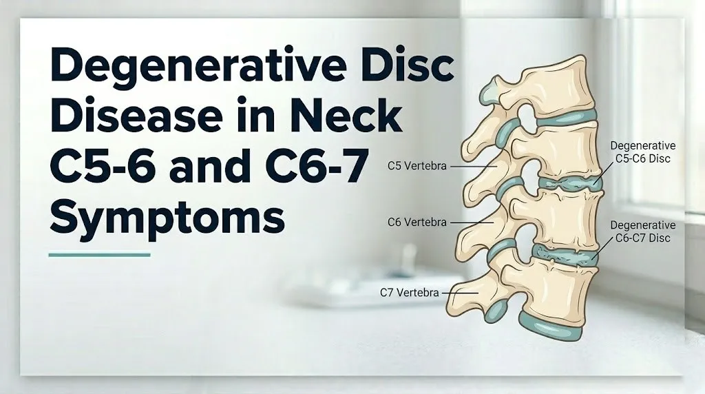
Think of the nucleus pulposus like a water-saturated sponge inside a fibrous casing. Under normal circumstances, the sponge absorbs spinal compression and distributes loads evenly. When MMP enzymes degrade the sponge material, it can no longer hold water under load. The casing flattens. The disc space narrows. And as the disc loses height, the neuroforaminal height (the vertical dimension of the bony channel housing the nerve root) also decreases, because the foramen’s height is partly determined by the disc height below it.
Once the nerve root is compressed in the narrowed foramen, a second mechanism activates. Compressed C6 or C7 dorsal root ganglion cells release substance P and calcitonin gene-related peptide (CGRP) into the surrounding tissues. These neuropeptides produce neurogenic inflammation, lowering the pain threshold in the nerve root and surrounding tissues and producing the characteristic burning, electric, or shooting quality of radicular arm pain that distinguishes nerve root compression from simple muscle strain.
The Journal of Neurosurgery Spine has published research documenting that both mechanical compression and neurochemical irritation contribute independently to radicular symptom severity, meaning that even modest disc material contact with the nerve root can produce severe pain when the inflammatory component is dominant.
Key Takeaway: Cervical disc degeneration produces radicular symptoms through two simultaneous mechanisms: mechanical compression of the nerve root in the narrowed neuroforamen AND neurochemical irritation from substance P and CGRP released by the compressed dorsal root ganglion, which is why the pain often feels burning and electric rather than simply pressure-like.
C5-C6 Degenerative Disc Disease Symptoms: The C6 Nerve Root
C5-C6 degenerative disc disease produces symptoms in the C6 nerve root distribution: burning or electric pain traveling from the neck down the outer (radial) side of the forearm into the thumb and index finger, with possible weakness of wrist extension and the brachioradialis muscle, and reduction of the brachioradialis deep tendon reflex.
The C6 nerve root exits the spinal canal through the C5-C6 neuroforamen. It travels distally through the brachial plexus to supply the radial surface of the forearm, the thumb, and the index finger. The dermatomal map for C6 is specific enough that a clinician can identify C6 root involvement from the distribution of a patient’s numbness alone: if you have altered sensation on the pad of your thumb and the radial side of your index finger, and the inner surface of your forearm from elbow to thumb, that is the C6 territory.
C5-C6 disc disease symptom pattern:
| Symptom Domain | C6 Nerve Root Presentation |
|---|---|
| Pain distribution | Neck, outer shoulder, radial forearm, thumb and index finger |
| Sensory change | Numbness or tingling in thumb, index finger, radial forearm |
| Motor weakness | Wrist extension (ECRL/ECRB), brachioradialis, biceps brachii (partial) |
| Reflex change | Reduced or absent brachioradialis reflex |
| Provocative factor | Neck extension or lateral bending toward affected side worsens pain |
Women with C5-C6 disc disease may report a broader, less well-defined pain pattern that extends into the periscapular and upper back region, which can lead to initial misattribution of the pain to shoulder pathology such as rotator cuff disease. The key distinguishing feature is that cervical radiculopathy pain is typically reproduced by neck position changes rather than by shoulder movement alone.
C6-C7 Degenerative Disc Disease Symptoms: The C7 Nerve Root
C6-C7 degenerative disc disease compresses the C7 nerve root in the C6-C7 neuroforamen and produces a distinct symptom pattern: pain traveling from the neck down the posterior upper arm into the middle finger and ring finger, weakness of the triceps brachii and finger extensors, and reduction or loss of the triceps deep tendon reflex.
The C7 nerve root territory is the largest dermatomal distribution of any cervical nerve root, covering the posterior arm, elbow, the dorsal (back) surface of the middle finger, and contributions to the ring finger. This broad territory means C7 radiculopathy can produce symptoms spanning from the posterior neck through the elbow region and into the middle finger, which sometimes misleads patients into believing multiple conditions are present when a single C7 root compression is responsible for the entire symptom chain.
The triceps brachii, the muscle responsible for elbow extension, receives its primary innervation from the C7 nerve root through the radial nerve. When C7 is compressed, the triceps reflex (elicited by tapping the triceps tendon with the elbow flexed) is reduced or absent on the affected side. This reflex change is one of the most reliable clinical signs of C7 root involvement and is specifically checked by any physician performing a neurological examination for cervical radiculopathy.
Quick Tip:
- C6 radiculopathy (C5-C6 disc): numbness in the thumb and index finger, weakened wrist extension and brachioradialis, reduced brachioradialis reflex
- C7 radiculopathy (C6-C7 disc): numbness in the middle finger, weakened triceps and finger extension, reduced triceps reflex
- If both levels are degenerated simultaneously, both distributions may be affected, producing a broader arm and hand symptom picture that spans thumb through middle finger
Older adults with C6-C7 disc disease may experience a less distinct dermatomal pattern because of the reduced precision of sensory testing in older nervous systems and because co-existing conditions such as carpal tunnel syndrome can produce overlapping finger numbness that complicates localization.
Neck and Shoulder Pain From C5-C6 and C6-C7 Disc Disease
Neck and shoulder pain from C5-C6 and C6-C7 degenerative disc disease originates from three distinct anatomical sources simultaneously: inflammatory nociceptor activation in the degenerating disc’s annulus fibrosus, mechanical irritation of the facet joints (zygapophyseal joints) at the same levels, and referred pain from the compressed C6 or C7 nerve root itself.
The annulus fibrosus of the intervertebral disc is richly innervated in its outer third by branches of the sinuvertebral nerve (the recurrent meningeal nerve). As disc degeneration progresses, annular tears allow inflammatory nucleus pulposus material to contact these pain fibers in the outer annulus, producing a deep, aching, poorly localized neck pain that is worse with prolonged postures (sitting at a computer, looking down at a phone) and partially relieved by lying flat, which reduces axial loading on the disc.
Simultaneously, the facet joints at C5-C6 and C6-C7 develop reactive arthropathy as disc height loss changes the load distribution between the disc and the facet joints. The facet joint capsule contains pain receptors innervated by the medial branch of the dorsal rami. When facet joint loading increases, these receptors generate a referred pain pattern that commonly extends into the periscapular region (between the shoulder blades), the posterior shoulder, and occasionally the posterior head, producing what patients describe as a constant, nagging ache in the neck and upper back area.
According to clinical guidelines from the North American Spine Society (NASS), the combination of discogenic and facet-mediated pain at C5-C6 and C6-C7 is one of the most frequently encountered presentations in spine specialty practice, and the two pain sources often require separate diagnostic and therapeutic approaches: discogenic pain responds differently to treatment than facet joint-mediated pain.
People who perform repetitive overhead work (painters, electricians, warehouse workers) or who maintain sustained cervical extension postures (looking overhead) develop accelerated facet joint loading at C5-C6 and C6-C7 due to the biomechanics of cervical extension, which closes the facet joint spaces and increases compressive loading on the joint capsule and articular cartilage.
Key Takeaway: Neck and shoulder pain from C5-C6 and C6-C7 disc disease typically involves three pain generators simultaneously (degenerating disc, inflamed facet joint, and nerve root), which is why the pain pattern is often diffuse and why single-injection treatments addressing only one generator frequently provide incomplete relief.
Arm and Hand Numbness From Cervical Disc Disease at C5-C6 and C6-C7
Arm and hand numbness from cervical disc disease at C5-C6 and C6-C7 reflects dermatomal sensory loss in the C6 or C7 territory, produced when disc herniation or osteophytic foraminal stenosis reduces sensory axon conduction capacity in the compressed nerve root.
The sensory axons of the C6 nerve root travel from the skin of the radial forearm, thumb, and index finger to the C6 dorsal root ganglion and then into the posterior horn of the spinal cord at the C5-C6 level. When these axons are compressed in the C5-C6 neuroforamen, their conduction velocity slows and eventually fails at the point of compression. The brain, receiving reduced or absent signal from that skin territory, interprets this signal reduction as numbness, tingling, or the “asleep” sensation that patients describe.
The pattern of numbness is clinically precise and reproducible. C6 compression (C5-C6 disc disease) produces numbness primarily in the thumb and index finger, occasionally extending up the radial aspect of the forearm. C7 compression (C6-C7 disc disease) produces numbness in the middle finger, sometimes extending to the ring finger and the dorsal surface of the hand between the middle and index fingers. When both discs are degenerating simultaneously and compressing both roots, numbness may span from the thumb through the middle finger.
The American Academy of Orthopaedic Surgeons (AAOS) notes that hand numbness that wakes patients from sleep, particularly in patterns consistent with nerve root dermatomal distribution, is one of the most clinically reliable indicators of cervical radiculopathy from disc compression, helping to distinguish it from carpal tunnel syndrome, which also produces nighttime hand numbness but in a different finger distribution (predominantly the thumb, index, and middle fingers together in a median nerve pattern, without the neck pain component).
People with diabetes mellitus may have pre-existing diabetic peripheral neuropathy producing baseline numbness in the hands. For this population, the appearance or worsening of numbness should not automatically be attributed to the pre-existing neuropathy without cervical imaging and EMG testing to assess whether cervical root compression is contributing to or causing the new symptom pattern.
Muscle Weakness From C5-C6 and C6-C7 Disc Disease
Muscle weakness from C5-C6 and C6-C7 disc disease reflects axonal damage or conduction block in the motor fibers of the C6 or C7 nerve root, producing specific and testable weakness patterns in the muscles those roots primarily innervate.
Motor weakness is the symptom that most clearly signals that nerve root compression has moved beyond the pain and sensory phase into actual neural dysfunction requiring clinical reassessment. Pain and tingling can coexist with normal nerve function. Measurable motor weakness indicates that enough axons in the affected root are compromised to produce detectable strength reduction in the corresponding muscle group.
C6 motor weakness (C5-C6 disc): the brachioradialis (elbow flexion from the neutral position), the extensor carpi radialis longus and brevis (wrist extension), and to a lesser degree the biceps brachii (which shares C5-C6 innervation). Clinically, patients notice difficulty opening jars, weakness when extending the wrist against resistance, or the forearm “giving out” when lifting objects with the palm turned inward.
C7 motor weakness (C6-C7 disc): the triceps brachii (elbow extension) and the finger extensors (extensor digitorum communis). Patients notice difficulty pushing themselves up from a chair using elbow extension, weakness when straightening the fingers against resistance, and occasionally a “drop wrist” sensation when the wrist extensors are also affected at C6-C7 due to shared C7 innervation.
| Nerve Root | Primary Weak Muscles | Functional Impact |
|---|---|---|
| C6 (C5-C6 disc) | Brachioradialis, wrist extensors (ECRL/ECRB) | Difficulty lifting, jar opening, wrist instability |
| C7 (C6-C7 disc) | Triceps brachii, finger extensors | Difficulty pushing up, extending fingers fully |
| Both C6 and C7 | Combined pattern above | Broader arm and hand weakness affecting multiple functional tasks |
Any progressive weakness, meaning weakness that is measurably worse week over week, warrants urgent evaluation by a board-certified neurosurgeon or orthopedic spine surgeon, because progressive motor deficit indicates ongoing nerve root or spinal cord compression that may result in permanent nerve damage if not addressed in a timely manner.
Headaches and Cervical DDD at C5-C6 and C6-C7
Headaches from degenerative disc disease at C5-C6 and C6-C7 are produced through cervicogenic headache mechanisms, where pain generated in the degenerating disc or facet joints at these levels is referred to the suboccipital, temporal, or frontal head regions via the trigeminocervical nucleus in the upper cervical spinal cord and brainstem.
The trigeminocervical nucleus is a region in the caudal brainstem and upper cervical spinal cord where sensory neurons from the trigeminal nerve (head and face) and from the upper cervical nerve roots converge. This convergence creates a neuroanatomical pathway through which pain signals originating from cervical spine structures, including the facet joints, discs, and posterior neck muscles at C5-C6 and C6-C7, can be perceived as pain in the head even though the source is in the neck.
The resulting cervicogenic headaches have specific clinical features that distinguish them from migraines and tension-type headaches. They are typically unilateral, starting at the base of the skull on the affected side, and radiating forward to the forehead, temple, or eye region. They are provoked or worsened by specific neck positions, particularly sustained neck flexion (looking down at screens) or extension. They may accompany, but are distinct from, the radicular arm symptoms of C6 or C7 root compression.
Research published in the Spine journal has documented that cervicogenic headache is estimated to account for approximately 15 to 20 percent of all chronic headaches in specialty headache clinic populations, and that cervical degenerative changes at the lower cervical levels, including C5-C6 and C6-C7, contribute to this syndrome through the trigeminocervical convergence mechanism described above.
Women experience cervicogenic headache from cervical degenerative disc disease at higher rates than men according to observational data, a pattern attributed partly to sex differences in pain sensitization pathways and partly to the higher prevalence of cervical muscle tension patterns in women with neck degenerative disease that contribute to the headache generator.
Key Takeaway: Headaches associated with C5-C6 and C6-C7 disc disease are not “stress headaches”: they are cervicogenic headaches produced by referred pain through the trigeminocervical nucleus, and they characteristically start at the base of the skull and worsen with specific neck positions, distinguishing them from primary headache disorders.
Cervical Myelopathy Symptoms From C5-C6 and C6-C7 Disc Disease
Cervical myelopathy from C5-C6 and C6-C7 disc disease occurs when central disc herniation or osteophytic ridging directly compresses the cervical spinal cord at these levels, damaging the corticospinal tracts and dorsal columns and producing bilateral neurological dysfunction: clumsiness of both hands, difficulty walking, and in advanced cases, bladder dysfunction.
Myelopathy is categorically different from radiculopathy and demands a different clinical urgency. While radiculopathy affects a single nerve root and produces unilateral arm and hand symptoms, myelopathy affects the spinal cord itself, which carries all motor and sensory information between the brain and the body below the level of compression. At C5-C6 and C6-C7, spinal cord compression affects the portions of the corticospinal tracts controlling both arms and both legs simultaneously.
The presenting symptoms of cervical myelopathy are often subtle initially, which is why the condition is frequently missed or attributed to aging or arthritis:
- Bilateral hand clumsiness: difficulty buttoning shirts, picking up small objects, writing, or performing fine motor tasks that were previously effortless
- Gait disturbance: a wide-based, unsteady walking pattern, described by patients as feeling “drunk” or as if the legs are not responding normally; this reflects dysfunction in the corticospinal tracts controlling lower limb coordination
- Lhermitte sign: an electric shock sensation traveling down the spine and into the arms or legs when the neck is flexed forward, produced by mechanical deformation of already-compressed dorsal column fibers
- Hoffmann sign: an involuntary flexion of the thumb and index finger when the middle fingernail is flicked downward, indicating upper motor neuron involvement from cord compression
- Bladder dysfunction: urgency, frequency, or difficulty initiating urination, reflecting autonomic pathway involvement in the cervical cord
According to the NASS clinical guidelines for degenerative cervical myelopathy, once myelopathic signs are present, the condition tends to deteriorate in a stepwise fashion without intervention, making early surgical decompression in appropriate candidates the recommended approach to prevent permanent cord damage.
Early Versus Advanced Symptoms of Cervical Disc Disease
Early-stage cervical disc disease at C5-C6 and C6-C7 typically produces intermittent neck pain and mild arm tingling that responds to postural changes, while advanced-stage disease produces constant radicular pain, objective muscle weakness, reflex changes, and in the most severe cases, myelopathic cord compression signs requiring neurosurgical evaluation.
The natural history of cervical DDD at these levels is not uniformly progressive. A proportion of patients experience stable symptoms for years or improve with conservative management. A smaller proportion deteriorate, particularly when osteophyte formation continues to narrow the neuroforamen or central canal.
| Stage | Primary Symptoms | Neurological Findings | Urgency |
|---|---|---|---|
| Early | Intermittent neck pain, mild arm tingling, positional aggravation | Normal reflex and strength | Primary care evaluation; physical therapy |
| Moderate | Persistent radicular arm pain, dermatomal numbness | Reflex reduction possible, no weakness | Spine specialist evaluation within weeks |
| Advanced radiculopathy | Constant radicular pain, weakness, dermatomal sensory loss | Clear reflex loss and muscle testing weakness | Spine specialist evaluation within days |
| Myelopathy | Bilateral hand dysfunction, gait change, bladder symptoms | Hoffmann sign, hyperreflexia below level, Lhermitte | Urgent neurosurgical evaluation |
The transition from radiculopathy to myelopathy represents a qualitative change in the clinical situation, not simply a worsening of the same problem. Any patient who notices bilateral hand symptoms, a change in walking pattern, or bladder changes in the context of known cervical DDD should treat this as an urgent evaluation need rather than a scheduled appointment matter.
Cervical DDD Symptoms by Population: Age, Sex, and Occupation
Cervical degenerative disc disease at C5-C6 and C6-C7 presents differently across age groups, biological sexes, and occupational exposures, and these differences alter both the symptom pattern and the appropriate clinical management approach.
Age: Younger adults (under 40) who develop C5-C6 or C6-C7 symptoms are more likely to have acute disc herniation (a fragment of nucleus pulposus material extruding through the annulus) as the dominant pathological mechanism, producing rapid-onset severe radicular pain. Older adults are more likely to have a mixed picture of disc degeneration, osteophyte formation, and facet arthropathy, producing a more gradual onset with prominent neck stiffness alongside the arm symptoms. Older adults are also at higher risk for concurrent myelopathy because osteophytic ridging develops over years and the cervical canal may be significantly narrowed before symptoms begin.
Biological sex: Women with C5-C6 and C6-C7 disc disease report higher pain intensity scores on the Visual Analog Scale compared to men with equivalent imaging findings, a pattern consistent with sex-based differences in central pain sensitization and in the descending pain modulation system. Women are also more likely to have a dominant neck-pain-and-headache presentation rather than the classic arm-dominant radiculopathy, which can delay the diagnosis of cervical disc disease when evaluating physicians anchor on the upper extremity symptom pattern as the diagnostic hallmark. Postmenopausal women have lower disc proteoglycan content at baseline, related to the loss of estrogen’s supportive effects on intervertebral disc matrix synthesis, making them prone to faster disc height loss after menopause.
Occupation: Professional drivers experience higher rates of C5-C6 and C6-C7 degeneration due to sustained cervical axial loading in the seated driving posture combined with vibration exposure. Overhead workers (electricians, painters) develop accelerated facet joint arthropathy at these levels from repetitive cervical extension. Computer workers with forward head posture shift the center of gravity of the head anterior to the cervical spine, increasing the effective mechanical load at C5-C6 and C6-C7.
Smoking: The American Academy of Orthopaedic Surgeons (AAOS) has identified smoking as an independent risk factor for accelerated cervical disc degeneration. Nicotine impairs vertebral endplate vascularity, reducing the passive diffusion of nutrients (oxygen and glucose) into the avascular disc from the endplate capillary bed, accelerating the degenerative cascade at all cervical disc levels including C5-C6 and C6-C7.
How Cervical Degenerative Disc Disease at C5-C6 and C6-C7 Is Diagnosed
Cervical degenerative disc disease at C5-C6 and C6-C7 is diagnosed through a combination of clinical history, neurological examination, MRI of the cervical spine (the primary imaging study), and when radiculopathy or myelopathy needs electrophysiological confirmation, electromyography (EMG) and nerve conduction velocity (NCV) testing.
The clinical history and neurological examination together provide the diagnostic direction before any imaging is ordered. A clinician asking about the specific distribution of arm pain, performing a dermatomal sensory examination, testing specific muscle strength bilaterally, eliciting deep tendon reflexes at the brachioradialis and triceps, checking the Hoffmann sign, and performing the Spurling’s test (applying axial compression to the head with the neck laterally bent and extended toward the affected side, which reproduces or worsens radicular pain when the corresponding foramen is narrowed) can localize the affected level with substantial clinical accuracy before an MRI scan is reviewed.
The diagnostic workup for C5-C6 and C6-C7 disc disease typically unfolds as follows:
- Clinical history: onset, distribution of neck, arm, and hand symptoms; aggravating and relieving positions; presence of bilateral symptoms or gait changes
- Neurological examination: motor strength testing of brachioradialis (C6), triceps (C7), wrist extensors; sensory examination of thumb/index (C6) and middle finger (C7); reflex testing of brachioradialis and triceps; Hoffmann sign for upper motor neuron involvement
- Spurling’s test: a positive result (reproduction of radicular arm pain) has high specificity for cervical radiculopathy at the corresponding level
- Cervical MRI: evaluates disc height loss, disc herniation, annular tears, osteophyte formation, neuroforaminal stenosis severity, and any evidence of cord signal change (T2 hyperintensity indicating myelopathy)
- CT myelogram: ordered when MRI is contraindicated (pacemaker, implants) or when surgical planning requires more precise bony anatomy detail
- EMG and NCV testing: differentiates cervical radiculopathy from peripheral entrapment neuropathies (carpal tunnel syndrome, cubital tunnel syndrome, peripheral neuropathy) and documents the electrophysiological severity of nerve root dysfunction
- Flexion-extension cervical radiographs: assesses for instability at the degenerated levels if surgical planning is anticipated
Reflexes and Neurological Signs in C5-C6 and C6-C7 Disc Disease
The brachioradialis reflex (C6 nerve root) and the triceps reflex (C7 nerve root) are the two most clinically informative reflex tests for confirming nerve root involvement at C5-C6 and C6-C7 respectively, and their reduction or absence provides objective neurological evidence of disc disease at the corresponding level beyond the patient’s subjective pain report.
Reflexes are mediated by monosynaptic arcs: the sensory and motor fibers of the nerve root both participate in the reflex loop at the same spinal level. When a disc compresses the C6 root at C5-C6, both the sensory afferent fibers and the motor efferent fibers of that root are affected, producing a weakened brachioradialis reflex that the examining clinician grades compared to the contralateral side.
The Hoffmann sign is the upper motor neuron reflex test for cervical myelopathy. It is elicited by flicking the dorsum of the middle fingernail downward while supporting the hand: a positive result is involuntary flexion of the thumb and index finger, indicating hyperexcitability of the motor system below the level of cord compression, which is a hallmark of corticospinal tract involvement. A positive Hoffmann sign in a patient with C5-C6 and C6-C7 disc disease suggests that the degenerative changes have produced cord compression rather than, or in addition to, simple nerve root compression.
The Lhermitte sign is assessed by having the patient flex the neck forward while seated: a positive result is an electric shock sensation traveling from the neck down into the arms or legs. This sign indicates that the dorsal columns of the cervical spinal cord are being mechanically deformed by flexion-induced movement of already-compressed cord tissue at C5-C6 or C6-C7.
People with pre-existing peripheral neuropathy (from diabetes, alcohol use, or chemotherapy) may show reduced reflexes bilaterally at baseline, making the clinical interpretation of a reduced brachioradialis or triceps reflex more complex. In these patients, the side-to-side asymmetry of the reflex is more diagnostically informative than the absolute reflex grade, and EMG testing is particularly important for separating radiculopathy from pre-existing peripheral nerve disease.
Key Takeaway: A reduced brachioradialis reflex on the affected side means C6 is compromised at C5-C6; a reduced triceps reflex means C7 is compromised at C6-C7; and a positive Hoffmann sign at either level means the spinal cord itself is under compression, which is a qualitatively more serious finding that changes the clinical urgency.
When Symptoms Indicate Surgery for Cervical DDD
Surgery for cervical degenerative disc disease at C5-C6 and C6-C7 is indicated when symptoms include progressive neurological deficit (worsening weakness or reflex loss), cervical myelopathy with cord signal change on MRI, or radiculopathy that has failed a minimum of six weeks of appropriate conservative management including physical therapy and targeted injections.
The most common surgical procedure for C5-C6 and C6-C7 disc disease is anterior cervical discectomy and fusion (ACDF), in which the degenerated disc is removed from the front of the neck, the neuroforamen is decompressed, and the vertebral bodies are fused with a bone graft and plate. Multiple controlled clinical trials published in the Journal of Neurosurgery Spine and the Spine journal have documented that ACDF produces reliable decompression of the C6 or C7 nerve root with high rates of radicular pain resolution and acceptable rates of adjacent segment degeneration over the long term.
Cervical disc arthroplasty (artificial disc replacement) is an alternative to fusion for selected patients with C5-C6 or C6-C7 single-level disc herniation who meet specific criteria (no significant facet joint arthropathy, no significant instability, appropriate bone density). The AAOS notes that arthroplasty preserves motion at the treated level and may reduce adjacent level degeneration compared to fusion, though long-term comparative data between the two approaches continues to accumulate.
The clinical threshold for surgery is not solely imaging-based. Patients who have herniated disc material visible on MRI but no neurological deficit and no significant pain disability do not necessarily require surgery. Conversely, patients with clinical myelopathy (cord compression with progressive neurological signs) should not wait through extended conservative management trials: the NASS guidelines specify that myelopathic patients with functional deficits or radiographic cord signal change should be offered surgical decompression as the primary treatment rather than a last resort.
Emergency Symptoms of Cervical Disc Disease at C5-C6 and C6-C7
Certain presentations associated with cervical disc disease at C5-C6 and C6-C7 represent spinal emergencies requiring immediate evaluation. Do not wait to see whether these symptoms resolve on their own.
Call 911 or go to the nearest emergency room immediately if you experience any of the following:
- Sudden onset of bilateral arm weakness or inability to raise both arms, indicating acute spinal cord compression at C5-C6 or C6-C7 requiring emergency decompression assessment
- Sudden inability to walk normally or sudden leg weakness developing in the context of known cervical disc disease, indicating acute cord compression below the level of the disc pathology
- Sudden loss of bladder control or inability to urinate, indicating autonomic pathway compression in the cervical cord that constitutes a neurological emergency
- Any neck injury (fall, motor vehicle accident, sporting impact) followed immediately by arm or leg weakness, numbness, or the inability to move any extremity, indicating potential acute traumatic disc herniation with cord compression
- Sudden onset of the Lhermitte sign (electric shocks down the spine with neck flexion) in someone who has never experienced this before, particularly if accompanied by any bilateral limb symptoms
- Rapidly progressive weakness, meaning strength that is measurably weaker day over day rather than week over week, in any arm or leg muscle distribution related to known cervical disc disease
These presentations can indicate acute cervical myelopathy requiring emergency surgical decompression or acute traumatic cord injury, and they require emergency medical assessment, not an urgent care appointment or a next-available spine clinic slot.
Frequently Asked Questions About Cervical DDD at C5-C6 and C6-C7
What does C5-C6 disc disease feel like?
C5-C6 disc disease produces pain, tingling, and sometimes numbness traveling from the neck down the outer (radial) side of the forearm into the thumb and index finger, following the C6 nerve root distribution.
The pain quality is often described as burning, electric, or shooting rather than aching, because it is produced by nerve root compression and the release of substance P and CGRP from the dorsal root ganglion, not from muscle strain.
Associated findings include weakness in wrist extension and the brachioradialis muscle, and a reduced brachioradialis reflex on the affected side compared to the other arm.
What are the symptoms of C6-C7 degenerative disc disease?
C6-C7 disc disease compresses the C7 nerve root and produces pain traveling from the neck down the posterior upper arm into the middle finger, with possible weakness in triceps extension and finger straightening.
The triceps deep tendon reflex is characteristically reduced or absent on the affected side, providing an objective clinical sign that distinguishes C7 radiculopathy from other causes of posterior arm and hand symptoms.
If both C5-C6 and C6-C7 discs are degenerated simultaneously, symptoms may span from the thumb through the middle finger in a combined C6 and C7 territory pattern.
Can C5-C6 and C6-C7 disc disease cause pain all the way down the arm into the fingers?
C5-C6 and C6-C7 disc disease frequently produces pain that travels from the neck all the way down the arm into specific fingers, following the dermatomal distribution of the compressed nerve root.
C6 root compression (C5-C6 disc) sends pain into the thumb and index finger, while C7 root compression (C6-C7 disc) sends pain into the middle finger, and when both levels are affected, the pain can span from the thumb through the middle finger.
This radicular pain pattern is produced by both mechanical compression of the nerve root axons and neurochemical irritation from inflammatory neuropeptides released by the compressed dorsal root ganglion.
How do I know if my neck disc disease is causing spinal cord compression?
Spinal cord compression from cervical disc disease produces bilateral symptoms affecting both hands or both legs, unlike nerve root compression which produces unilateral arm symptoms.
Specific signs include clumsiness of both hands performing fine motor tasks, an unsteady or wide-based walking pattern, a positive Hoffmann sign (detected by a clinician), or electric shock sensations traveling down the spine when the neck is bent forward (Lhermitte sign).
Any of these bilateral or myelopathic symptoms in a person with known C5-C6 or C6-C7 disc disease warrants urgent evaluation by a board-certified neurosurgeon or orthopedic spine surgeon, not a scheduled routine appointment.
Is C5-C6 and C6-C7 disc disease serious?
C5-C6 and C6-C7 disc disease ranges from mildly bothersome (intermittent neck pain without neurological deficit) to medically serious (cervical myelopathy with cord compression requiring surgical intervention).
The seriousness is determined not by imaging appearance alone but by the clinical findings: pain with normal strength and reflexes warrants conservative management, while weakness, reflex loss, bilateral symptoms, gait changes, or bladder dysfunction indicates the more serious end of the spectrum requiring specialist evaluation.
The North American Spine Society guidelines identify progressive neurological deficit and myelopathic signs as the key thresholds that shift management from conservative to surgical consideration.
Can degenerative disc disease in the neck get better without surgery?
Cervical radiculopathy from C5-C6 and C6-C7 disc disease resolves or substantially improves without surgery in a majority of appropriately selected patients over weeks to months with physical therapy, activity modification, and targeted injections.
Research cited by the AAOS documents that 75 to 90 percent of patients with cervical radiculopathy improve with conservative management within 12 weeks, though the rate and completeness of recovery varies with the severity of nerve root compression and the presence of objective neurological deficit.
Cervical myelopathy from cord compression, however, does not typically improve with conservative management and generally requires surgical decompression to prevent further neurological deterioration.
Knowing Your Level Makes the Difference
A person with C5-C6 and C6-C7 disc disease who understands that their thumb and index finger numbness reflects C6 root involvement is equipped to have a completely different conversation with their spine specialist than someone who simply describes “hand tingling.” That anatomical precision matters for diagnosis, for treatment decisions, and for recognizing when symptoms are progressing in a concerning direction.
Monitor your symptoms for two specific changes: worsening weakness in the specific muscle groups described for your affected level, and any bilateral symptoms affecting both arms or both legs. Either of those changes warrants contacting a board-certified neurosurgeon or orthopedic spine surgeon promptly, not waiting for your next scheduled visit.
The symptoms of C5-C6 and C6-C7 disc disease are specific enough to be matched to anatomy, and that match is exactly what gives you the clinical clarity to act at the right time.