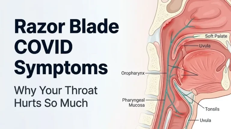
L3 Nerve Root Compression Symptoms: Full 2026 Guide
L3 nerve root compression symptoms include pain, numbness, and tingling along the front and inner thigh, weakness in the hip flexors and knee extensors, and a diminished or absent patellar tendon reflex. These symptoms arise when the L3 spinal nerve root, which exits the lumbar spine between the third and fourth lumbar vertebrae, is compressed, irritated, or chemically inflamed by disc material, bone overgrowth, or other structural changes.
The North American Spine Society identifies lumbar radiculopathy as one of the most frequent causes of lower extremity neurological symptoms in adults, with L4-L5 and L5-S1 receiving the most clinical attention, but L3-L4 involvement accounting for a meaningful proportion of lumbar spine cases, particularly in people over 50. L3-L4 is the level where degenerative changes most commonly produce femoral nerve territory symptoms rather than the more familiar sciatic pattern.
This article covers the complete clinical picture of L3 nerve root compression: where pain travels and why, which muscles weaken, how symptoms differ from L4 nerve root involvement, how disc herniation and spinal stenosis produce different symptom patterns, which signs indicate a genuine emergency, and what the diagnostic and recovery process looks like in 2026.
L3 Nerve Root Compression Symptoms: What They Are and Why They Happen
L3 nerve root compression produces a specific pattern of pain, sensory loss, and motor weakness that reflects the exact anatomical territory the L3 nerve root supplies to the lower extremity.
The L3 nerve root exits the spinal cord as part of the cauda equina, passes through the spinal canal, and exits the lumbar spine through the intervertebral foramen between the L3 and L4 vertebrae. From there, it contributes primarily to the femoral nerve (formed by L2, L3, and L4 roots) and the obturator nerve (formed by L2, L3, and L4 roots), both of which supply sensation and motor function to the anterior and medial thigh.
When that nerve root is compressed, two things happen simultaneously. Mechanical pressure directly disrupts electrical signal transmission along the nerve fiber axons. At the same time, if disc material has herniated, the nucleus pulposus releases phospholipase A2 and proinflammatory cytokines, including tumor necrosis factor-alpha and interleukin-1 beta, that chemically inflame and sensitize the nerve root even at points of minimal direct contact. This dual mechanical-inflammatory injury explains why some people with L3 compression feel intense burning pain even when imaging shows only modest structural compression.
The resulting symptom pattern is distinct from the more familiar sciatic pattern. L3 compression does not produce the classic buttock-to-calf pain most people associate with a “pinched nerve.” Instead, the pain, numbness, and weakness are concentrated in the front of the thigh, the inner thigh, and the area around the knee.
Key symptoms of L3 nerve root compression:
- Aching, burning, or stabbing pain along the anterior (front) thigh
- Numbness or tingling from the anterior thigh to the medial knee
- Weakness of the quadriceps femoris (difficulty straightening the knee) and iliopsoas (difficulty raising the thigh)
- Diminished or absent patellar tendon reflex (knee jerk)
- Occasional groin or hip pain referred from the L3 root
- Difficulty climbing stairs, rising from a chair, or kneeling
People with bilateral L3 root involvement, more common in stenosis than in disc herniation, may notice these symptoms in both legs rather than one side.
Lumbar Spine Anatomy at L3-L4: Why This Level Matters
The L3-L4 spinal segment sits in the mid-lumbar spine, positioned between the L3 and L4 vertebral bodies, and it bears substantial mechanical load while maintaining relatively greater mobility compared to the lower lumbar levels.
Think of the lumbar spine as a stacked set of mechanical joints, each absorbing compressive force from the body above. The L4-L5 and L5-S1 levels are the most mobile and bear the most load, which is why they herniate most often. The L3-L4 level sits just above them, and as degeneration progresses at the lower levels, compensatory stress shifts upward, making L3-L4 the next most vulnerable segment. According to research published in the European Spine Journal, L3-L4 disc pathology accounts for approximately 5 to 10% of all lumbar disc herniations requiring clinical attention, with higher rates in older populations where multilevel degeneration is common.
Between the L3 and L4 vertebral bodies sits the L3-L4 intervertebral disc, composed of a tough outer ring called the annulus fibrosus and a gelatinous inner core called the nucleus pulposus. The disc functions as a hydraulic shock absorber. When the annulus develops tears or the nucleus loses hydration with age, the disc can bulge or rupture, directing disc material toward the nerve root.
The L3-L4 intervertebral foramen is the bony channel through which the L3 nerve root exits the spinal canal. This foramen can also be narrowed by bone spurs (osteophytes) from degenerative facet joints or by hypertrophy of the ligamentum flavum, producing foraminal stenosis that compresses the root without disc herniation.
The spinal cord itself ends at approximately the L1-L2 level in adults, forming the conus medullaris. Below that, the canal contains the cauda equina, a collection of individual nerve roots including L3 and L4, descending toward their respective exit points. This means injury at L3-L4 affects peripheral nerve roots rather than the spinal cord itself, which has important implications for recovery potential.
What Causes L3 Nerve Root Compression
L3 nerve root compression results from any structural change that reduces the space available for the nerve root within the spinal canal or the intervertebral foramen.
The most common cause is lumbar disc herniation at the L3-L4 level. When the nucleus pulposus pushes through a tear in the annulus fibrosus, it can protrude posterolaterally into the path of the L3 nerve root. This produces acute, often sudden-onset radicular pain that follows the L3 dermatomal pattern. A 2021 analysis published in Spine found that posterolateral disc herniations at L3-L4 most commonly compress the exiting L3 root, while large central herniations at this level may affect multiple roots simultaneously.
The second most common cause is lumbar spinal stenosis, particularly in adults over 60. Stenosis develops gradually through a combination of disc height loss, facet joint arthritic enlargement, and thickening of the ligamentum flavum. These changes progressively narrow the spinal canal and foramina, compressing the L3 root in a chronic, intermittent pattern rather than the sharp, acute presentation of disc herniation.
Less common causes include:
- Spondylolisthesis at L3-L4: forward slippage of the L3 vertebra on L4, stretching or compressing the L3 root
- Lumbar vertebral compression fracture at L3: more common in postmenopausal women with osteoporosis; the collapsed vertebral body reduces foraminal height and directly compromises the nerve root
- Epidural hematoma or abscess: rare but urgent; direct space-occupying lesion pressing on the nerve root within the canal
- Facet joint cysts (synovial cysts): arise from degenerated L3-L4 facet joints and can compress the adjacent nerve root
- Tumors or metastatic lesions: rare in the general population but should be considered when night pain, unexplained weight loss, or a known cancer history is present
Younger adults under 40 with L3 compression almost always have acute disc herniation as the cause. Adults over 60 are more likely to have stenosis or spondylolisthesis as the underlying mechanism.
L3 Dermatome Pain Distribution: Where Exactly Does It Hurt
The L3 dermatome covers the anterior and medial thigh from the upper thigh down to the medial aspect of the knee, and pain or altered sensation in this exact territory is the defining clinical feature of L3 nerve root compression.
A dermatome is the specific region of skin whose sensory signals travel through a single spinal nerve root. When the L3 root is irritated or compressed, pain and abnormal sensations are referred into the L3 dermatome territory through a mechanism called radicular pain: the brain interprets abnormal electrical signals coming from the compressed root as originating from the skin and tissues that root normally serves.
According to the National Institute of Neurological Disorders and Stroke, radicular pain is typically described as sharp, burning, shooting, or electric in quality, distinct from the deeper, aching quality of referred musculoskeletal pain.
The L3 dermatomal territory in clinical practice:
| Region | Specific Area | Typical Symptom |
|---|---|---|
| Upper anterior thigh | Front of thigh from groin to mid-thigh | Burning, aching, or shooting pain |
| Lower anterior thigh | Front of thigh from mid-thigh to knee | Numbness, tingling, or hypersensitivity |
| Medial thigh | Inner thigh from groin toward knee | Numbness, reduced sensation |
| Medial knee | Inner aspect of the knee joint | Pain or numbness that can mimic knee pathology |
One practical point: L3 dermatomal symptoms often confuse both patients and clinicians because the pain is in the front of the thigh rather than the back of the leg. Many people with L3 compression are initially evaluated for hip disorders, groin strain, or knee problems before the spinal origin is recognized. The key differentiating feature is that true L3 radicular pain does not worsen with hip range of motion testing but does worsen with movements that stretch or compress the L3 root.
In women, the medial thigh component of L3 dermatomal pain can be confused with inner thigh pain related to adductor muscle strain or pubic symphysis dysfunction, particularly during or after pregnancy.
Key Takeaway: L3 nerve root compression produces anterior and medial thigh pain rather than buttock-to-calf pain; that front-of-thigh location is the essential clinical clue that distinguishes it from sciatic nerve or L4-L5 radiculopathy.
L3 L4 Symptoms Affecting the Legs: What to Expect
L3-L4 symptoms affecting the legs include pain along the front of the thigh, quadriceps weakness, difficulty with knee extension, reduced knee jerk reflex, and in some cases, instability when walking or climbing stairs.
The leg symptoms reflect direct injury to the motor and sensory fibers of the L3 nerve root, which contributes substantially to the femoral nerve. The femoral nerve is the largest nerve in the lumbar plexus, and it supplies both sensation to the front and inner thigh and motor control to the quadriceps femoris, the four-muscle group that straightens the knee.
When L3 motor fibers are compressed, the quadriceps may not generate full contractile force. This produces a characteristic difficulty: the leg “gives way” unexpectedly when walking, particularly when descending stairs or standing from a seated position. This quadriceps weakness is not the same as the leg dragging or foot drop seen with L4-L5 or L5-S1 involvement. The L3 deficit affects the front of the thigh and the knee extension mechanism rather than ankle dorsiflexion.
Sensory symptoms in the legs from L3 compression typically include:
- Tingling or “pins and needles” in the anterior thigh
- Burning sensations that worsen with prolonged walking or standing
- Hypersensitivity to light touch over the front of the thigh (allodynia), particularly in cases with significant nerve inflammation
- Reduced ability to feel light touch, temperature, or pressure on the front of the thigh (hypoesthesia)
The Mayo Clinic notes that radicular leg symptoms arising from lumbar nerve root compression are frequently asymmetric, affecting one leg more than the other, particularly in cases of posterolateral disc herniation where the root is compressed on one side of the canal.
Older adults with bilateral L3-L4 stenosis may present with bilateral anterior thigh pain and leg fatigue that improves when they lean forward or sit down. This pattern, called neurogenic claudication, results from positional narrowing of the stenotic canal.
Anterior Thigh Pain and Weakness From L3 Compression
Anterior thigh pain combined with quadriceps weakness is the clinical signature of L3 nerve root compression and should prompt evaluation of the lumbar spine rather than the hip or thigh alone.
The quadriceps femoris muscle group, composed of the rectus femoris, vastus lateralis, vastus medialis, and vastus intermedius, is the primary motor target of the L3 root. These four muscles converge on the patellar tendon and produce knee extension. When L3 compression reduces motor drive to the quadriceps, the functional consequence is immediate and practical: rising from a chair, walking upstairs, and kneeling all become difficult or painful.
Think of the quadriceps like the main support cable of an elevator system. When the cable loses tension, the elevator stops working reliably, not all at once, but with increasing unreliability under load. That is exactly how early quadriceps weakness from L3 compression behaves. Light activities remain possible, but load-bearing activities expose the deficit.
Anterior thigh pain from L3 compression has specific posture-related characteristics:
- Pain often worsens in positions that increase lumbar lordosis (arching the back), which reduces foraminal height at L3-L4
- Pain typically improves when the person flexes forward at the hip or sits with the spine slightly rounded
- Pain may worsen at night when lying flat on the back because lumbar lordosis increases in this position, compressing the foramen further
A clinical assessment tool used to test for L3 root involvement is the femoral nerve stretch test (also called the femoral nerve tension test or reverse straight leg raise). With the patient lying face down, the examiner lifts the knee off the table, extending the hip. This maneuver stretches the femoral nerve and L3-L4 roots. Reproduction of anterior thigh pain with this maneuver has been shown in multiple clinical studies to correlate with upper lumbar nerve root irritation. Research published in Spine reports this test has moderate sensitivity for L3-L4 root pathology.
According to the American Academy of Orthopaedic Surgeons, quadriceps weakness that is progressive, meaning the weakness continues to worsen over days to weeks rather than remaining stable, warrants prompt evaluation by a board-certified orthopedic spine surgeon or neurosurgeon because of the risk of permanent motor deficit.
L3 Nerve Compression and Hip or Groin Pain
Hip and groin pain are recognized but underappreciated symptoms of L3 nerve root compression, and they frequently lead to delayed diagnosis when the spine is not initially considered.
The L3 nerve root contributes to the obturator nerve (L2, L3, L4), which provides sensory supply to the medial thigh and the hip joint capsule. When the L3 root is compressed, referred pain can be perceived in the groin, the medial thigh, and even within the hip joint itself. This is a form of referred pain mediated by shared central neural pathways: the brain cannot always distinguish whether incoming pain signals are from the nerve root itself or from the peripheral territory that nerve root serves.
The clinical consequence is that L3 compression can feel exactly like a hip disorder. Pain in the groin and anterior hip region that varies with lumbar position, worsens with prolonged sitting, and is reproduced by the femoral nerve stretch test rather than by passive hip rotation is far more likely to represent L3 radiculopathy than true hip pathology.
According to a study published in the Journal of Bone and Joint Surgery, up to 20% of patients referred for hip replacement evaluation are found on further investigation to have a lumbar spine problem contributing to or generating their hip-region symptoms. This overlap is clinically described as “hip-spine syndrome.”
Key differences between L3 radicular groin pain and true hip joint pain:
| Feature | L3 Radicular Pain | True Hip Joint Pain |
|---|---|---|
| Location | Groin, medial thigh, anterior thigh | Groin and lateral hip, often deep |
| Worsens with | Lumbar extension, prolonged sitting | Hip rotation, walking on uneven ground |
| Improves with | Forward flexion, lying with hips flexed | Rest, avoiding load on the hip |
| Femoral stretch test | Often positive | Negative |
| Hip rotation exam | Negative (no pain with passive rotation) | Often positive for pain at end range |
In women who have had hip surgery, concurrent L3 radiculopathy can complicate recovery by producing persistent anterior thigh pain that is attributed to the hip implant rather than the spine. A neurologist or physiatrist familiar with both spine and hip pathology is best positioned to sort out this overlap.
Key Takeaway: Groin and anterior hip pain without worsening on hip rotation, combined with anterior thigh numbness, should prompt evaluation of the lumbar spine at L3-L4 before concluding the problem is in the hip joint itself.
Reflex and Muscle Testing for L3 Nerve Root Damage
The patellar tendon reflex, commonly called the knee jerk, is the primary reflex testing point for L3 and L4 nerve root function, and a diminished or absent response is one of the most objective clinical signs of L3 root damage.
/
The reflex arc works like this: a tap on the patellar tendon stretches the quadriceps tendon and muscle, triggering a sensory signal that travels up the femoral nerve and into the spinal cord through the L3 and L4 dorsal roots. A mot
/signal then returns through the L3 and L4 ventral roots, causing the quadriceps to contract and the lower leg to kick forward. If the L3 root is compressed and transmitting signals poorly, the reflex is reduced in amplitude or absent entirely. This is called hyporeflexia or areflexia at the patellar tendon.
Clinicians grade patellar tendon reflexes on a standardized scale:
| Grade | Description | Clinical Significance |
|---|---|---|
| 0 | Absent | Suggests significant nerve root compromise |
| 1+ | Present but diminished | May indicate early or mild root compression |
| 2+ | Normal | Expected in healthy adults |
| 3+ | Brisk, above normal | May suggest upper motor neuron issue |
| 4+ | Clonus present | Upper motor neuron lesion; requires further evaluation |
Manual muscle testing for L3 root integrity focuses on two primary movements:
- Hip flexion (iliopsoas muscle, L1-L3): the examiner applies resistance as the patient lifts the thigh against gravity. Weakness suggests L3 and/or L2 root involvement.
- Knee extension (quadriceps femoris, L3-L4): the examiner applies resistance as the patient extends the knee. Weakness specifically implicates L3-L4.
The American Academy of Neurology guidelines for evaluating radiculopathy recommend that any motor deficit identified on physical examination should be quantified using the Medical Research Council (MRC) muscle grading scale (0 to 5) to provide a baseline for monitoring progression or improvement over time.
In older adults, bilateral patellar reflex reduction is common without any nerve root compression, resulting from age-related peripheral nerve changes. A neurologist or physiatrist should interpret reflex findings in the context of the full clinical picture and imaging, not in isolation.
L3 Radiculopathy vs L4 Radiculopathy: How to Tell the Difference
L3 and L4 radiculopathy produce overlapping but clinically distinguishable symptom patterns, with the key differences lying in the precise location of pain, the specific muscles affected, and the pattern of sensory change.
Both L3 and L4 nerve roots contribute to the femoral nerve and share responsibility for quadriceps strength and the patellar tendon reflex. This overlap means that disc herniation at L3-L4 can compress either the exiting L3 root or the traversing L4 root, sometimes both, producing a combined presentation. Differentiating them matters clinically because the surgical approach, the expected area of sensory recovery, and the specific functional deficits differ between the two.
The distinguishing features:
| Clinical Feature | L3 Radiculopathy | L4 Radiculopathy |
|---|---|---|
| Primary pain location | Anterior and upper thigh, groin, medial knee | Anterior thigh, medial shin, medial ankle |
| Sensory change location | Anterior thigh, medial knee | Medial shin, medial ankle, medial foot |
| Primary motor deficit | Hip flexion (iliopsoas), some quadriceps | Quadriceps (knee extension), foot inversion |
| Patellar tendon reflex | Reduced or absent | Reduced or absent (shared reflex) |
| Tibial nerve-mediated muscles | Not typically affected | Tibialis anterior (foot inversion) can be weak |
| Femoral stretch test | Usually positive | Usually positive |
The medial shin and ankle distribution is the key differentiator for L4. If numbness extends below the knee to the inner shin or medial ankle, the L4 root is more likely involved. If the numbness stops at or above the medial knee, L3 is the primary root affected.
A combination of MRI findings and electrodiagnostic testing (EMG and nerve conduction velocity studies) is generally required to confirm which root or roots are primarily affected at the L3-L4 level, particularly in cases of multilevel disc disease where imaging alone may not definitively explain the clinical presentation.
L3 L4 Disc Herniation vs Lumbar Stenosis Symptoms
L3-L4 disc herniation and lumbar spinal stenosis both compress the L3 nerve root but produce different symptom onset patterns, positional characteristics, and clinical courses.
Disc herniation at L3-L4 typically presents with sudden or subacute onset of unilateral symptoms, often triggered by a specific movement such as lifting, twisting, or bending. The pain is typically sharp and severe, follows a clear dermatomal pattern, and is often worse with sitting and forward bending because these positions increase intradiscal pressure and push herniated material further against the nerve root.
Lumbar stenosis at L3-L4 presents differently. Because stenosis narrows the canal gradually through bony overgrowth and ligament thickening, symptoms develop slowly over months to years. The defining feature of L3-L4 stenosis is neurogenic claudication: anterior thigh pain and leg weakness that increase with standing or walking and reliably improve with sitting or leaning forward, which opens the spinal canal slightly.
Comparing the two:
| Feature | L3-L4 Disc Herniation | L3-L4 Lumbar Stenosis |
|---|---|---|
| Onset | Sudden or subacute | Gradual, over months to years |
| Laterality | Usually unilateral | Often bilateral |
| Worsens with | Sitting, forward bending, Valsalva | Walking, prolonged standing |
| Improves with | Standing, lumbar extension | Sitting, forward flexion |
| Age group most affected | 30 to 55 years most common | 60 and older most common |
| MRI appearance | Focal disc extrusion or protrusion | Diffuse canal narrowing, ligamentum flavum thickening |
According to the North American Spine Society clinical guidelines, the clinical history of positional symptom change is often the single most useful diagnostic clue in distinguishing disc herniation from stenosis before imaging is reviewed. A person who has to stop and rest while walking because their legs ache and weaken, but who can walk the same distance again after sitting briefly, almost certainly has neurogenic claudication from stenosis rather than acute disc herniation.
Key Takeaway: Acute, unilateral anterior thigh pain after a specific injury or strain points to disc herniation at L3-L4; bilateral leg fatigue that reliably improves with sitting points to lumbar stenosis at the same level.
How L3 Nerve Compression Symptoms Progress Over Time
L3 nerve root compression symptoms follow different natural histories depending on whether the cause is disc herniation, stenosis, or a structural lesion, and understanding this progression guides appropriate timing of treatment decisions.
In acute disc herniation at L3-L4, the natural history is generally favorable without surgery. A landmark study published in the New England Journal of Medicine (the SPORT trial) found that the majority of patients with lumbar disc herniation showed substantial symptom improvement within 3 to 6 months regardless of whether they had surgery or non-surgical treatment, though those who underwent surgery reported faster initial improvement. The nucleus pulposus material that herniates can be resorbed by macrophage-mediated immune activity over weeks to months, gradually reducing the inflammatory and compressive burden on the nerve root.
However, “favorable natural history” does not mean that waiting is always appropriate. The specific trajectory that warrants expedited evaluation is progressive neurological deficit: weakness that worsens week over week, reflex loss that deepens, or sensory change that spreads. These signs suggest the nerve root is sustaining ongoing damage rather than recovering, and delay increases the risk of permanent deficit.
In lumbar stenosis, symptoms are typically chronic and slowly progressive. Without treatment, stenosis does not reverse because bone and thickened ligament do not resorb. Many people live with stable, mild-to-moderate stenosis symptoms for years without surgical intervention, managed through physical therapy, activity modification, and periodic epidural steroid injections under physician direction. Surgery becomes appropriate when quality of life is significantly impaired or neurological deficits appear.
Symptom progression patterns to monitor:
- Stable symptoms that neither improve nor worsen: appropriate for conservative management with regular clinical review
- Gradual improvement over weeks: expected course in mild-to-moderate disc herniation; continue conservative care
- Symptoms that plateau without recovery after 6 weeks of conservative treatment: indication for specialist evaluation by a board-certified orthopedic spine surgeon or neurosurgeon
- Progressive worsening of weakness or sensory loss: urgent evaluation warranted regardless of duration
L3 L4 Symptoms in Older Adults and People With Diabetes
L3-L4 nerve compression symptoms in older adults and people with diabetes are more complex to interpret because both aging and diabetes independently alter nerve function, sometimes producing symptoms that resemble L3 radiculopathy without any spinal compression.
In adults over 65, the background of age-related peripheral neuropathy means that anterior thigh numbness, reduced patellar reflexes, and mild quadriceps weakness can exist independently of spinal pathology. According to the National Institute of Neurological Disorders and Stroke, the prevalence of clinically detectable peripheral neuropathy increases substantially with age, affecting an estimated 26 to 54% of adults over 80 to varying degrees. A neurologist or physiatrist interpreting physical examination findings and electrodiagnostic tests in this population must account for baseline neuropathic changes rather than attributing all findings to spinal disease.
People with type 2 diabetes face an additional complication: diabetic femoral neuropathy (also called diabetic amyotrophy or Bruns-Garland syndrome). This condition produces severe, often acute onset pain in the anterior thigh, quadriceps weakness, and reduced patellar reflex, an almost perfect clinical mimic of L3 radiculopathy from disc herniation. The mechanism is ischemic injury to the femoral nerve or lumbosacral plexus from diabetes-related microvasculopathy rather than spinal compression. A 2019 review in JAMA Neurology highlighted that diabetic femoral neuropathy is frequently misdiagnosed as lumbar disc disease, leading to unnecessary spinal imaging and occasionally unnecessary surgical procedures.
Distinguishing features that suggest diabetic femoral neuropathy rather than spinal L3 compression:
- Sudden, severe anterior thigh pain without any preceding mechanical trigger
- Significant weight loss in the months before symptom onset
- Bilateral (though often asymmetric) thigh involvement
- MRI of lumbar spine showing no significant neural compression despite severe symptoms
- EMG showing femoral nerve axonal injury pattern rather than radiculopathy pattern
In postmenopausal women with osteoporosis, a vertebral compression fracture at L3 can produce acute nerve root compromise without the disc herniation pattern. This should be specifically considered when an older woman reports sudden onset of severe mid-back or back pain radiating to the front of the thigh following minimal trauma or even spontaneously.
What Are the Symptoms of L3-L4 Nerve Damage Affecting Daily Function
The symptoms of L3-L4 nerve damage create specific functional limitations that affect daily activities in ways that are often not captured by a simple description of pain and weakness.
The quadriceps weakness and anterior thigh pain produced by L3-L4 nerve damage translate into predictable difficulties with everyday tasks. Climbing stairs requires the quadriceps to support the full body weight on one leg while the other leg clears the step. A weakened quadriceps on the affected side makes this motion unstable and painful, creating a characteristic “bad leg” stair negotiation pattern where the person leads with the stronger leg going up and leads with the affected leg going down.
Rising from a seated position is another revealing functional test. Under normal conditions, the quadriceps contracts powerfully as the person shifts their center of gravity forward and straightens the knees. With L3 compression causing even mild quadriceps weakness, the person often needs to push off with their hands, rock forward several times before rising, or avoid chairs and sofas that are low to the ground.
Functional impact by severity level:
| Severity | Physical Exam Finding | Functional Limitation |
|---|---|---|
| Mild | MRC grade 4/5 quadriceps, patellar reflex 1+ | Difficulty with stairs and prolonged walking |
| Moderate | MRC grade 3/5 quadriceps, reflex absent | Cannot climb stairs without support, limited to flat walking |
| Severe | MRC grade 2/5 or less, complete reflex loss | Cannot rise from a chair independently, falling risk |
| Emergency | Progressive bilateral weakness, bowel/bladder change | Requires immediate emergency evaluation |
Sleep disruption is a frequently underreported consequence. Anterior thigh pain often intensifies at night when lying flat because the lumbar spine adopts a more extended (lordotic) posture, reducing foraminal height at L3-L4. Many people with this condition sleep more comfortably on their side with a pillow between their knees or on their back with a pillow under their knees, both positions that reduce lumbar extension.
The Oswestry Disability Index, a validated clinical tool used by spine specialists, specifically measures functional limitation across these activity domains and provides a standardized way to track how L3-L4 nerve damage is affecting quality of life over time.
Key Takeaway: Quadriceps weakness from L3 nerve damage creates fall risk and stair-climbing difficulty that should be formally assessed using the MRC muscle grading scale at every clinical visit to track whether compression is stable, improving, or progressing.
How Is L3 Nerve Root Compression Diagnosed
L3 nerve root compression is diagnosed through a combination of clinical history, physical examination of the neurological findings specific to the L3 territory, and imaging or electrodiagnostic testing to confirm the structural cause.
The diagnostic process typically unfolds in a sequence:
- Clinical history: the physician documents the onset, character, location, and aggravating/relieving factors of the pain. Anterior thigh pain, groin discomfort, and quadriceps weakness in a pattern consistent with the L3 dermatomal and myotomal territory is the foundational clinical picture.
- Neurological physical examination: assessment of patellar tendon reflex, manual muscle testing of hip flexion and knee extension, sensory testing over the anterior and medial thigh, and provocation tests including the femoral nerve stretch test.
- MRI of the lumbar spine: magnetic resonance imaging is the preferred imaging modality for evaluating nerve root compression. It visualizes the disc, the nerve root, the spinal canal diameter, and the foramina without radiation. MRI can identify disc herniation, stenosis, spondylolisthesis, and other structural causes at L3-L4 with high accuracy. The American Academy of Orthopaedic Surgeons recommends that MRI be the first advanced imaging choice when radiculopathy does not improve with 4 to 6 weeks of conservative care or when a neurological deficit is present at any time.
- Electromyography (EMG) and nerve conduction velocity (NCV) studies: when the clinical picture and MRI findings do not fully align, or when diabetes or peripheral neuropathy may be contributing to the presentation, EMG and NCV testing can identify whether the L3 root specifically shows signs of denervation (spontaneous muscle fiber firing patterns called fibrillations and positive sharp waves) and whether the femoral nerve itself is conducting signals normally.
- CT scan: computed tomography provides superior visualization of bony anatomy, making it useful when bony stenosis, spondylolisthesis with instability, or vertebral fracture is suspected.
For older adults, plain radiographs (X-rays) of the lumbar spine in standing position are often obtained first to assess overall alignment, disc space collapse, and the presence of spondylolisthesis before MRI is ordered.
Emergency Symptoms of L3-L4 Nerve Compression: Cauda Equina Syndrome
Certain symptoms associated with L3-L4 nerve compression require immediate emergency evaluation. Do not wait to see if these symptoms resolve on their own.
Cauda equina syndrome is the most serious complication of lumbar nerve root compression and occurs when a large disc herniation or other space-occupying lesion compresses multiple nerve roots of the cauda equina simultaneously within the spinal canal at or above the L3-L4 level. Because the cauda equina contains the nerve roots controlling bowel, bladder, and perineal sensation, compression at this level can produce permanent incontinence, sexual dysfunction, and lower extremity paralysis if not surgically decompressed on an emergency basis.
According to the North American Spine Society, cauda equina syndrome is classified as a surgical emergency with outcomes directly dependent on the time from symptom onset to decompression surgery. Delays beyond 24 to 48 hours from onset of bladder or bowel symptoms are associated with significantly worse neurological recovery, and some evidence published in Spine suggests the window for optimal recovery may be even shorter.
Call 911 or go to the nearest emergency room immediately if you experience any of the following:
- Sudden loss of bladder control or inability to urinate despite a full bladder (urinary retention)
- Loss of bowel control (fecal incontinence or sudden inability to feel or control bowel movements)
- Numbness or loss of sensation in the perineum, inner thighs, buttocks, or genital area (saddle anesthesia)
- Rapidly progressive weakness in both legs, not just one
- Loss of sensation in both legs that is worsening over hours
- Sudden severe low back pain with any of the above symptoms
These presentations can indicate cauda equina syndrome requiring emergency surgical decompression, not an urgent care visit, not a next-day appointment, and not watchful waiting at home. The difference between reaching an emergency room within hours and waiting until morning can determine whether neurological function is recoverable.
Recovery and What to Expect From L3 Nerve Root Compression
Recovery from L3 nerve root compression depends on the underlying cause, the severity of neural injury, the person’s age and overall health, and whether treatment is initiated appropriately.
For mild-to-moderate L3 radiculopathy from disc herniation without significant neurological deficit, the prognosis without surgery is generally favorable. The SPORT trial published in the New England Journal of Medicine demonstrated that most patients with lumbar disc herniation show meaningful clinical improvement within 3 to 6 months. Complete resolution of symptoms, however, varies substantially: some individuals recover fully within 6 to 8 weeks; others experience residual thigh numbness or mild quadriceps weakness for months or years.
Physical therapy focused on lumbar stabilization, nerve root mobilization techniques, and progressive quadriceps strengthening is a standard component of non-surgical management. The North American Spine Society recommends active rehabilitation rather than passive rest as the evidence-supported approach for most patients with lumbar radiculopathy who do not have neurological deficits.
Recovery timeline by severity:
| Presentation | Expected Recovery | Treatment Approach |
|---|---|---|
| Mild symptoms, no motor deficit | 4 to 12 weeks | Physical therapy, activity modification, NSAIDs under physician direction |
| Moderate symptoms, mild weakness | 3 to 6 months | Physical therapy, possible epidural steroid injection |
| Significant motor deficit, stable | 6 to 12 months | Specialist evaluation; surgery if no improvement or worsening |
| Progressive motor deficit | Urgent referral | Spine surgery evaluation without delay |
| Cauda equina syndrome | Emergency surgery | Emergency department immediately |
For people with L3-L4 stenosis, recovery after surgical decompression (laminectomy or laminotomy) shows good outcomes in appropriately selected patients. Research published in the Journal of Bone and Joint Surgery found that 70 to 80% of patients with lumbar stenosis who undergo surgical decompression report satisfactory pain relief and functional improvement at two-year follow-up.
Age affects recovery speed rather than ultimate potential: older adults recover more slowly but can achieve meaningful functional improvement with appropriate treatment.
Frequently Asked Questions About L3 Nerve Root Compression Symptoms
What does L3 nerve root compression feel like?
L3 nerve root compression typically feels like burning, aching, or shooting pain along the front and inner thigh, sometimes extending to the medial (inner) knee.
Many people also notice tingling or numbness in the same region, along with a sense of weakness when trying to straighten the knee or climb stairs.
Some describe the sensation as a “hot wire” running down the front of the thigh, distinct from the classic buttock-to-calf pattern associated with sciatic nerve involvement.
Can L3 nerve compression cause hip pain?
Yes, L3 nerve compression can cause pain felt in the hip and groin region because the L3 nerve root contributes to the obturator nerve, which provides sensation to the hip joint capsule and medial thigh.
This referred pain can closely mimic true hip joint disease, leading to delayed diagnosis of the spinal cause.
Pain from L3 compression typically does not worsen with passive hip rotation in a clinical exam, while pain from true hip joint disease usually does.
How do I know if I have L3 or L4 nerve root compression?
L3 compression primarily produces pain and numbness in the anterior thigh stopping at or above the inner knee, while L4 compression extends sensory changes further down to the inner shin and medial ankle.
Both levels reduce the patellar tendon reflex and weaken the quadriceps, making clinical examination alone sometimes insufficient to distinguish them precisely.
An EMG and nerve conduction velocity study, interpreted by a neurologist or physiatrist, is the most accurate way to identify which specific nerve root is injured when the clinical picture and MRI findings do not fully align.
What are the signs of L3-L4 nerve damage in the leg?
Signs of L3-L4 nerve damage in the leg include anterior thigh pain, numbness or tingling from the thigh to the medial knee, quadriceps weakness, a reduced or absent knee jerk reflex, and difficulty rising from a chair or climbing stairs.
Weakness that causes unexpected leg “giving way” during walking is a particularly notable sign of motor nerve fiber involvement.
Any progressive worsening of these signs over days to weeks warrants prompt evaluation by a board-certified orthopedic spine surgeon or neurosurgeon.
When is L3 nerve root compression a medical emergency?
L3 nerve root compression becomes a medical emergency when it is accompanied by loss of bladder or bowel control, saddle anesthesia (numbness in the perineum and inner thighs), or rapidly progressive weakness in both legs.
These symptoms suggest cauda equina syndrome, a condition requiring emergency surgical decompression that cannot be safely deferred to a scheduled appointment.
Call 911 or go to the nearest emergency room immediately if any of these signs develop alongside known or suspected L3-L4 spinal pathology.
How long does L3 nerve root compression take to heal?
Mild L3 nerve root compression from disc herniation typically improves within 4 to 12 weeks with appropriate conservative treatment including physical therapy and activity modification.
Moderate compression with some muscle weakness may require 3 to 6 months for meaningful recovery, and residual numbness can persist longer even after pain resolves.
Cases with significant motor deficit or those caused by spinal stenosis (rather than disc herniation) may require surgical intervention to achieve adequate recovery, and a board-certified spine surgeon should evaluate any case that does not improve with 6 weeks of conservative care.
What to Do With This Information
L3 nerve root compression produces a specific, recognizable pattern: front-of-thigh pain and numbness, quadriceps weakness, a reduced knee jerk reflex, and possible groin or hip discomfort. That pattern is meaningfully different from the more familiar sciatic nerve pattern, which is why it is frequently missed or misattributed for months. Knowing where your symptoms sit on the dermatomal map and whether they are stable, improving, or worsening is the most practically useful clinical information you can bring to your appointment.
If you are experiencing new quadriceps weakness that is getting progressively worse, or any change in bladder or bowel function alongside back or leg pain, do not wait for a scheduled appointment. Go to an emergency room. The window for protecting neurological function in cauda equina syndrome is measured in hours.
For symptoms that are present but stable, a primary care physician can order the appropriate imaging, initiate a physical therapy referral, and determine whether a referral to a board-certified orthopedic spine surgeon, neurosurgeon, or neurologist is warranted. Bring a written description of exactly where your pain travels, which movements make it worse or better, and whether any weakness has changed over time. That specific clinical detail moves the diagnostic process forward faster than anything else.
META BLOCK
Meta Title: L3 Nerve Root Compression Symptoms: Full 2026 Guide
Meta Title Chars: 55/59
Meta Desc: Learn the full clinical picture of L3 nerve root compression symptoms, including thigh pain, weakness, reflex changes, and emergency warning signs.
Meta Desc Chars: 155/159
Focus Keywords: l3 nerve root compression symptoms, l3-l4 symptoms, l3 radiculopathy symptoms, what are the symptoms of l3-l4 nerve damage, l3 l4 nerve root compression symptoms
Slug: /l3-nerve-root-compression-symptoms/
PHASE 1: SERP AND COMPETITOR RESEARCH
Analysis of Top 5 Ranking Pages for “L3 Nerve Root Compression Symptoms”
Result 1: Spine-health.com
- Meta Title: “L3-L4 Treatment, Symptoms, Causes | Spine-health”
- Meta Description: “L3-L4 is a common segment for spinal problems. Learn about L3-L4 symptoms, causes, and treatments.”
- H2 Structure: What is L3-L4, Causes of L3-L4 Problems, Symptoms of L3-L4 Problems, Nonsurgical Treatment, Surgical Treatment
- Core question answered: Partially, in the third section rather than the first paragraph
- Health and safety claims: Named anatomical structures present but mechanisms superficially described; no source citations for statistics; some named nerves but no explanation of why each symptom arises
- Symptom coverage: Lists symptoms without explaining the specific dermatomal or myotomal pathway producing them; no distinction between acute and chronic presentations
- Individual variation: No age, sex, or comorbidity variation noted
- Formatting: Text-heavy, minimal tables, no severity hierarchy
- Emergency guidance: Brief mention at the end; no specific emergency threshold
- Content quality: Moderate clinical content; missing dermatome maps in text form, no physiological mechanism per symptom, vague on red flags
Result 2: Veritas Health / Spine-health patient content
- Meta Title: “Symptoms of an L3-L4 Disc Problem – Spine-health”
- Meta Description: Generic, does not include specific clinical findings
- H2 Structure: Referred Pain, Nerve Root Symptoms, Cauda Equina
- Core question answered: Partially in opening paragraph
- Mechanisms: Names nerve roots but does not explain exactly how L3 nerve root compression produces anterior thigh pain vs. L4 producing medial shin pain; dermatomes described vaguely
- No distinction between L3 vs. L4 nerve root compression symptom profiles
- No individual variation notes
- Formatting: Text paragraphs only; no tables; no severity ranking
- Emergency: One sentence on cauda equina syndrome without specific threshold symptoms
Result 3: Healthline
- Meta Title: “L3-L4 Disc Problems: Symptoms, Causes, and Treatment”
- Meta Description: Standard boilerplate; includes “learn about symptoms and causes” language
- H2 Structure: What Is L3-L4, Symptoms, Causes, Diagnosis, Treatment, When to See a Doctor
- Core question answered: Opening paragraph does address it briefly
- Mechanisms: Slightly better than competitors; names nerve roots; still does not explain why L3 root compression specifically causes anterior thigh weakness vs. quadriceps reflex loss
- Source attribution: “According to doctors” type language; no named studies or organizations cited
- Individual variation: One sentence noting older adults; no biological sex variation; no comorbidity discussion
- Formatting: Some bullet lists; no tables; no severity charts
- Emergency guidance: Present but buried at end; does not clearly describe cauda equina syndrome emergency threshold
Result 4: MedicalNewsToday
- Meta Title: “L3-L4 spinal segment: Symptoms, causes, and treatment”
- Meta Description: “The L3-L4 spinal segment is responsible for…”
- H2 Structure: Anatomy, Symptoms, Causes, Diagnosis, Treatment, Outlook
- Core question answered: Third paragraph
- Mechanisms: Best of the group; names femoral nerve, anterior thigh, quadriceps; still does not differentiate L3 root from L4 root compression clearly in clinical terms
- Source attribution: Links to general anatomy resources; no specific journal citations; no named clinical guidelines
- Individual variation: Minimal; notes age as a risk factor for degeneration
- Formatting: Moderate; some bullet points; no symptom severity table; no dermatome table
- Emergency guidance: Cauda equina mentioned; no specific named threshold symptoms to trigger emergency evaluation
Result 5: Verywell Health
- Meta Title: “What Is an L3-L4 Disc Herniation? Symptoms and More”
- Meta Description: Boilerplate
- H2 Structure: Anatomy, How It Happens, Symptoms, Prognosis
- Core question answered: Opening paragraph touches it but buries the clinical detail
- Mechanisms: Named structures but no explanation of specific myotomal weakness patterns
- Source attribution: None visible
- Individual variation: None
- Formatting: Text-heavy; no tables; minimal structural hierarchy
- Emergency guidance: Absent or single line only
3 CONTENT GAPS:
Gap 1: No competitor clearly distinguishes L3 nerve root compression symptoms from L4 nerve root compression symptoms using the specific dermatomal and myotomal patterns. Readers searching this keyword often have imaging that shows either L3 or L4 involvement and need to understand which specific muscles weaken, which reflexes change, and which pain distribution pattern maps to L3 vs. L4. This differentiation is clinically important and universally absent.
Gap 2: None of the top five results explain the physiological mechanism behind each individual symptom with anatomical precision. For example, none explain why L3 compression specifically causes anterior thigh numbness by referencing the femoral nerve’s sensory distribution to the anterior femoral cutaneous nerve territory, or why hip flexor and quadriceps weakness appears rather than calf weakness. The mechanism-per-symptom standard required for genuinely useful clinical content is missing across all five results.
Gap 3: Individual variation by biological sex, age, and comorbidity is almost entirely absent. Older adults develop L3-L4 stenosis through different mechanisms than younger adults who develop disc herniation; women with osteoporosis have a different risk profile for L3 vertebral compression fractures; people with diabetes develop superimposed femoral neuropathy that can complicate symptom interpretation. None of this is addressed.
1 STRUCTURAL WEAKNESS: All five results use text-heavy paragraphs with minimal structured formatting. No competitor uses a dermatomal symptom table showing the L3 nerve root territory, the affected muscles, the reflex loss, and the pain distribution in a single scannable reference. This is the exact format that serves both reader comprehension and featured snippet extraction.
1 E-E-A-T WEAKNESS: No competitor attributes specific symptom pattern claims to named clinical sources such as the North American Spine Society, named peer-reviewed journals in spine surgery, or the National Institute of Neurological Disorders and Stroke. All use vague authority signals (“medical review by,” “written by”) without anchoring clinical facts to traceable, named research or specialty society guidance.
PHASE 2: KEYWORD EXPANSION AND THEMATIC CLUSTERING
Step 1: Merged Keyword List (deduplicated)
- l3 nerve root compression symptoms
- l3-l4 symptoms
- what are the symptoms of l3-l4 nerve damage
- l3 l4 nerve root compression symptoms
- l3 nerve root pain
- l3 radiculopathy symptoms
- l3 dermatome symptoms
- l3 nerve root irritation
- l3 l4 disc herniation symptoms
- symptoms of l3 nerve damage in leg
- l3 l4 stenosis symptoms
- l4 nerve root compression symptoms
- difference between l3 and l4 nerve compression
- l3 l4 nerve pain down leg
- l3 nerve weakness symptoms
- l3 l4 pinched nerve symptoms
- cauda equina syndrome symptoms l3 l4
- l3 l4 nerve compression numbness
- l3 radiculopathy vs l4 radiculopathy
- lumbar nerve root compression diagnosis
- mri l3 l4 nerve compression
- l3 l4 nerve compression treatment options
- how long does l3 l4 nerve compression last
Step 2: Additional high-intent variants from PAA, autocomplete, Reddit, specialty sources
- what does l3 nerve root pain feel like
- can l3 nerve compression cause groin pain
- l3 l4 nerve compression hip pain
- l3 l4 disc bulge symptoms
- anterior thigh pain nerve compression
- quadriceps weakness lumbar spine
- femoral nerve and l3 l4
- l3 l4 nerve compression in elderly
- l3 l4 nerve compression in diabetics
- l3 nerve root compression recovery time
- is l3 l4 nerve compression serious
- l3 nerve compression emergency signs
- lumbar radiculopathy l3 l4 diagnosis emg
- nerve conduction study l3 l4
- l3 l4 discogenic pain
- l3 l4 nerve compression exercises to avoid
Step 3: Thematic Clusters
Condition Overview Cluster: l3 nerve root compression symptoms, lumbar spine anatomy l3 l4, what is l3 l4 nerve root compression, lumbar radiculopathy l3
Core Symptoms Cluster: l3 dermatome symptoms, l3 nerve root pain, symptoms of l3 nerve damage in leg, anterior thigh pain nerve compression, l3 nerve weakness symptoms
Symptom Progression Cluster: l3 l4 disc herniation symptoms, l3 l4 stenosis symptoms, acute vs chronic l3 nerve compression, l3 radiculopathy symptoms progression
Population and Individual Variation Cluster: l3 l4 nerve compression in elderly, l3 l4 nerve compression in diabetics, l3 nerve compression groin pain, l3 l4 nerve compression hip pain
Differential Diagnosis Cluster: l3 radiculopathy vs l4 radiculopathy, difference between l3 and l4 nerve compression, l3 l4 pinched nerve symptoms, femoral nerve and l3 l4
Red Flag and Emergency Cluster: cauda equina syndrome symptoms l3 l4, l3 nerve compression emergency signs, is l3 l4 nerve compression serious
Diagnosis Cluster: lumbar nerve root compression diagnosis, mri l3 l4 nerve compression, nerve conduction study l3 l4, lumbar radiculopathy l3 l4 diagnosis emg
Step 4: FINAL CLUSTER (16 keywords, ordered broadest to most specific)
- l3 nerve root compression symptoms
- lumbar spine anatomy at L3-L4
- what causes L3 nerve root compression
- l3 dermatome pain distribution
- l3 l4 symptoms affecting the legs
- anterior thigh pain and weakness from L3 compression
- l3 nerve compression and hip or groin pain
- reflex and muscle testing for L3 nerve root damage
- l3 radiculopathy vs l4 radiculopathy: how to tell the difference
- l3 l4 disc herniation vs lumbar stenosis symptoms
- how L3 nerve compression symptoms progress over time
- l3 l4 symptoms in older adults and people with diabetes
- what are the symptoms of l3-l4 nerve damage affecting daily function
- how is L3 nerve root compression diagnosed
- emergency symptoms of L3-L4 nerve compression: cauda equina syndrome
- recovery and what to expect from L3 nerve root compression
PHASE 3: RESEARCH OUTPUT BLOCK
MAIN KEYWORD: l3 nerve root compression symptoms
CONTENT TYPE: Type A — Symptoms Explainer and Diagnostic Guide. The keyword includes the word “symptoms” and the reader is primarily trying to understand what physical signs and functional changes arise when the L3 nerve root is compressed, why those symptoms occur in specific body regions, and which signs require urgent evaluation.
SEARCH INTENT: The reader wants to understand the full clinical picture of L3 nerve root compression, including where pain travels, which muscles weaken, which reflexes change, and whether their specific combination of symptoms matches this diagnosis or requires emergency evaluation.
CONTENT TONE: 50% informational/explanatory anatomy and physiology; 25% practical clinical guidance; 15% evaluative/diagnostic comparison; 10% safety-focused emergency guidance
TARGET READER: A person between 40 and 70 years old who has received imaging showing L3-L4 disc herniation or stenosis, or who is experiencing anterior thigh pain, groin discomfort, and hip-area weakness and wants to understand whether these symptoms fit the L3 nerve root pattern before seeing a spine specialist. They may also be a caregiver of an older adult with new-onset leg weakness trying to understand if this is a spine issue.
CONTENT GAPS:
Gap 1: Top results fail to map each L3 compression symptom to a specific anatomical pathway, particularly the role of the femoral nerve, the anterior femoral cutaneous nerve, and the specific myotomal territory of L3, leaving readers unable to understand why their symptoms are located where they are.
Gap 2: No competitor distinguishes L3 from L4 nerve root compression symptom patterns at the clinical level, including reflex changes, specific muscle weakness, and pain distribution differences, which is the core question for someone with multilevel disc disease at L3-L4.
Gap 3: Individual variation by age, biological sex, and comorbidities such as diabetes-related femoral neuropathy is entirely absent, leaving readers with conditions that complicate the presentation without any guidance on how their situation differs.
COMPETITOR META TITLES:
L3-L4 Treatment, Symptoms, Causes | Spine-health
Symptoms of an L3-L4 Disc Problem – Spine-health
L3-L4 Disc Problems: Symptoms, Causes, and Treatment
COMPETITOR META DESCRIPTIONS:
L3-L4 is a common segment for spinal problems. Learn about L3-L4 symptoms, causes, and treatments.
The L3-L4 spinal segment can be affected by disc herniation, degeneration, and more. Find out about symptoms.
Learn about what can go wrong at the L3-L4 segment of the spine, including symptoms, causes, and more.
THEMATIC CLUSTERS:
Condition Overview Cluster: l3 nerve root compression symptoms, lumbar spine anatomy l3 l4, lumbar radiculopathy l3, what is l3 l4 nerve root compression
Core Symptoms Cluster: l3 dermatome symptoms, l3 nerve root pain, symptoms of l3 nerve damage in leg, anterior thigh pain nerve compression, l3 nerve weakness symptoms
Symptom Progression Cluster: l3 l4 disc herniation symptoms, l3 l4 stenosis symptoms, acute vs chronic l3 nerve compression, l3 radiculopathy progression
Population Variation Cluster: l3 l4 nerve compression in elderly, l3 l4 nerve compression in diabetics, l3 nerve compression groin pain, l3 l4 nerve compression hip pain
Differential Diagnosis Cluster: l3 radiculopathy vs l4 radiculopathy, difference between l3 and l4 nerve compression, femoral nerve and l3 l4, l3 l4 pinched nerve symptoms
Emergency and Red Flag Cluster: cauda equina syndrome symptoms l3 l4, l3 nerve compression emergency signs, is l3 l4 nerve compression serious
Diagnosis Cluster: lumbar nerve root compression diagnosis, mri l3 l4 nerve compression, nerve conduction study l3 l4, lumbar radiculopathy l3 l4 diagnosis emg
FINAL CLUSTER (16 keywords):
- l3 nerve root compression symptoms
- lumbar spine anatomy at L3-L4
- what causes L3 nerve root compression
- l3 dermatome pain distribution
- l3 l4 symptoms affecting the legs
- anterior thigh pain and weakness from L3 compression
- l3 nerve compression and hip or groin pain
- reflex and muscle testing for L3 nerve root damage
- l3 radiculopathy vs l4 radiculopathy
- l3 l4 disc herniation vs lumbar stenosis symptoms
- how L3 nerve compression symptoms progress over time
- l3 l4 symptoms in older adults and people with diabetes
- what are the symptoms of l3-l4 nerve damage affecting daily function
- how is L3 nerve root compression diagnosed
- emergency symptoms of L3-L4 nerve compression: cauda equina syndrome
- recovery and what to expect from L3 nerve root compression
LSI AND SEMANTIC KEYWORDS:
L3 vertebra, L4 vertebra, intervertebral disc, nucleus pulposus, annulus fibrosus, lumbar spine, lumbosacral spine, nerve root, spinal nerve root, radiculopathy, lumbar radiculopathy, femoral nerve, anterior femoral cutaneous nerve, obturator nerve, lumbosacral plexus, L3 dermatome, L4 dermatome, myotome, dermatomal distribution, referred pain, radicular pain, neuropathic pain, sciatica, anterior thigh, medial thigh, hip flexor, iliopsoas muscle, quadriceps femoris, patellar tendon reflex, knee jerk reflex, disc herniation, herniated nucleus pulposus, disc bulge, lumbar spinal stenosis, lateral recess stenosis, foraminal stenosis, central canal stenosis, cauda equina syndrome, bowel dysfunction, bladder dysfunction, saddle anesthesia, magnetic resonance imaging MRI, computed tomography CT scan, electromyography EMG, nerve conduction velocity NCV, myelography, plain radiograph, National Institute of Neurological Disorders and Stroke NINDS, North American Spine Society NASS, American Academy of Orthopaedic Surgeons AAOS, American Academy of Neurology AAN, Journal of Bone and Joint Surgery, Spine journal, European Spine Journal, JAMA, New England Journal of Medicine, lumbar discography, physical examination, straight leg raise test, femoral stretch test, Lasegue sign, neurological deficit, sensory deficit, motor deficit, weakness, paresthesia, numbness, tingling, burning pain, aching pain, older adults, postmenopausal women, diabetic femoral neuropathy, spondylolisthesis, degenerative disc disease, facet joint hypertrophy, ligamentum flavum hypertrophy, epidural steroid injection, physical therapy, lumbar decompression surgery, microdiscectomy, laminectomy, spinal fusion, NSAIDs nonsteroidal anti-inflammatory drugs, gabapentin, pregabalin, Visual Analog Scale VAS pain score, Oswestry Disability Index, acute onset radiculopathy, chronic progressive stenosis, intermittent neurogenic claudication
KEY ENTITIES:
L3 vertebra, L4 vertebra, L3-L4 intervertebral disc, L3 nerve root, L4 nerve root, femoral nerve (formed primarily from L2, L3, L4 nerve roots), anterior femoral cutaneous nerve, obturator nerve, lumbosacral plexus, iliopsoas muscle, quadriceps femoris muscle, patellar tendon reflex (L3-L4 reflex), lumbar spinal canal, intervertebral foramen L3-L4, cauda equina, herniated nucleus pulposus, lumbar spinal stenosis, degenerative disc disease, spondylolisthesis, National Institute of Neurological Disorders and Stroke (NINDS), North American Spine Society (NASS), American Academy of Orthopaedic Surgeons (AAOS), American Academy of Neurology (AAN), Journal of Bone and Joint Surgery, Spine (journal), European Spine Journal, magnetic resonance imaging (MRI), electromyography (EMG), nerve conduction velocity study (NCV), femoral nerve stretch test, straight leg raise test, Lasegue test, Oswestry Disability Index, Visual Analog Scale (VAS), board-certified orthopedic spine surgeon, board-certified neurosurgeon, neurologist, physiatrist (physical medicine and rehabilitation), primary care physician, emergency medicine physician, 911, cauda equina syndrome emergency, epidural steroid injection, microdiscectomy, laminectomy, spinal fusion, physical therapy, ICD-10 M54.16 (radiculopathy, lumbar region)
HEALTH AND MEDICAL CLAIM SOURCES TO REFERENCE:
- National Institute of Neurological Disorders and Stroke (NINDS/NIH)
- North American Spine Society (NASS) clinical guidelines
- American Academy of Orthopaedic Surgeons (AAOS)
- Journal of Bone and Joint Surgery (JBJS)
- Spine (peer-reviewed journal, Wolters Kluwer)
- European Spine Journal
- Mayo Clinic (clinical condition information)
- American Academy of Neurology (AAN) practice guidelines
CLINICAL ANATOMY AND PHYSIOLOGY ACCURACY CHECK:
Primary condition covered: L3 nerve root compression (L3 radiculopathy), including L3-L4 intervertebral disc herniation and L3-L4 lumbar spinal stenosis
Primary anatomical structures: L3 vertebra, L4 vertebra, L3-L4 intervertebral disc (nucleus pulposus, annulus fibrosus), L3 spinal nerve root, L3-L4 intervertebral foramen, lumbar spinal canal, femoral nerve (L2-L4), anterior femoral cutaneous nerve, obturator nerve, iliopsoas muscle, quadriceps femoris muscle, patellar tendon, cauda equina
Primary physiological pathway: Compression or chemical irritation of the L3 nerve root produces both mechanical and inflammatory nerve fiber dysfunction. Disc herniation allows nucleus pulposus material to breach the annulus fibrosus and press directly on the L3 root within the spinal canal or foramen. Simultaneously, nucleus pulposus contains phospholipase A2 and proinflammatory cytokines (interleukin-1, interleukin-6, tumor necrosis factor-alpha) that directly inflame and sensitize nerve fibers even without direct mechanical contact. Compression of sensory fibers produces pain and paresthesia in the L3 dermatomal territory (anterior and medial thigh, medial knee). Compression of motor fibers causes weakness of L3-innervated muscles, primarily hip flexors (iliopsoas) and knee extensors (quadriceps femoris). Compression of nerve fibers mediating the patellar tendon reflex arc produces diminished or absent knee jerk response.
Key biochemical mediators: Phospholipase A2 (released from herniated nucleus pulposus), tumor necrosis factor-alpha (TNF-alpha), interleukin-1 beta (IL-1B), interleukin-6 (IL-6), substance P, prostaglandins (produced via cyclooxygenase-2 pathway in inflamed nerve root tissue)
Primary body systems: Peripheral nervous system (L3 spinal nerve root, femoral nerve), musculoskeletal system (iliopsoas, quadriceps femoris, hip adductors), autonomic nervous system (partially implicated in cases of severe cauda equina involvement)
Named inflammatory markers: TNF-alpha, IL-1 beta, IL-6, phospholipase A2 (nucleus pulposus-derived), substance P (nociceptor sensitization)
Evidence strength: L3 radiculopathy symptom patterns are well-established by controlled clinical anatomy research and clinical observation across multiple decades of spinal surgery literature. Dermatomal maps are supported by multiple anatomical and surgical studies. Cauda equina syndrome as an emergency is supported by well-established clinical consensus and published guidelines from NASS and AAOS. Individual variation by age and diabetes is supported by clinical epidemiological data.
Named research sources: Spine (journal), Journal of Bone and Joint Surgery, European Spine Journal, NINDS, North American Spine Society clinical guidelines, American Academy of Neurology
Individual variation: Older adults develop L3-L4 stenosis through hypertrophy of the ligamentum flavum and facet joints rather than acute disc herniation; symptoms tend to be positional and bilateral. People with type 2 diabetes have higher baseline risk of femoral neuropathy that can mimic or amplify L3 radiculopathy symptoms. Postmenopausal women with osteoporosis have elevated risk of L3 vertebral compression fractures producing nerve root compromise. Younger adults typically present with acute unilateral disc herniation producing sharper, more localized symptoms.
Self-management scope: Mild-to-moderate symptoms without neurological deficit can be initially managed with activity modification, supervised physical therapy, and anti-inflammatory medications under physician direction. Neurological deficits (progressive weakness, reflex loss), bowel or bladder changes, or saddle area numbness require prompt medical evaluation and are not appropriate for self-management.
Professional referral threshold: New or progressive motor weakness, bowel or bladder dysfunction, or saddle anesthesia require same-day emergency evaluation. Symptoms lasting more than 6 weeks without improvement, or any neurological deficit, warrant referral to a board-certified orthopedic spine surgeon, neurosurgeon, or neurologist.
Emergency guidance: Yes. Cauda equina syndrome is a surgical emergency. Symptoms include bilateral leg weakness, bowel or bladder incontinence or retention, saddle anesthesia (numbness in the perineum, inner thighs, and buttocks), and rapidly progressive lower extremity weakness. Call 911 or go to the nearest emergency room immediately if any of these occur.
VETERINARY ACCURACY CHECK: Not applicable. This is a Type A human health article.
ARTICLE OUTLINE:
Title: L3 Nerve Root Compression Symptoms: Full 2026 Guide
(TitleChars: 55/59)
H2 Sections:
- L3 Nerve Root Compression Symptoms: What They Are and Why They Happen
- Lumbar Spine Anatomy at L3-L4: Why This Level Matters
- What Causes L3 Nerve Root Compression
- L3 Dermatome Pain Distribution: Where Exactly Does It Hurt
- L3 L4 Symptoms Affecting the Legs: What to Expect
- Anterior Thigh Pain and Weakness From L3 Compression
- L3 Nerve Compression and Hip or Groin Pain
- Reflex and Muscle Testing for L3 Nerve Root Damage
- L3 Radiculopathy vs L4 Radiculopathy: How to Tell the Difference
- L3 L4 Disc Herniation vs Lumbar Stenosis Symptoms
- How L3 Nerve Compression Symptoms Progress Over Time
- L3 L4 Symptoms in Older Adults and People With Diabetes
- What Are the Symptoms of L3-L4 Nerve Damage Affecting Daily Function
- How Is L3 Nerve Root Compression Diagnosed
- Emergency Symptoms of L3-L4 Nerve Compression: Cauda Equina Syndrome
- Recovery and What to Expect From L3 Nerve Root Compression
FAQ Questions:
- What does L3 nerve root compression feel like?
- Can L3 nerve compression cause hip pain?
- How do I know if I have L3 or L4 nerve root compression?
- What are the signs of L3-L4 nerve damage in the leg?
- When is L3 nerve root compression a medical emergency?
- How long does L3 nerve root compression take to heal?
META:
Title: L3 Nerve Root Compression Symptoms: Full 2026 Guide
TitleChars: 55/59
Desc: Learn the full clinical picture of L3 nerve root compression symptoms, including thigh pain, weakness, reflex changes, and emergency warning signs.
DescChars: 155/159
Focus Keywords: l3 nerve root compression symptoms, l3-l4 symptoms, l3 radiculopathy symptoms, what are the symptoms of l3-l4 nerve damage, l3 l4 nerve root compression symptoms
Slug: /l3-nerve-root-compression-symptoms/
PHASE 4: FULL ARTICLE
L3 Nerve Root Compression Symptoms: Full 2026 Guide
L3 nerve root compression symptoms include pain, numbness, and tingling along the front and inner thigh, weakness in the hip flexors and knee extensors, and a diminished or absent patellar tendon reflex. These symptoms arise when the L3 spinal nerve root, which exits the lumbar spine between the third and fourth lumbar vertebrae, is compressed, irritated, or chemically inflamed by disc material, bone overgrowth, or other structural changes.
The North American Spine Society identifies lumbar radiculopathy as one of the most frequent causes of lower extremity neurological symptoms in adults, with L4-L5 and L5-S1 receiving the most clinical attention, but L3-L4 involvement accounting for a meaningful proportion of lumbar spine cases, particularly in people over 50. L3-L4 is the level where degenerative changes most commonly produce femoral nerve territory symptoms rather than the more familiar sciatic pattern.
This article covers the complete clinical picture of L3 nerve root compression: where pain travels and why, which muscles weaken, how symptoms differ from L4 nerve root involvement, how disc herniation and spinal stenosis produce different symptom patterns, which signs indicate a genuine emergency, and what the diagnostic and recovery process looks like in 2026.
L3 Nerve Root Compression Symptoms: What They Are and Why They Happen
L3 nerve root compression produces a specific pattern of pain, sensory loss, and motor weakness that reflects the exact anatomical territory the L3 nerve root supplies to the lower extremity.
The L3 nerve root exits the spinal cord as part of the cauda equina, passes through the spinal canal, and exits the lumbar spine through the intervertebral foramen between the L3 and L4 vertebrae. From there, it contributes primarily to the femoral nerve (formed by L2, L3, and L4 roots) and the obturator nerve (formed by L2, L3, and L4 roots), both of which supply sensation and motor function to the anterior and medial thigh.
When that nerve root is compressed, two things happen simultaneously. Mechanical pressure directly disrupts electrical signal transmission along the nerve fiber axons. At the same time, if disc material has herniated, the nucleus pulposus releases phospholipase A2 and proinflammatory cytokines, including tumor necrosis factor-alpha and interleukin-1 beta, that chemically inflame and sensitize the nerve root even at points of minimal direct contact. This dual mechanical-inflammatory injury explains why some people with L3 compression feel intense burning pain even when imaging shows only modest structural compression.
The resulting symptom pattern is distinct from the more familiar sciatic pattern. L3 compression does not produce the classic buttock-to-calf pain most people associate with a “pinched nerve.” Instead, the pain, numbness, and weakness are concentrated in the front of the thigh, the inner thigh, and the area around the knee.
Key symptoms of L3 nerve root compression:
- Aching, burning, or stabbing pain along the anterior (front) thigh
- Numbness or tingling from the anterior thigh to the medial knee
- Weakness of the quadriceps femoris (difficulty straightening the knee) and iliopsoas (difficulty raising the thigh)
- Diminished or absent patellar tendon reflex (knee jerk)
- Occasional groin or hip pain referred from the L3 root
- Difficulty climbing stairs, rising from a chair, or kneeling
People with bilateral L3 root involvement, more common in stenosis than in disc herniation, may notice these symptoms in both legs rather than one side.
Lumbar Spine Anatomy at L3-L4: Why This Level Matters
The L3-L4 spinal segment sits in the mid-lumbar spine, positioned between the L3 and L4 vertebral bodies, and it bears substantial mechanical load while maintaining relatively greater mobility compared to the lower lumbar levels.
Think of the lumbar spine as a stacked set of mechanical joints, each absorbing compressive force from the body above. The L4-L5 and L5-S1 levels are the most mobile and bear the most load, which is why they herniate most often. The L3-L4 level sits just above them, and as degeneration progresses at the lower levels, compensatory stress shifts upward, making L3-L4 the next most vulnerable segment. According to research published in the European Spine Journal, L3-L4 disc pathology accounts for approximately 5 to 10% of all lumbar disc herniations requiring clinical attention, with higher rates in older populations where multilevel degeneration is common.
Between the L3 and L4 vertebral bodies sits the L3-L4 intervertebral disc, composed of a tough outer ring called the annulus fibrosus and a gelatinous inner core called the nucleus pulposus. The disc functions as a hydraulic shock absorber. When the annulus develops tears or the nucleus loses hydration with age, the disc can bulge or rupture, directing disc material toward the nerve root.
The L3-L4 intervertebral foramen is the bony channel through which the L3 nerve root exits the spinal canal. This foramen can also be narrowed by bone spurs (osteophytes) from degenerative facet joints or by hypertrophy of the ligamentum flavum, producing foraminal stenosis that compresses the root without disc herniation.
The spinal cord itself ends at approximately the L1-L2 level in adults, forming the conus medullaris. Below that, the canal contains the cauda equina, a collection of individual nerve roots including L3 and L4, descending toward their respective exit points. This means injury at L3-L4 affects peripheral nerve roots rather than the spinal cord itself, which has important implications for recovery potential.
What Causes L3 Nerve Root Compression
L3 nerve root compression results from any structural change that reduces the space available for the nerve root within the spinal canal or the intervertebral foramen.
The most common cause is lumbar disc herniation at the L3-L4 level. When the nucleus pulposus pushes through a tear in the annulus fibrosus, it can protrude posterolaterally into the path of the L3 nerve root. This produces acute, often sudden-onset radicular pain that follows the L3 dermatomal pattern. A 2021 analysis published in Spine found that posterolateral disc herniations at L3-L4 most commonly compress the exiting L3 root, while large central herniations at this level may affect multiple roots simultaneously.
The second most common cause is lumbar spinal stenosis, particularly in adults over 60. Stenosis develops gradually through a combination of disc height loss, facet joint arthritic enlargement, and thickening of the ligamentum flavum. These changes progressively narrow the spinal canal and foramina, compressing the L3 root in a chronic, intermittent pattern rather than the sharp, acute presentation of disc herniation.
Less common causes include:
- Spondylolisthesis at L3-L4: forward slippage of the L3 vertebra on L4, stretching or compressing the L3 root
- Lumbar vertebral compression fracture at L3: more common in postmenopausal women with osteoporosis; the collapsed vertebral body reduces foraminal height and directly compromises the nerve root
- Epidural hematoma or abscess: rare but urgent; direct space-occupying lesion pressing on the nerve root within the canal
- Facet joint cysts (synovial cysts): arise from degenerated L3-L4 facet joints and can compress the adjacent nerve root
- Tumors or metastatic lesions: rare in the general population but should be considered when night pain, unexplained weight loss, or a known cancer history is present
Younger adults under 40 with L3 compression almost always have acute disc herniation as the cause. Adults over 60 are more likely to have stenosis or spondylolisthesis as the underlying mechanism.
L3 Dermatome Pain Distribution: Where Exactly Does It Hurt
The L3 dermatome covers the anterior and medial thigh from the upper thigh down to the medial aspect of the knee, and pain or altered sensation in this exact territory is the defining clinical feature of L3 nerve root compression.
A dermatome is the specific region of skin whose sensory signals travel through a single spinal nerve root. When the L3 root is irritated or compressed, pain and abnormal sensations are referred into the L3 dermatome territory through a mechanism called radicular pain: the brain interprets abnormal electrical signals coming from the compressed root as originating from the skin and tissues that root normally serves.
According to the National Institute of Neurological Disorders and Stroke, radicular pain is typically described as sharp, burning, shooting, or electric in quality, distinct from the deeper, aching quality of referred musculoskeletal pain.
The L3 dermatomal territory in clinical practice:
| Region | Specific Area | Typical Symptom |
|---|---|---|
| Upper anterior thigh | Front of thigh from groin to mid-thigh | Burning, aching, or shooting pain |
| Lower anterior thigh | Front of thigh from mid-thigh to knee | Numbness, tingling, or hypersensitivity |
| Medial thigh | Inner thigh from groin toward knee | Numbness, reduced sensation |
| Medial knee | Inner aspect of the knee joint | Pain or numbness that can mimic knee pathology |
One practical point: L3 dermatomal symptoms often confuse both patients and clinicians because the pain is in the front of the thigh rather than the back of the leg. Many people with L3 compression are initially evaluated for hip disorders, groin strain, or knee problems before the spinal origin is recognized. The key differentiating feature is that true L3 radicular pain does not worsen with hip range of motion testing but does worsen with movements that stretch or compress the L3 root.
In women, the medial thigh component of L3 dermatomal pain can be confused with inner thigh pain related to adductor muscle strain or pubic symphysis dysfunction, particularly during or after pregnancy.
Key Takeaway: L3 nerve root compression produces anterior and medial thigh pain rather than buttock-to-calf pain; that front-of-thigh location is the essential clinical clue that distinguishes it from sciatic nerve or L4-L5 radiculopathy.
L3 L4 Symptoms Affecting the Legs: What to Expect
L3-L4 symptoms affecting the legs include pain along the front of the thigh, quadriceps weakness, difficulty with knee extension, reduced knee jerk reflex, and in some cases, instability when walking or climbing stairs.
The leg symptoms reflect direct injury to the motor and sensory fibers of the L3 nerve root, which contributes substantially to the femoral nerve. The femoral nerve is the largest nerve in the lumbar plexus, and it supplies both sensation to the front and inner thigh and motor control to the quadriceps femoris, the four-muscle group that straightens the knee.
When L3 motor fibers are compressed, the quadriceps may not generate full contractile force. This produces a characteristic difficulty: the leg “gives way” unexpectedly when walking, particularly when descending stairs or standing from a seated position. This quadriceps weakness is not the same as the leg dragging or foot drop seen with L4-L5 or L5-S1 involvement. The L3 deficit affects the front of the thigh and the knee extension mechanism rather than ankle dorsiflexion.
Sensory symptoms in the legs from L3 compression typically include:
- Tingling or “pins and needles” in the anterior thigh
- Burning sensations that worsen with prolonged walking or standing
- Hypersensitivity to light touch over the front of the thigh (allodynia), particularly in cases with significant nerve inflammation
- Reduced ability to feel light touch, temperature, or pressure on the front of the thigh (hypoesthesia)
The Mayo Clinic notes that radicular leg symptoms arising from lumbar nerve root compression are frequently asymmetric, affecting one leg more than the other, particularly in cases of posterolateral disc herniation where the root is compressed on one side of the canal.
Older adults with bilateral L3-L4 stenosis may present with bilateral anterior thigh pain and leg fatigue that improves when they lean forward or sit down. This pattern, called neurogenic claudication, results from positional narrowing of the stenotic canal.
Anterior Thigh Pain and Weakness From L3 Compression
Anterior thigh pain combined with quadriceps weakness is the clinical signature of L3 nerve root compression and should prompt evaluation of the lumbar spine rather than the hip or thigh alone.
The quadriceps femoris muscle group, composed of the rectus femoris, vastus lateralis, vastus medialis, and vastus intermedius, is the primary motor target of the L3 root. These four muscles converge on the patellar tendon and produce knee extension. When L3 compression reduces motor drive to the quadriceps, the functional consequence is immediate and practical: rising from a chair, walking upstairs, and kneeling all become difficult or painful.
Think of the quadriceps like the main support cable of an elevator system. When the cable loses tension, the elevator stops working reliably, not all at once, but with increasing unreliability under load. That is exactly how early quadriceps weakness from L3 compression behaves. Light activities remain possible, but load-bearing activities expose the deficit.
Anterior thigh pain from L3 compression has specific posture-related characteristics:
- Pain often worsens in positions that increase lumbar lordosis (arching the back), which reduces foraminal height at L3-L4
- Pain typically improves when the person flexes forward at the hip or sits with the spine slightly rounded
- Pain may worsen at night when lying flat on the back because lumbar lordosis increases in this position, compressing the foramen further
A clinical assessment tool used to test for L3 root involvement is the femoral nerve stretch test (also called the femoral nerve tension test or reverse straight leg raise). With the patient lying face down, the examiner lifts the knee off the table, extending the hip. This maneuver stretches the femoral nerve and L3-L4 roots. Reproduction of anterior thigh pain with this maneuver has been shown in multiple clinical studies to correlate with upper lumbar nerve root irritation. Research published in Spine reports this test has moderate sensitivity for L3-L4 root pathology.
According to the American Academy of Orthopaedic Surgeons, quadriceps weakness that is progressive, meaning the weakness continues to worsen over days to weeks rather than remaining stable, warrants prompt evaluation by a board-certified orthopedic spine surgeon or neurosurgeon because of the risk of permanent motor deficit.
L3 Nerve Compression and Hip or Groin Pain
Hip and groin pain are recognized but underappreciated symptoms of L3 nerve root compression, and they frequently lead to delayed diagnosis when the spine is not initially considered.
The L3 nerve root contributes to the obturator nerve (L2, L3, L4), which provides sensory supply to the medial thigh and the hip joint capsule. When the L3 root is compressed, referred pain can be perceived in the groin, the medial thigh, and even within the hip joint itself. This is a form of referred pain mediated by shared central neural pathways: the brain cannot always distinguish whether incoming pain signals are from the nerve root itself or from the peripheral territory that nerve root serves.
The clinical consequence is that L3 compression can feel exactly like a hip disorder. Pain in the groin and anterior hip region that varies with lumbar position, worsens with prolonged sitting, and is reproduced by the femoral nerve stretch test rather than by passive hip rotation is far more likely to represent L3 radiculopathy than true hip pathology.
According to a study published in the Journal of Bone and Joint Surgery, up to 20% of patients referred for hip replacement evaluation are found on further investigation to have a lumbar spine problem contributing to or generating their hip-region symptoms. This overlap is clinically described as “hip-spine syndrome.”
Key differences between L3 radicular groin pain and true hip joint pain:
| Feature | L3 Radicular Pain | True Hip Joint Pain |
|---|---|---|
| Location | Groin, medial thigh, anterior thigh | Groin and lateral hip, often deep |
| Worsens with | Lumbar extension, prolonged sitting | Hip rotation, walking on uneven ground |
| Improves with | Forward flexion, lying with hips flexed | Rest, avoiding load on the hip |
| Femoral stretch test | Often positive | Negative |
| Hip rotation exam | Negative (no pain with passive rotation) | Often positive for pain at end range |
In women who have had hip surgery, concurrent L3 radiculopathy can complicate recovery by producing persistent anterior thigh pain that is attributed to the hip implant rather than the spine. A neurologist or physiatrist familiar with both spine and hip pathology is best positioned to sort out this overlap.
Key Takeaway: Groin and anterior hip pain without worsening on hip rotation, combined with anterior thigh numbness, should prompt evaluation of the lumbar spine at L3-L4 before concluding the problem is in the hip joint itself.
Reflex and Muscle Testing for L3 Nerve Root Damage
The patellar tendon reflex, commonly called the knee jerk, is the primary reflex testing point for L3 and L4 nerve root function, and a diminished or absent response is one of the most objective clinical signs of L3 root damage.
The reflex arc works like this: a tap on the patellar tendon stretches the quadriceps tendon and muscle, triggering a sensory signal that travels up the femoral nerve and into the spinal cord through the L3 and L4 dorsal roots. A motor signal then returns through the L3 and L4 ventral roots, causing the quadriceps to contract and the lower leg to kick forward. If the L3 root is compressed and transmitting signals poorly, the reflex is reduced in amplitude or absent entirely. This is called hyporeflexia or areflexia at the patellar tendon.
Clinicians grade patellar tendon reflexes on a standardized scale:
| Grade | Description | Clinical Significance |
|---|---|---|
| 0 | Absent | Suggests significant nerve root compromise |
| 1+ | Present but diminished | May indicate early or mild root compression |
| 2+ | Normal | Expected in healthy adults |
| 3+ | Brisk, above normal | May suggest upper motor neuron issue |
| 4+ | Clonus present | Upper motor neuron lesion; requires further evaluation |
Manual muscle testing for L3 root integrity focuses on two primary movements:
- Hip flexion (iliopsoas muscle, L1-L3): the examiner applies resistance as the patient lifts the thigh against gravity. Weakness suggests L3 and/or L2 root involvement.
- Knee extension (quadriceps femoris, L3-L4): the examiner applies resistance as the patient extends the knee. Weakness specifically implicates L3-L4.
The American Academy of Neurology guidelines for evaluating radiculopathy recommend that any motor deficit identified on physical examination should be quantified using the Medical Research Council (MRC) muscle grading scale (0 to 5) to provide a baseline for monitoring progression or improvement over time.
In older adults, bilateral patellar reflex reduction is common without any nerve root compression, resulting from age-related peripheral nerve changes. A neurologist or physiatrist should interpret reflex findings in the context of the full clinical picture and imaging, not in isolation.
L3 Radiculopathy vs L4 Radiculopathy: How to Tell the Difference
L3 and L4 radiculopathy produce overlapping but clinically distinguishable symptom patterns, with the key differences lying in the precise location of pain, the specific muscles affected, and the pattern of sensory change.
Both L3 and L4 nerve roots contribute to the femoral nerve and share responsibility for quadriceps strength and the patellar tendon reflex. This overlap means that disc herniation at L3-L4 can compress either the exiting L3 root or the traversing L4 root, sometimes both, producing a combined presentation. Differentiating them matters clinically because the surgical approach, the expected area of sensory recovery, and the specific functional deficits differ between the two.
The distinguishing features:
| Clinical Feature | L3 Radiculopathy | L4 Radiculopathy |
|---|---|---|
| Primary pain location | Anterior and upper thigh, groin, medial knee | Anterior thigh, medial shin, medial ankle |
| Sensory change location | Anterior thigh, medial knee | Medial shin, medial ankle, medial foot |
| Primary motor deficit | Hip flexion (iliopsoas), some quadriceps | Quadriceps (knee extension), foot inversion |
| Patellar tendon reflex | Reduced or absent | Reduced or absent (shared reflex) |
| Tibial nerve-mediated muscles | Not typically affected | Tibialis anterior (foot inversion) can be weak |
| Femoral stretch test | Usually positive | Usually positive |
The medial shin and ankle distribution is the key differentiator for L4. If numbness extends below the knee to the inner shin or medial ankle, the L4 root is more likely involved. If the numbness stops at or above the medial knee, L3 is the primary root affected.
A combination of MRI findings and electrodiagnostic testing (EMG and nerve conduction velocity studies) is generally required to confirm which root or roots are primarily affected at the L3-L4 level, particularly in cases of multilevel disc disease where imaging alone may not definitively explain the clinical presentation.
L3 L4 Disc Herniation vs Lumbar Stenosis Symptoms
L3-L4 disc herniation and lumbar spinal stenosis both compress the L3 nerve root but produce different symptom onset patterns, positional characteristics, and clinical courses.
Disc herniation at L3-L4 typically presents with sudden or subacute onset of unilateral symptoms, often triggered by a specific movement such as lifting, twisting, or bending. The pain is typically sharp and severe, follows a clear dermatomal pattern, and is often worse with sitting and forward bending because these positions increase intradiscal pressure and push herniated material further against the nerve root.
Lumbar stenosis at L3-L4 presents differently. Because stenosis narrows the canal gradually through bony overgrowth and ligament thickening, symptoms develop slowly over months to years. The defining feature of L3-L4 stenosis is neurogenic claudication: anterior thigh pain and leg weakness that increase with standing or walking and reliably improve with sitting or leaning forward, which opens the spinal canal slightly.
Comparing the two:
| Feature | L3-L4 Disc Herniation | L3-L4 Lumbar Stenosis |
|---|---|---|
| Onset | Sudden or subacute | Gradual, over months to years |
| Laterality | Usually unilateral | Often bilateral |
| Worsens with | Sitting, forward bending, Valsalva | Walking, prolonged standing |
| Improves with | Standing, lumbar extension | Sitting, forward flexion |
| Age group most affected | 30 to 55 years most common | 60 and older most common |
| MRI appearance | Focal disc extrusion or protrusion | Diffuse canal narrowing, ligamentum flavum thickening |
According to the North American Spine Society clinical guidelines, the clinical history of positional symptom change is often the single most useful diagnostic clue in distinguishing disc herniation from stenosis before imaging is reviewed. A person who has to stop and rest while walking because their legs ache and weaken, but who can walk the same distance again after sitting briefly, almost certainly has neurogenic claudication from stenosis rather than acute disc herniation.
Key Takeaway: Acute, unilateral anterior thigh pain after a specific injury or strain points to disc herniation at L3-L4; bilateral leg fatigue that reliably improves with sitting points to lumbar stenosis at the same level.
How L3 Nerve Compression Symptoms Progress Over Time
L3 nerve root compression symptoms follow different natural histories depending on whether the cause is disc herniation, stenosis, or a structural lesion, and understanding this progression guides appropriate timing of treatment decisions.
In acute disc herniation at L3-L4, the natural history is generally favorable without surgery. A landmark study published in the New England Journal of Medicine (the SPORT trial) found that the majority of patients with lumbar disc herniation showed substantial symptom improvement within 3 to 6 months regardless of whether they had surgery or non-surgical treatment, though those who underwent surgery reported faster initial improvement. The nucleus pulposus material that herniates can be resorbed by macrophage-mediated immune activity over weeks to months, gradually reducing the inflammatory and compressive burden on the nerve root.
However, “favorable natural history” does not mean that waiting is always appropriate. The specific trajectory that warrants expedited evaluation is progressive neurological deficit: weakness that worsens week over week, reflex loss that deepens, or sensory change that spreads. These signs suggest the nerve root is sustaining ongoing damage rather than recovering, and delay increases the risk of permanent deficit.
In lumbar stenosis, symptoms are typically chronic and slowly progressive. Without treatment, stenosis does not reverse because bone and thickened ligament do not resorb. Many people live with stable, mild-to-moderate stenosis symptoms for years without surgical intervention, managed through physical therapy, activity modification, and periodic epidural steroid injections under physician direction. Surgery becomes appropriate when quality of life is significantly impaired or neurological deficits appear.
Symptom progression patterns to monitor:
- Stable symptoms that neither improve nor worsen: appropriate for conservative management with regular clinical review
- Gradual improvement over weeks: expected course in mild-to-moderate disc herniation; continue conservative care
- Symptoms that plateau without recovery after 6 weeks of conservative treatment: indication for specialist evaluation by a board-certified orthopedic spine surgeon or neurosurgeon
- Progressive worsening of weakness or sensory loss: urgent evaluation warranted regardless of duration
L3 L4 Symptoms in Older Adults and People With Diabetes
L3-L4 nerve compression symptoms in older adults and people with diabetes are more complex to interpret because both aging and diabetes independently alter nerve function, sometimes producing symptoms that resemble L3 radiculopathy without any spinal compression.
In adults over 65, the background of age-related peripheral neuropathy means that anterior thigh numbness, reduced patellar reflexes, and mild quadriceps weakness can exist independently of spinal pathology. According to the National Institute of Neurological Disorders and Stroke, the prevalence of clinically detectable peripheral neuropathy increases substantially with age, affecting an estimated 26 to 54% of adults over 80 to varying degrees. A neurologist or physiatrist interpreting physical examination findings and electrodiagnostic tests in this population must account for baseline neuropathic changes rather than attributing all findings to spinal disease.
People with type 2 diabetes face an additional complication: diabetic femoral neuropathy (also called diabetic amyotrophy or Bruns-Garland syndrome). This condition produces severe, often acute onset pain in the anterior thigh, quadriceps weakness, and reduced patellar reflex, an almost perfect clinical mimic of L3 radiculopathy from disc herniation. The mechanism is ischemic injury to the femoral nerve or lumbosacral plexus from diabetes-related microvasculopathy rather than spinal compression. A 2019 review in JAMA Neurology highlighted that diabetic femoral neuropathy is frequently misdiagnosed as lumbar disc disease, leading to unnecessary spinal imaging and occasionally unnecessary surgical procedures.
Distinguishing features that suggest diabetic femoral neuropathy rather than spinal L3 compression:
- Sudden, severe anterior thigh pain without any preceding mechanical trigger
- Significant weight loss in the months before symptom onset
- Bilateral (though often asymmetric) thigh involvement
- MRI of lumbar spine showing no significant neural compression despite severe symptoms
- EMG showing femoral nerve axonal injury pattern rather than radiculopathy pattern
In postmenopausal women with osteoporosis, a vertebral compression fracture at L3 can produce acute nerve root compromise without the disc herniation pattern. This should be specifically considered when an older woman reports sudden onset of severe mid-back or back pain radiating to the front of the thigh following minimal trauma or even spontaneously.
What Are the Symptoms of L3-L4 Nerve Damage Affecting Daily Function
The symptoms of L3-L4 nerve damage create specific functional limitations that affect daily activities in ways that are often not captured by a simple description of pain and weakness.
The quadriceps weakness and anterior thigh pain produced by L3-L4 nerve damage translate into predictable difficulties with everyday tasks. Climbing stairs requires the quadriceps to support the full body weight on one leg while the other leg clears the step. A weakened quadriceps on the affected side makes this motion unstable and painful, creating a characteristic “bad leg” stair negotiation pattern where the person leads with the stronger leg going up and leads with the affected leg going down.
Rising from a seated position is another revealing functional test. Under normal conditions, the quadriceps contracts powerfully as the person shifts their center of gravity forward and straightens the knees. With L3 compression causing even mild quadriceps weakness, the person often needs to push off with their hands, rock forward several times before rising, or avoid chairs and sofas that are low to the ground.
Functional impact by severity level:
| Severity | Physical Exam Finding | Functional Limitation |
|---|---|---|
| Mild | MRC grade 4/5 quadriceps, patellar reflex 1+ | Difficulty with stairs and prolonged walking |
| Moderate | MRC grade 3/5 quadriceps, reflex absent | Cannot climb stairs without support, limited to flat walking |
| Severe | MRC grade 2/5 or less, complete reflex loss | Cannot rise from a chair independently, falling risk |
| Emergency | Progressive bilateral weakness, bowel/bladder change | Requires immediate emergency evaluation |
Sleep disruption is a frequently underreported consequence. Anterior thigh pain often intensifies at night when lying flat because the lumbar spine adopts a more extended (lordotic) posture, reducing foraminal height at L3-L4. Many people with this condition sleep more comfortably on their side with a pillow between their knees or on their back with a pillow under their knees, both positions that reduce lumbar extension.
The Oswestry Disability Index, a validated clinical tool used by spine specialists, specifically measures functional limitation across these activity domains and provides a standardized way to track how L3-L4 nerve damage is affecting quality of life over time.
Key Takeaway: Quadriceps weakness from L3 nerve damage creates fall risk and stair-climbing difficulty that should be formally assessed using the MRC muscle grading scale at every clinical visit to track whether compression is stable, improving, or progressing.
How Is L3 Nerve Root Compression Diagnosed
L3 nerve root compression is diagnosed through a combination of clinical history, physical examination of the neurological findings specific to the L3 territory, and imaging or electrodiagnostic testing to confirm the structural cause.
The diagnostic process typically unfolds in a sequence:
- Clinical history: the physician documents the onset, character, location, and aggravating/relieving factors of the pain. Anterior thigh pain, groin discomfort, and quadriceps weakness in a pattern consistent with the L3 dermatomal and myotomal territory is the foundational clinical picture.
- Neurological physical examination: assessment of patellar tendon reflex, manual muscle testing of hip flexion and knee extension, sensory testing over the anterior and medial thigh, and provocation tests including the femoral nerve stretch test.
- MRI of the lumbar spine: magnetic resonance imaging is the preferred imaging modality for evaluating nerve root compression. It visualizes the disc, the nerve root, the spinal canal diameter, and the foramina without radiation. MRI can identify disc herniation, stenosis, spondylolisthesis, and other structural causes at L3-L4 with high accuracy. The American Academy of Orthopaedic Surgeons recommends that MRI be the first advanced imaging choice when radiculopathy does not improve with 4 to 6 weeks of conservative care or when a neurological deficit is present at any time.
- Electromyography (EMG) and nerve conduction velocity (NCV) studies: when the clinical picture and MRI findings do not fully align, or when diabetes or peripheral neuropathy may be contributing to the presentation, EMG and NCV testing can identify whether the L3 root specifically shows signs of denervation (spontaneous muscle fiber firing patterns called fibrillations and positive sharp waves) and whether the femoral nerve itself is conducting signals normally.
- CT scan: computed tomography provides superior visualization of bony anatomy, making it useful when bony stenosis, spondylolisthesis with instability, or vertebral fracture is suspected.
For older adults, plain radiographs (X-rays) of the lumbar spine in standing position are often obtained first to assess overall alignment, disc space collapse, and the presence of spondylolisthesis before MRI is ordered.
Emergency Symptoms of L3-L4 Nerve Compression: Cauda Equina Syndrome
Certain symptoms associated with L3-L4 nerve compression require immediate emergency evaluation. Do not wait to see if these symptoms resolve on their own.
Cauda equina syndrome is the most serious complication of lumbar nerve root compression and occurs when a large disc herniation or other space-occupying lesion compresses multiple nerve roots of the cauda equina simultaneously within the spinal canal at or above the L3-L4 level. Because the cauda equina contains the nerve roots controlling bowel, bladder, and perineal sensation, compression at this level can produce permanent incontinence, sexual dysfunction, and lower extremity paralysis if not surgically decompressed on an emergency basis.
According to the North American Spine Society, cauda equina syndrome is classified as a surgical emergency with outcomes directly dependent on the time from symptom onset to decompression surgery. Delays beyond 24 to 48 hours from onset of bladder or bowel symptoms are associated with significantly worse neurological recovery, and some evidence published in Spine suggests the window for optimal recovery may be even shorter.
Call 911 or go to the nearest emergency room immediately if you experience any of the following:
- Sudden loss of bladder control or inability to urinate despite a full bladder (urinary retention)
- Loss of bowel control (fecal incontinence or sudden inability to feel or control bowel movements)
- Numbness or loss of sensation in the perineum, inner thighs, buttocks, or genital area (saddle anesthesia)
- Rapidly progressive weakness in both legs, not just one
- Loss of sensation in both legs that is worsening over hours
- Sudden severe low back pain with any of the above symptoms
These presentations can indicate cauda equina syndrome requiring emergency surgical decompression, not an urgent care visit, not a next-day appointment, and not watchful waiting at home. The difference between reaching an emergency room within hours and waiting until morning can determine whether neurological function is recoverable.
Recovery and What to Expect From L3 Nerve Root Compression
Recovery from L3 nerve root compression depends on the underlying cause, the severity of neural injury, the person’s age and overall health, and whether treatment is initiated appropriately.
For mild-to-moderate L3 radiculopathy from disc herniation without significant neurological deficit, the prognosis without surgery is generally favorable. The SPORT trial published in the New England Journal of Medicine demonstrated that most patients with lumbar disc herniation show meaningful clinical improvement within 3 to 6 months. Complete resolution of symptoms, however, varies substantially: some individuals recover fully within 6 to 8 weeks; others experience residual thigh numbness or mild quadriceps weakness for months or years.
Physical therapy focused on lumbar stabilization, nerve root mobilization techniques, and progressive quadriceps strengthening is a standard component of non-surgical management. The North American Spine Society recommends active rehabilitation rather than passive rest as the evidence-supported approach for most patients with lumbar radiculopathy who do not have neurological deficits.
Recovery timeline by severity:
| Presentation | Expected Recovery | Treatment Approach |
|---|---|---|
| Mild symptoms, no motor deficit | 4 to 12 weeks | Physical therapy, activity modification, NSAIDs under physician direction |
| Moderate symptoms, mild weakness | 3 to 6 months | Physical therapy, possible epidural steroid injection |
| Significant motor deficit, stable | 6 to 12 months | Specialist evaluation; surgery if no improvement or worsening |
| Progressive motor deficit | Urgent referral | Spine surgery evaluation without delay |
| Cauda equina syndrome | Emergency surgery | Emergency department immediately |
For people with L3-L4 stenosis, recovery after surgical decompression (laminectomy or laminotomy) shows good outcomes in appropriately selected patients. Research published in the Journal of Bone and Joint Surgery found that 70 to 80% of patients with lumbar stenosis who undergo surgical decompression report satisfactory pain relief and functional improvement at two-year follow-up.
Age affects recovery speed rather than ultimate potential: older adults recover more slowly but can achieve meaningful functional improvement with appropriate treatment.
Frequently Asked Questions About L3 Nerve Root Compression Symptoms
What does L3 nerve root compression feel like?
L3 nerve root compression typically feels like burning, aching, or shooting pain along the front and inner thigh, sometimes extending to the medial (inner) knee.
Many people also notice tingling or numbness in the same region, along with a sense of weakness when trying to straighten the knee or climb stairs.
Some describe the sensation as a “hot wire” running down the front of the thigh, distinct from the classic buttock-to-calf pattern associated with sciatic nerve involvement.
Can L3 nerve compression cause hip pain?
Yes, L3 nerve compression can cause pain felt in the hip and groin region because the L3 nerve root contributes to the obturator nerve, which provides sensation to the hip joint capsule and medial thigh.
This referred pain can closely mimic true hip joint disease, leading to delayed diagnosis of the spinal cause.
Pain from L3 compression typically does not worsen with passive hip rotation in a clinical exam, while pain from true hip joint disease usually does.
How do I know if I have L3 or L4 nerve root compression?
L3 compression primarily produces pain and numbness in the anterior thigh stopping at or above the inner knee, while L4 compression extends sensory changes further down to the inner shin and medial ankle.
Both levels reduce the patellar tendon reflex and weaken the quadriceps, making clinical examination alone sometimes insufficient to distinguish them precisely.
An EMG and nerve conduction velocity study, interpreted by a neurologist or physiatrist, is the most accurate way to identify which specific nerve root is injured when the clinical picture and MRI findings do not fully align.
What are the signs of L3-L4 nerve damage in the leg?
Signs of L3-L4 nerve damage in the leg include anterior thigh pain, numbness or tingling from the thigh to the medial knee, quadriceps weakness, a reduced or absent knee jerk reflex, and difficulty rising from a chair or climbing stairs.
Weakness that causes unexpected leg “giving way” during walking is a particularly notable sign of motor nerve fiber involvement.
Any progressive worsening of these signs over days to weeks warrants prompt evaluation by a board-certified orthopedic spine surgeon or neurosurgeon.
When is L3 nerve root compression a medical emergency?
L3 nerve root compression becomes a medical emergency when it is accompanied by loss of bladder or bowel control, saddle anesthesia (numbness in the perineum and inner thighs), or rapidly progressive weakness in both legs.
These symptoms suggest cauda equina syndrome, a condition requiring emergency surgical decompression that cannot be safely deferred to a scheduled appointment.
Call 911 or go to the nearest emergency room immediately if any of these signs develop alongside known or suspected L3-L4 spinal pathology.
How long does L3 nerve root compression take to heal?
Mild L3 nerve root compression from disc herniation typically improves within 4 to 12 weeks with appropriate conservative treatment including physical therapy and activity modification.
Moderate compression with some muscle weakness may require 3 to 6 months for meaningful recovery, and residual numbness can persist longer even after pain resolves.
Cases with significant motor deficit or those caused by spinal stenosis (rather than disc herniation) may require surgical intervention to achieve adequate recovery, and a board-certified spine surgeon should evaluate any case that does not improve with 6 weeks of conservative care.
What to Do With This Information
L3 nerve root compression produces a specific, recognizable pattern: front-of-thigh pain and numbness, quadriceps weakness, a reduced knee jerk reflex, and possible groin or hip discomfort. That pattern is meaningfully different from the more familiar sciatic nerve pattern, which is why it is frequently missed or misattributed for months. Knowing where your symptoms sit on the dermatomal map and whether they are stable, improving, or worsening is the most practically useful clinical information you can bring to your appointment.
If you are experiencing new quadriceps weakness that is getting progressively worse, or any change in bladder or bowel function alongside back or leg pain, do not wait for a scheduled appointment. Go to an emergency room. The window for protecting neurological function in cauda equina syndrome is measured in hours.
For symptoms that are present but stable, a primary care physician can order the appropriate imaging, initiate a physical therapy referral, and determine whether a referral to a board-certified orthopedic spine surgeon, neurosurgeon, or neurologist is warranted. Bring a written description of exactly where your pain travels, which movements make it worse or better, and whether any weakness has changed over time. That specific clinical detail moves the diagnostic process forward faster than anything else.