Pulled Stomach Muscle in Women: Symptoms, Causes, Recovery 2026
You pulled a stomach muscle. It hurts when you move, cough, laugh, or even sit up in bed. As a woman, you have the added concern of wondering whether that sharp pull near your lower belly is really just a muscle or whether it signals something involving your reproductive organs, your digestive tract, or a surgical issue you should not ignore. The answer starts with understanding exactly what a pulled abdominal muscle feels like in a female body, why it happens, and what the symptoms tell you about the severity of the injury.
Abdominal muscle strains are among the most frequently self-misdiagnosed injuries in women because the abdominal wall sits directly over structures that produce pain in overlapping regions. According to the National Institutes of Health, musculoskeletal abdominal wall pain accounts for a substantial minority of abdominal pain presentations in primary care, yet it is often overlooked in favor of gastrointestinal or gynecological investigation. This means many women undergo unnecessary testing for a muscle strain that a focused physical exam could identify.
This article walks you through the full symptom picture of a pulled stomach muscle specifically in women. You will learn what each symptom feels like, the anatomy responsible, how pregnancy and female hormones change the injury picture, how to distinguish muscle pain from organ pain, and exactly when to call your doctor or go to the emergency room. Everything here is grounded in named anatomy, clinical evidence, and the real presentation patterns that primary care physicians, obstetrician-gynecologists, and sports medicine doctors see in practice.
What Does a Pulled Stomach Muscle Feel Like in a Woman
A pulled stomach muscle in a woman produces sharp, localized pain in the abdominal wall that intensifies with movement, coughing, sneezing, or engaging the abdominal muscles. The pain is distinctly somatic, meaning it comes from the muscle and connective tissue itself rather than from an internal organ, and you can often point to the exact spot with one finger.
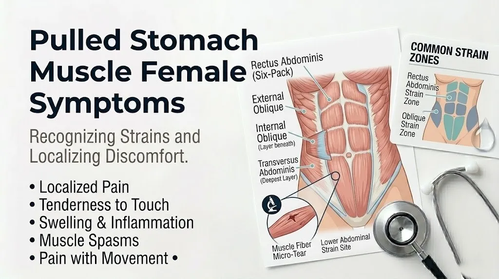
The abdominal wall comprises four paired muscles. The rectus abdominis runs vertically down the front, the familiar “six-pack” muscle. The external obliques angle diagonally down the sides. The internal obliques lie beneath them running the opposite diagonal. The deepest layer, the transversus abdominis, wraps horizontally around your midsection like a corset. When any of these muscle fibers tear from sudden force or overstretching, the damaged tissue releases prostaglandins and substance P directly at the injury site, activating pain-sensing nociceptors that fire with every contraction.
The sensation is immediate if the tear happens during a specific movement. You might feel a sudden sharp pull or pop in your abdomen while lifting something heavy, twisting quickly, or pushing through a hard cough. Over the following hours, the area becomes tender to touch, and even small movements like rolling over in bed or transitioning from sitting to standing can provoke a sharp reminder that the muscle is injured. The pain localizes to the abdominal wall rather than spreading deep into the pelvis or radiating through your back in a pattern that would suggest spine or organ involvement.
Strained Abdominal Muscle Symptoms
Strained abdominal muscle symptoms include localized tenderness, pain that worsens with muscle contraction, visible or palpable swelling, muscle spasm, and reduced ability to move the trunk freely. These symptoms follow a predictable pattern based on the severity of the muscle fiber disruption.
The tenderness you feel when pressing on the strained area reflects the localized inflammatory response. Damaged muscle cells release intracellular contents that trigger bradykinin production and mast cell activation, sensitizing nerve endings so that even light pressure feels painful. This tenderness is typically well-defined. You can trace the tender zone with your fingertips, and the pain stays within the muscle belly rather than crossing into areas that would suggest organ involvement.
| Symptom | What You Feel | Clinical Explanation |
|---|---|---|
| Localized tenderness | Pain when pressing on a specific spot | Nociceptor sensitization from prostaglandin release at the tear site |
| Pain with contraction | Sharp pull when sitting up, coughing, or laughing | Torn muscle fibers stretch further during contraction, triggering mechanoreceptor pain signals |
| Swelling | Puffiness or fullness over the muscle | Localized vasodilation and capillary leakage produce interstitial edema at the injury |
| Muscle spasm | Involuntary tight knot or hardening of the muscle | Reflexive neuromuscular guarding to splint the injured fibers |
| Reduced trunk movement | Difficulty twisting, bending, or straightening up | Pain inhibition and mechanical disruption limit active range of motion |
The specific pattern helps distinguish muscle strain from other conditions. Pain that changes with position but not with abdominal muscle contraction specifically suggests the problem may not be muscular. True abdominal wall pain should reproduce exactly when you contract the muscle against resistance, such as lifting your head and shoulders slightly off the bed while someone presses on the tender area. This is called Carnett’s sign, a clinical test that helps confirm the abdominal wall as the pain source.
Pulled Muscle in Abdomen Symptoms
Pulled muscle in abdomen symptoms range from mild discomfort with certain movements to severe pain that limits nearly all trunk activity. The grading system that clinicians use sorts muscle strains into three distinct levels that directly match what you will experience.
A grade 1 strain involves micro-tearing of fewer than 5 percent of muscle fibers. The pain is mild and does not significantly limit movement. You might notice a dull ache or pulling sensation during specific activities like a hard sneeze or a quick turn, but the muscle still functions through its full range. There is no palpable defect or visible bruising. Most women with grade 1 strains continue daily activities with only mild awareness that something feels “off” in the abdominal wall.
A grade 2 strain involves partial tearing of a larger proportion of muscle fibers. The pain is moderate to sharp. Movement becomes noticeably limited. You will likely feel pain when trying to sit up from a lying position, twist to reach something, or laugh unexpectedly. A grade 2 strain often produces visible swelling and a tender knot where the muscle fibers have partially torn and surrounding muscle has gone into protective spasm. According to the American Academy of Orthopaedic Surgeons, grade 2 strains generally require 2 to 6 weeks for functional recovery, though full tissue remodeling continues beyond that window.
A grade 3 strain involves a complete muscle rupture. The pain is severe at the moment of injury, sometimes followed by a deceptive reduction in pain as the torn ends retract and stop pulling on each other. A palpable gap or divot may be felt in the muscle belly where the rupture occurred. Movement of the trunk is substantially impaired. Grade 3 strains involving the abdominal wall are rare outside of high-force trauma or contact sports, but when they occur, they require urgent orthopedic or general surgical evaluation.
Abdominal Muscle Tear Symptoms
Abdominal muscle tear symptoms extend beyond the immediate pain to include visible bruising, a palpable defect in the muscle, and functional weakness that persists after the acute pain subsides. A true tear, as opposed to a simple strain, disrupts enough muscle and vascular tissue to produce these external markers.
Bruising develops when torn muscle fibers bleed into the surrounding tissue. The blood tracks along fascial planes, and depending on which muscle is torn, you may see discoloration appearing away from the exact injury site. Rectus abdominis tears can produce bruising that appears below the belly button or toward the pubic bone even though the tear occurred higher up. The bruise color shifts from dark purple or red in the first 48 hours to greenish and then yellowish over the following week as hemoglobin breaks down into biliverdin and then bilirubin.
A palpable defect feels like a divot or gap within the muscle when you gently press along the abdominal wall. This gap represents the space where torn muscle ends have separated. Small defects in the deeper obliques are difficult to feel, but a defect in the rectus abdominis close to the surface can be clearly identified. If you feel a gap that bulges outward rather than inward when you contract your abdominal muscles, you may be feeling a hernia rather than a muscle defect. Herniated tissue pushes through a fascial opening, while torn muscle simply separates, though the two can coexist.
Functional weakness continues even when the sharp pain has dulled. You might notice that one side of your abdomen does not engage properly when you try to sit up or that twisting to one side feels unstable. This weakness reflects the physical disruption of the muscle-tendon unit. The muscle cannot generate full contractile force because some of its fibers are no longer in continuity.
Key Takeaway: A true muscle tear produces visible bruising and a palpable gap or weakness, not just pain. Bruising that tracks downward, a divot you can feel in the muscle, or persistent functional weakness after the acute pain settles signals a grade 2 or 3 injury that needs a physical exam by a primary care physician or sports medicine doctor.
Ab Strain Symptoms
Ab strain symptoms include pain that follows a clear mechanical pattern, tenderness concentrated along the muscle course rather than diffusely across the abdomen, and symptoms that improve with rest and worsen predictably with use. Understanding the mechanical pattern helps separate muscle pain from other abdominal complaints.
The pain of an abdominal strain is consistently mechanical. It hurts when the muscle works and eases when the muscle rests in a neutral position. Lying flat on your back with knees bent typically reduces strain pain because this position slackens the rectus abdominis and reduces tension on the oblique muscles. Conversely, any action that requires the abdominal muscles to contract or stretch produces pain reliably. A 2023 review published in the British Journal of Sports Medicine confirmed that mechanical symptom behavior, meaning pain that consistently responds to loading and unloading, is a hallmark feature that distinguishes musculoskeletal pain from visceral pain.
Tenderness in an ab strain traces along the muscle fibers rather than spreading diffusely. If your external oblique is strained, tenderness follows the diagonal line from the lower ribs toward the opposite hip. If your rectus abdominis is strained, tenderness runs vertically along the midline or slightly off to one side. Internal organ pain, by contrast, tends to be more diffuse and harder to localize with a single finger. The somatic nerve supply to the abdominal wall provides precise localization, while the autonomic nerve supply to internal organs produces referred and less precisely localized pain.
Symptoms improve with rest in a way that organ pain does not. Appendicitis does not improve because you lie still, though lying still might reduce movement-related jostling. A strained abdominal muscle genuinely feels better after a period of relative rest in a comfortable position, then flares again when you resume activity. This rest-activity pattern is consistent across muscle strains throughout the body and applies equally to the abdominal wall.
Muscle Strain in Stomach Symptoms
Muscle strain in stomach symptoms involve pain that you can reproduce by tensing the muscle, a visible or palpable tight band within the muscle, and discomfort that intensifies hours after the initial injury as the inflammatory process fully develops. The stomach is not the organ but the colloquial term for the anterior abdominal region, and strain here follows muscle injury physiology.
Reproducibility is the clinical hallmark. When your doctor or physical therapist asks you to perform a partial sit-up or to lift your head against gentle resistance, a strained abdominal muscle will produce the exact pain you have been feeling at home. This reproducibility localizes the problem to the abdominal wall. Organ pain does not consistently reproduce with abdominal wall contraction. According to Cleveland Clinic clinical guidance, a positive Carnett’s sign, where abdominal wall contraction intensifies the pain while pressure is applied to the tender spot, strongly suggests a musculoskeletal rather than intra-abdominal source.
The tight band you feel within the muscle represents localized muscle spasm. Injured muscle tissue triggers a spinal reflex arc that causes the surrounding healthy muscle fibers to contract and splint the damaged area. This protective mechanism reduces movement at the injury site but also produces the hard, tender knot you can feel. The spasm may persist for days after the initial injury, gradually releasing as the acute inflammatory phase transitions into repair.
Pain intensification over the first 24 to 48 hours is normal for a muscle strain. The inflammatory process takes time to fully develop. Prostaglandin synthesis increases, capillary permeability rises, and immune cells infiltrate the damaged tissue. What felt like a minor pull at the moment of injury can become quite painful by the next morning. This delayed intensification pattern is typical of musculoskeletal injury and does not necessarily mean the injury has worsened. However, pain that continues escalating beyond 72 hours or that begins to spread beyond the abdominal wall into the flank or pelvic region warrants evaluation.
Torn Abdominal Muscle Symptoms
Torn abdominal muscle symptoms include severe sharp pain at the moment of injury, a sensation of something giving way or popping, substantial bruising that develops within hours, and difficulty maintaining upright posture without support. A torn muscle, as distinct from a strained muscle, involves a macroscopic disruption of muscle tissue.
The pop or tearing sensation at the moment of injury is a clinical feature that patients consistently report with grade 2 and grade 3 tears. This sensation occurs because the muscle fibers, which normally slide smoothly past each other within their fascial sheaths, suddenly rupture under excessive tensile load. The sound and sensation are produced by the mechanical failure of the muscle tissue itself, not by joint cavitation as with a knuckle crack. When a woman reports hearing or feeling a distinct pop in her abdominal wall during a forceful movement and then develops immediate pain and dysfunction, the likelihood of a clinically meaningful tear is high.
Substantial bruising develops because muscle tissue is highly vascular. The rectus abdominis receives its blood supply from the superior and inferior epigastric arteries. When muscle fibers tear, these small arterial branches can be disrupted, allowing blood to pool within the muscle belly and track along tissue planes. In women, lower abdominal wall bruising from a torn rectus abdominis must be distinguished from gynecological or uterine sources of bleeding, though the association with a specific injury event usually makes the distinction clear. A rectus sheath hematoma, where bleeding collects within the connective tissue envelope that contains the rectus abdominis, can produce a palpable, tender mass and may require imaging to differentiate from other abdominal masses.
Difficulty maintaining upright posture reflects the loss of anterior abdominal wall integrity. The abdominal muscles provide critical trunk support. When the rectus abdominis or major portions of the oblique muscles are torn, standing fully upright becomes painful and mechanically difficult. Women with a torn abdominal muscle often walk slightly hunched or lean toward the injured side to reduce tension on the torn fibers. This postural compensation is a visible clinical sign that distinguishes a significant tear from a minor strain.
Abdomen Strain Symptoms
Abdomen strain symptoms in women involve a constellation of sensations including pulling, burning, stabbing, and aching that vary across the healing timeline and can be influenced by female hormonal status, particularly estrogen levels that affect soft tissue compliance and pain perception.
The pulling sensation dominates the early phase. As torn muscle fibers begin the repair process, scar tissue forms a temporary bridge between the separated ends. This early scar tissue is less elastic than healthy muscle. When you move, the scar bridge pulls taut before the surrounding healthy muscle reaches its full stretch, producing a distinct pulling sensation that is sharper than a stretch but less intense than the original injury pain. Research published in the Journal of Orthopaedic and Sports Physical Therapy indicates that this pulling sensation typically resolves as the scar tissue remodels over weeks to months, assuming graduated loading is applied appropriately.
The burning sensation some women report likely reflects the involvement of small sensory nerve fibers that course through the muscle tissue. Muscle injury can irritate or entrap these fine nerve branches, producing a neuropathic-quality burning pain in addition to the mechanical aching. In female patients, estrogen modulates peripheral nerve sensitivity. Fluctuations in estrogen across the menstrual cycle can produce subtle changes in how pain is perceived, which may explain why some women notice their healing strain feels more bothersome at certain points in their cycle. This is not psychological amplification but a real physiological effect of estrogen on nociceptive processing.
Postmenopausal women with lower circulating estrogen levels may experience different pain characteristics and potentially slower soft tissue healing. Estrogen receptors are present in skeletal muscle and connective tissue, and while the effect of estrogen on muscle repair is still being actively researched, observational data suggests that postmenopausal women may have a prolonged inflammatory phase after muscle injury compared to premenopausal women. A primary care physician or sports medicine doctor can factor this into recovery expectations.
Key Takeaway: The pulling and burning sensations that accompany abdominal muscle strain healing are normal phases of tissue repair and nerve irritation. Women may notice that pain perception fluctuates with menstrual cycle phases due to estrogen effects on nerve sensitivity, a real physiological phenomenon rather than a sign the injury has worsened.
Symptoms of Pulled Abdominal Muscle in Females
Symptoms of a pulled abdominal muscle in females include all the general strain symptoms plus specific considerations related to female pelvic anatomy, reproductive organ proximity, and the effect of relaxin and estrogen on abdominal wall connective tissue compliance and healing.
The anatomical proximity of the lower abdominal muscles to the uterus, ovaries, and round ligaments creates potential symptom confusion unique to female patients. The round ligaments of the uterus attach from the uterine fundus to the labia majora, passing through the inguinal canal. When these ligaments stretch or spasm, they produce sharp groin and lower abdominal pain that can closely mimic a lower abdominal muscle strain. The key distinction is that round ligament pain is provoked by position changes that move the uterus, such as rolling over quickly or standing up suddenly, rather than by abdominal muscle contraction against resistance. A woman who notices sharp lower abdominal pain when coughing can test whether the pain intensifies further when she consciously tightens her abdominal muscles. If it does, the abdominal wall is the more likely source.
Estrogen influences the structural properties of the abdominal wall connective tissue. Higher estrogen levels are associated with greater connective tissue compliance, meaning the tissue stretches more before reaching the point of failure. This provides some protective effect against muscle strain in premenopausal women compared to men of similar age and fitness level. However, pregnancy introduces relaxin, a hormone produced by the corpus luteum and later the placenta. Relaxin significantly increases connective tissue laxity throughout the body, including the abdominal wall, linea alba, and pelvic ligaments. This increased laxity makes pregnant women more vulnerable to abdominal muscle strain even from movements that would not typically cause injury.
The female pelvic floor also factors into abdominal strain recovery. The abdominal muscles and pelvic floor muscles function as an integrated unit. When abdominal wall injury causes you to alter your movement patterns and breath-holding strategies, the pelvic floor may compensate in ways that produce pelvic pressure or discomfort. Postpartum women recovering from both abdominal strain and pelvic floor changes after childbirth face a more complex recovery pathway than a male athlete with the same grade of abdominal muscle strain.
Pulled Stomach Muscle Female Lower Abdomen
A pulled stomach muscle in the female lower abdomen presents with pain below the navel that worsens with sitting up, coughing, or lifting, and can be confused with ovarian, uterine, bladder, or round ligament pain. Localizing the pain to the abdominal wall rather than deeper pelvic structures requires specific clinical maneuvers and careful attention to symptom triggers.
The lower abdominal wall below the navel is predominantly formed by the lower portions of the rectus abdominis, the internal oblique and transversus abdominis aponeuroses, and the pyramidalis muscle when present. The rectus abdominis inserts on the pubic symphysis and pubic crest. Strain of the lower rectus fibers produces pain just above the pubic bone, in the region where many women also experience menstrual cramping, bladder discomfort, or early pregnancy sensations. The transversus abdominis and internal oblique contribute to the posterior rectus sheath above the arcuate line and pass anterior to the rectus below it, meaning lower abdominal strains can involve complex fascial interactions.
Distinguishing lower abdominal wall pain from pelvic organ pain requires attention to what makes the pain better or worse. Muscle pain eases in positions that relax the abdominal wall. Lying supine with a pillow under the knees, sitting slightly reclined, or standing with one foot elevated on a step all reduce tension on the lower abdominal muscles. Ovarian cyst pain, endometriosis-related pain, and bladder pain do not predictably respond to these positional changes in the same way. Menstrual cramping follows a cyclical pattern independent of mechanical loading, though abdominal muscle contraction can secondarily intensify the sensation of uterine cramping that is already present.
A practical at-home assessment sequence can help clarify the source:
- Lie flat on your back with knees bent and feet flat. Palpate the tender area gently. Note the pain level.
- Tighten your abdominal muscles by gently lifting your head and shoulders an inch off the surface, as if starting a small crunch. If the tenderness intensifies sharply with this contraction, the abdominal wall is the likely source.
- Relax and note whether the pain diminishes within 30 seconds of resting in the comfortable supine position. Muscle pain will ease. Organ pain will remain relatively constant.
- Repeat the sequence on a different day at a different point in your menstrual cycle if possible. Consistent mechanical pain behavior across different cycle phases strongly suggests a musculoskeletal source.
Abdominal Muscle Strain During Pregnancy
Abdominal muscle strain during pregnancy occurs more easily because of hormonally mediated connective tissue laxity, mechanical overload from uterine expansion, and altered abdominal muscle geometry that changes the line of pull and force distribution across the abdominal wall.
The hormone relaxin rises substantially during pregnancy, peaking in the first trimester and again near term. Relaxin remodels collagen in connective tissues throughout the body, increasing their distensibility. The linea alba, the connective tissue band that runs down the midline of the abdomen between the two rectus abdominis bellies, becomes more compliant under relaxin’s influence. This increased compliance is necessary for the abdominal wall to accommodate the growing uterus, but it reduces the tensile strength of the abdominal connective tissue and makes muscle strains more likely during activities that would not have caused injury before pregnancy.
The mechanical environment of the abdominal muscles changes progressively throughout pregnancy. As the uterus expands, the rectus abdominis muscles are stretched and displaced laterally in many women, a condition called diastasis recti. The muscle bellies no longer sit adjacent to each other at the midline but separate, connected only by the stretched linea alba. This separation alters the angle of muscle fiber pull and reduces the efficiency of force transfer across the abdominal wall. Simple movements like rolling over in bed, which previously engaged intact abdominal musculature smoothly, now place asymmetric load on the separated muscles and their connective tissue attachments.
According to the American College of Obstetricians and Gynecologists, abdominal wall pain during pregnancy should always be evaluated by an obstetrician-gynecologist or midwife because the potential causes span from benign muscle strain to conditions that require urgent management. Pregnant women should not independently assume abdominal pain is muscular without a clinical assessment. The evaluation typically includes checking uterine tone, fetal well-being, and cervical status while also examining the abdominal wall. Treatment for confirmed muscle strain during pregnancy emphasizes activity modification, positional support, and physical therapy with practitioners experienced in prenatal musculoskeletal care. Ice, rest, and gentle movement within comfortable ranges are generally considered safe.
| Pregnancy Stage | Abdominal Wall Change | Strain Risk Implication |
|---|---|---|
| First trimester | Rising relaxin levels, uterine growth begins | Increased connective tissue laxity, lower threshold for strain with sudden movement |
| Second trimester | Rapid uterine expansion, rectus displacement begins | Altered muscle geometry, diastasis recti may start, asymmetric loading risk |
| Third trimester | Maximal abdominal wall stretch, significant diastasis common | Highest strain risk, even low-force movements can overload stretched muscles |
| Postpartum immediate | Loss of uterine volume, connective tissue still lax | Abdominal wall temporarily unstable, muscles shortened from chronic stretch, re-injury risk high |
Pulled Stomach Muscle Postpartum Recovery
Pulled stomach muscle postpartum recovery requires addressing both the acute muscle strain and the broader abdominal wall changes of pregnancy and delivery, including diastasis recti if present, pelvic floor coordination, and the residual effects of relaxin on connective tissue compliance.
The postpartum abdominal wall is not simply a non-pregnant abdominal wall with a strain added on top. After delivery, the abdominal muscles are structurally and neurologically altered. They have been stretched to their physiological limit for months. The rectus abdominis may still be separated at the linea alba. The neuromuscular control patterns that coordinate abdominal contraction with breathing and pelvic floor engagement have been disrupted. A strain that occurs during postpartum activity, such as lifting the baby, a car seat, or a laundry basket, lands on a system that is already working from a mechanically disadvantaged starting point.
Connective tissue laxity persists postpartum for weeks to months. Relaxin levels drop sharply after delivery of the placenta, but the tissue remodeling that relaxin initiated does not reverse immediately. Collagen cross-linking gradually restores connective tissue tensile strength over the postpartum period, with individual variation depending on breastfeeding status, as lactation maintains a different hormonal environment, and genetic factors affecting collagen metabolism. The American Journal of Obstetrics and Gynecology has published research indicating that connective tissue recovery after pregnancy extends well beyond the traditional six-week postpartum visit, with full restoration of pre-pregnancy tensile properties taking many months in some women.
Recovery from a postpartum abdominal muscle strain should be guided by a physical therapist with expertise in postpartum rehabilitation when the strain is more than mild. The rehabilitation must address not only the strained muscle but the integrated abdominal wall, pelvic floor, diaphragm, and spinal stabilizer system. Returning too quickly to full activity, including high-impact exercise or heavy lifting, risks re-injury and can also worsen diastasis recti or pelvic floor dysfunction. A gradual, progressive loading program that respects the unique postpartum physiology produces better outcomes than following a standard muscle strain protocol designed for the general population.
Key Takeaway: Postpartum abdominal muscle strain recovery is not the same as standard abdominal strain recovery. The muscles, connective tissue, and neuromuscular control systems are in a postpartum state that requires specialized rehabilitation. Work with a women’s health or pelvic health physical therapist rather than following generic return-to-exercise timelines.
How to Tell If Abdominal Pain Is Muscular or Internal Female
You can distinguish muscular abdominal wall pain from internal organ pain in women by assessing whether the pain changes with abdominal muscle contraction, localizes to one finger-point spot, and improves with rest in a position that relaxes the abdominal wall. Internal organ pain typically remains constant or follows an internal pattern independent of abdominal wall movement.
The abdominal wall test, based on Carnett’s sign, is the most reliable clinical tool for differentiation. Lie on your back with knees bent. Locate the precise tender spot. Press on it and note the pain level. Now lift your head and shoulders slightly off the surface, tensing your abdominal muscles while maintaining pressure on the spot. If the pain intensifies significantly, the source is likely the abdominal wall. If the pain stays the same or actually eases because you are distracted by the effort, the source is more likely internal. A positive Carnett’s sign has been validated in multiple clinical studies, with research published in the Journal of Orthopaedic and Sports Physical Therapy confirming its utility for identifying abdominal wall pain.
Pain character and triggers provide additional information. Muscular pain is sharp or pulling and directly tied to movement, coughing, or sneezing. Internal organ pain tends to be crampy, colicky, burning, or pressure-like and may follow a pattern related to eating, bowel movements, urination, or the menstrual cycle rather than solely to mechanical loading of the abdominal wall. Pain from the ovary or fallopian tube, for example, is typically felt deeper in the pelvis and may radiate to the lower back or inner thigh. Pain from the bladder is felt in the suprapubic midline and changes with bladder filling and emptying. Pain from the gastrointestinal tract often relates to meals or bowel patterns. A careful symptom diary tracking pain timing with movement, meals, bladder function, and menstrual cycle phase can reveal the pattern and guide clinical evaluation.
| Feature | Muscular Abdominal Wall Pain | Internal Organ Pain |
|---|---|---|
| Pain location precision | Can point with one finger | Diffuse, hard to localize precisely |
| Effect of abdominal contraction | Intensifies pain | No consistent change |
| Effect of rest in neutral position | Pain eases | Pain remains relatively constant |
| Relation to meals or bowel movements | None | Often related |
| Relation to menstrual cycle | None (except possibly pain perception) | May follow cyclical pattern |
| Visible swelling or bruising | Possible with grade 2-3 strain | Rare unless underlying mass |
| Carnett’s sign | Positive (pain increases with contraction) | Negative |
Even when symptoms strongly suggest a muscular source, any abdominal pain that is severe, persistent beyond two weeks, or accompanied by fever, vomiting, unexplained weight loss, or change in bowel or bladder function requires medical evaluation. A woman with lower abdominal pain and a positive Carnett’s sign may still have both a muscle strain and an independent gynecological or gastrointestinal condition. The presence of a muscle strain does not rule out internal pathology.
Abdominal Muscle Strain vs Hernia in Women
An abdominal muscle strain and an abdominal wall hernia in women both produce localized pain and tenderness, but a hernia typically produces a palpable bulge that becomes more prominent with standing, coughing, or straining, while a simple muscle strain does not. The differentiation matters because a hernia can involve trapped tissue that requires surgical evaluation.
An abdominal wall hernia occurs when abdominal contents, usually a portion of intestine or intra-abdominal fat, push through a defect or weak spot in the fascial layer of the abdominal wall. In women, hernias can occur at the umbilicus, in the groin at the inguinal canal, at the femoral canal just below the inguinal ligament, or at previous surgical incision sites. The key physical finding that distinguishes a hernia from a muscle strain is the presence of a reducible or non-reducible bulge that changes with position and with Valsalva maneuver, which is bearing down as if having a bowel movement.
The bulge of a hernia feels different from the tender knot of a strained muscle. A muscle strain knot is firm muscle in spasm that does not change size dramatically when you cough. A hernia bulge is soft tissue protruding through a fascial opening that often pushes further outward when intra-abdominal pressure increases with coughing or straining. You may be able to gently push the hernia contents back through the fascial defect, causing the bulge to temporarily disappear. A muscle knot cannot be pushed back in because it is the muscle itself, not contents moving through an opening.
Women have specific hernia considerations. Femoral hernias, while less common than inguinal hernias overall, occur more frequently in women than in men. A femoral hernia presents as a bulge in the upper thigh just below the groin crease and can cause pain that radiates down the inner thigh. This pain pattern can be mistaken for a groin muscle strain. Inguinal hernias in women may involve the round ligament of the uterus rather than intestine, producing groin pain without a prominent bulge that is easy to detect. According to surgical guidelines published by the American College of Surgeons, any suspected hernia with pain that becomes suddenly severe, a bulge that becomes firm and cannot be pushed back in, or associated nausea and vomiting requires immediate surgical evaluation because these findings suggest incarcerated or strangulated hernia, a surgical emergency.
Emergency Symptoms: When to Call 911 or Go to the ER
Certain symptoms associated with what might initially seem like a pulled stomach muscle require immediate emergency evaluation. Do not wait to see if these resolve on their own.
Call 911 or go to the nearest emergency room immediately if you experience:
- Sudden severe abdominal pain with a rigid, board-like abdomen that does not relax when you try to breathe gently. This can indicate internal bleeding, organ perforation, or a large rectus sheath hematoma compressing vital structures.
- An expanding, tender mass in the abdominal wall that is growing larger over minutes to hours, especially if you recently started or changed anticoagulant medication. This can indicate an expanding rectus sheath hematoma that requires urgent surgical management.
- Abdominal pain accompanied by lightheadedness, rapid heart rate, cold clammy skin, or fainting. These are signs of internal blood loss and possible hemorrhagic shock.
- A known or suspected hernia that becomes suddenly painful, firm, and cannot be pushed back in, especially with nausea or vomiting. This can indicate incarcerated or strangulated hernia, a surgical emergency.
- Severe abdominal pain in a pregnant woman beyond the first trimester, whether or not it seems muscular. Placental abruption, uterine rupture, and other obstetric emergencies can present with abdominal pain that may initially be attributed to muscular causes.
- Abdominal wall pain after blunt trauma, such as a car accident or fall, where internal organ injury must be ruled out even if the abdominal wall is also strained.
These presentations can indicate serious internal pathology that requires emergency medical assessment, not an urgent care appointment or a phone call to your doctor’s office. Abdominal pain that crosses the threshold from manageable to emergency-level typically does so for structural reasons that cannot be managed with rest and ice at home.
When to See a Doctor for Pulled Stomach Muscle
A primary care physician or sports medicine physician should evaluate a pulled stomach muscle when pain is severe, function remains limited beyond one to two weeks, a palpable defect or bulge is present, or you are pregnant or postpartum. Specific circumstances dictate which provider type is appropriate and how urgently you should be seen.
Schedule an appointment with your primary care physician in the following situations. Pain from a suspected muscle strain that has not meaningfully improved after 7 to 10 days of appropriate self-care including relative rest and activity modification warrants evaluation. Pain that interferes with your ability to work, care for children, or perform daily activities should be assessed rather than tolerated indefinitely. Any palpable lump, bulge, or defect in the abdominal wall that you can feel but that does not meet emergency criteria still requires a physical exam to rule out hernia or significant fascial injury. Women who are pregnant or within the first year postpartum should have abdominal wall pain evaluated by their obstetrician-gynecologist or primary care physician rather than self-managing, because pregnancy-related conditions can present with abdominal wall pain.
A sports medicine physician or physical therapist evaluation is appropriate for grade 2 strains and for any strain in a woman who wants to return to athletics, high-intensity exercise, or physically demanding work. These providers can assess muscle function, identify biomechanical contributors to the strain, and design a progressive return-to-activity program that reduces re-injury risk. A board-certified orthopedic surgeon should evaluate suspected grade 3 ruptures with a palpable gap or significant functional impairment.
When you go to the appointment, bring specific information. Note exactly when the injury occurred and what movement produced it. Describe the pain location in terms of distance from the navel, pubic bone, or rib margin. Report what movements reproduce the pain most reliably and what positions relieve it. Mention whether you have noticed any visible bruising, swelling, or a bulge. List any medications you take, especially anticoagulants or antiplatelet drugs, because these affect bleeding risk within the muscle. For menstruating women, note where you are in your cycle and whether you have noticed any relationship between pain intensity and cycle phase.
Key Takeaway: Give a muscle strain 7 to 10 days of appropriate self-care to show clear improvement. If it does not, or if you feel a bulge, defect, or have significant functional limitation, see your primary care physician or sports medicine doctor. Pregnant and postpartum women should have abdominal wall pain evaluated rather than assuming it is a simple strain.
Frequently Asked Questions About Pulled Stomach Muscle in Women
What are the first signs of a pulled stomach muscle in a woman?
The first signs are sharp localized pain in the abdominal wall that occurs during a specific movement like coughing, twisting, or lifting.
You may feel a pulling or popping sensation at the moment of injury followed by tenderness when pressing on the area.
The pain reliably intensifies when you engage your abdominal muscles by sitting up, laughing, or coughing.
How long does a pulled abdominal muscle take to heal in females?
A grade 1 abdominal muscle strain typically heals within 1 to 2 weeks with appropriate relative rest and gradual return to activity.
A grade 2 strain generally requires 2 to 6 weeks for functional recovery, though full tissue remodeling continues for months.
Postpartum women and postmenopausal women may experience somewhat longer healing timelines due to hormonal influences on connective tissue and muscle repair.
Can a pulled stomach muscle feel like period cramps?
A pulled lower abdominal muscle can produce an aching sensation above the pubic bone that some women describe as similar to menstrual cramping.
However, muscle strain pain changes predictably with movement and abdominal contraction, while menstrual cramping follows a cyclical pattern independent of mechanical loading.
The two can coexist, and a careful symptom diary tracking pain timing with movement versus cycle phase helps differentiate the sources.
How do I know if my abdominal pain is muscle or organ related?
Test whether abdominal muscle contraction increases the pain by gently lifting your head and shoulders while lying down with someone pressing on the tender spot.
If the pain intensifies with this contraction, known as a positive Carnett’s sign, the source is likely the abdominal wall rather than an internal organ.
Organ pain typically remains constant regardless of abdominal wall contraction and may relate to meals, bladder filling, or menstrual cycle rather than solely to movement.
Can you pull a stomach muscle during pregnancy?
Yes, abdominal muscle strains occur more easily during pregnancy because the hormone relaxin increases connective tissue laxity and the expanding uterus alters abdominal muscle geometry.
Pregnant women should always have abdominal pain evaluated by their obstetrician-gynecologist or midwife rather than assuming it is muscular, because pregnancy-related conditions can present similarly.
Treatment emphasizes activity modification, positional support, and physical therapy rather than medication, with ice and rest considered safe for confirmed muscle strain.
Should I go to the ER for a pulled stomach muscle?
Go to the ER if you have sudden severe abdominal pain with a rigid board-like abdomen, an expanding mass in the abdominal wall, or pain with lightheadedness or rapid heart rate.
A known hernia that becomes suddenly painful, firm, and cannot be pushed back in also requires immediate emergency evaluation for possible strangulation.
A typical grade 1 or mild grade 2 muscle strain without these features does not require an ER visit and can be evaluated by a primary care physician within a few days if needed.
The distinction between a pulled stomach muscle and more serious abdominal or pelvic conditions is one that every woman should feel equipped to make. Your body gives you specific information: where the pain is, what makes it flare, what calms it down, and whether you can feel a structural change in the abdominal wall. Learning to read those signals and knowing the specific thresholds for calling your doctor or going to the ER puts you in a position of informed self-advocacy rather than anxious guesswork.
If your symptoms align with a muscular strain and are manageable at home, give yourself the rest and graduated movement that muscle tissue needs to repair. If anything about your pain pattern does not fit the mechanical, reproducible, abdominal wall picture described here, or if you are pregnant or postpartum, see your primary care physician or obstetrician-gynecologist for a physical exam. The right diagnosis is the one that keeps you safe while getting you back to the activities and people you care about.