Fabry Disease Symptoms: Full 2026 Clinical Guide and Signs
Fabry disease symptoms arise because a deficient enzyme called alpha-galactosidase A fails to break down a fatty substance called globotriaosylceramide (Gb3), which then accumulates inside the lysosomes of cells throughout the body, progressively damaging the kidneys, heart, nervous system, skin, eyes, and blood vessels. The symptom pattern is broad, often misattributed to more common conditions, and produces a median diagnostic delay of more than a decade in most patients.
According to the National Organization for Rare Disorders (NORD), Fabry disease affects an estimated 1 in 40,000 males in its classic form, though newborn screening data suggests the true prevalence may be considerably higher when atypical variants are included. The disease is X-linked, meaning males with a pathogenic GLA gene variant experience the full enzymatic deficiency, while females with one abnormal copy have highly variable symptom severity depending on which cells express the deficient enzyme.
This article covers the complete clinical picture: the molecular mechanism behind each symptom, how symptoms differ between childhood, adulthood, males, and females, the critical emergency presentations that require immediate action, and exactly how the diagnosis is confirmed.
Fabry Disease Symptoms: The Clinical Overview
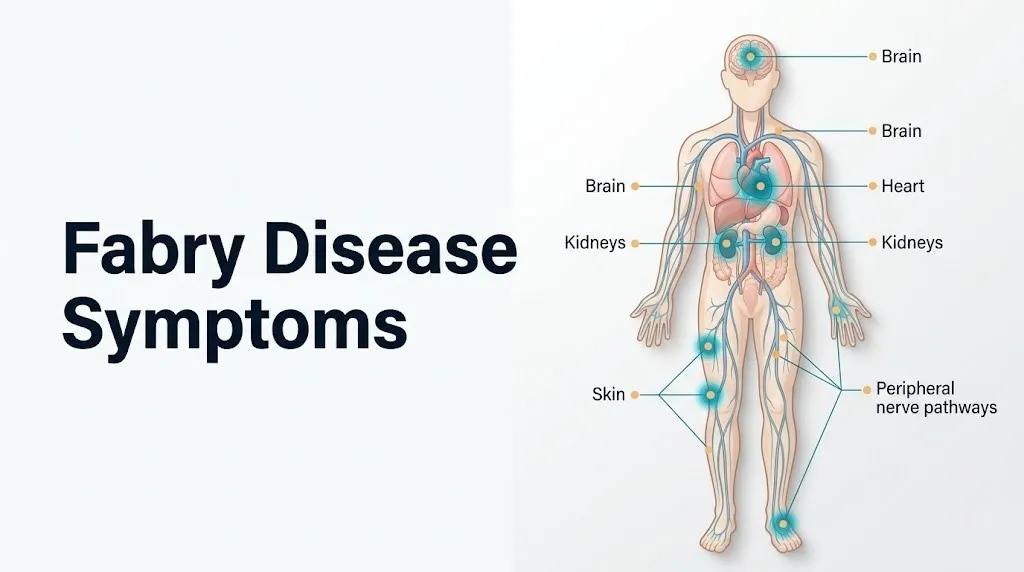
Fabry disease symptoms span multiple organ systems and include burning neuropathic pain in the hands and feet, skin lesions called angiokeratomas, reduced or absent sweating, gastrointestinal cramping, progressive kidney disease, heart muscle thickening, stroke, and corneal opacities visible under a slit lamp.
The symptom picture differs substantially depending on age, biological sex, and the specific GLA mutation involved. In hemizygous males with classic disease, symptoms typically begin in childhood or adolescence with neuropathic pain and progress through early adulthood to involve the kidneys, heart, and cerebrovascular system. In heterozygous females, the onset and severity vary widely due to the variable pattern of X-chromosome inactivation across tissues.
Here is the full clinical symptom inventory with the primary organ system responsible for each:
| Symptom | Organ System | Onset Phase | Seen in Males | Seen in Females |
|---|---|---|---|---|
| Burning pain in hands and feet (acroparesthesia) | Peripheral nervous system | Childhood/adolescence | Almost universal | Up to 70% of females |
| Angiokeratomas (skin lesions) | Integumentary system | Adolescence/early adulthood | Very frequent | Less frequent |
| Hypohidrosis (reduced sweating) | Autonomic nervous system | Childhood | Frequent | Variable |
| Gastrointestinal cramping, diarrhea | Enteric autonomic nervous system | Childhood/adolescence | Frequent | Frequent |
| Cornea verticillata | Ophthalmologic | Childhood/adulthood | Frequent | Frequent |
| Proteinuria, progressive nephropathy | Renal system | Early adulthood | Frequent | Variable |
| Left ventricular hypertrophy | Cardiovascular system | Middle adulthood | Frequent | Frequent in older females |
| Ischemic stroke or TIA | Cerebrovascular system | Young to middle adulthood | Frequent | Frequent |
| Sensorineural hearing loss, tinnitus | Auditory system | Variable | Reported | Reported |
| Fatigue, exercise intolerance | Multisystem | Throughout | Common | Common |
The National Institute of Neurological Disorders and Stroke (NINDS) notes that Fabry disease is one of the most frequently misdiagnosed rare diseases, with patients commonly receiving incorrect diagnoses of rheumatoid arthritis (for the joint and extremity pain), multiple sclerosis (for the neurological symptoms), irritable bowel syndrome (for the gastrointestinal symptoms), or essential hypertension (for the renal and vascular involvement).
What Is Fabry Disease and How Does It Cause Symptoms
Fabry disease is an X-linked lysosomal storage disorder caused by pathogenic variants in the GLA gene located at chromosomal locus Xq22.1, resulting in deficient or absent activity of the lysosomal enzyme alpha-galactosidase A, which is responsible for catabolizing the glycosphingolipid globotriaosylceramide (Gb3).
Think of lysosomes as the recycling centers inside every cell. Alpha-galactosidase A is the specific recycling machine designed to dismantle Gb3, a fatty molecule that the cell continuously produces as a byproduct of normal membrane metabolism. When that machine is broken, Gb3 builds up inside the recycling center with each passing week. The recycling center swells. Eventually it ruptures. The cellular debris that results triggers inflammation, apoptosis (programmed cell death), and organ dysfunction.
The cell types that accumulate Gb3 most rapidly and are most vulnerable to the resulting damage are: glomerular podocytes in the kidney, dorsal root ganglion neurons in the peripheral nervous system, vascular endothelial cells lining blood vessels throughout the body, cardiomyocytes in the heart muscle, and the epithelial cells of the cornea. This explains why those specific organ systems are the ones that produce Fabry disease’s defining symptoms.
Beyond Gb3, accumulation also produces the deacylated metabolite globotriaosylsphingosine (lyso-Gb3), which is more water-soluble, circulates systemically in the blood, and has direct toxic effects on neurons and endothelial cells that extend beyond where Gb3 itself accumulates. Lyso-Gb3 is now recognized as both a key pathogenic mediator and the primary plasma biomarker used to monitor disease activity.
Individuals with GLA variants that produce some residual alpha-galactosidase A activity (typically missense variants rather than nonsense or frameshift mutations) develop what is called the atypical or cardiac-variant form of Fabry disease. These individuals may not have the classic childhood neuropathic pain or skin findings but develop cardiac involvement, often in the fifth or sixth decade of life, without a prior diagnosis.
The Alpha-Galactosidase A Enzyme Deficiency Mechanism
Alpha-galactosidase A (alpha-Gal A) is a lysosomal acid hydrolase that cleaves the terminal alpha-galactosyl residue from Gb3, and when its activity is absent or below the threshold needed to process the cellular Gb3 load, Gb3 accumulates in a progressively damaging lysosomal storage pattern that is irreversible without therapeutic intervention.
The enzyme is encoded by the GLA gene, which contains 7 exons. Over 1,000 distinct pathogenic variants have been identified across this gene, ranging from large deletions that eliminate enzyme production entirely, to single nucleotide substitutions (missense variants) that produce a misfolded enzyme with reduced but not zero catalytic activity. The American College of Medical Genetics and Genomics (ACMG) classifies GLA variants by their pathogenicity based on the combined weight of functional assay data, population frequency, and clinical correlation.
At the cellular level, the deficient alpha-Gal A means Gb3 entering the lysosome through endocytosis cannot be processed. Lysosomal Gb3 inclusions accumulate, visible under electron microscopy as lamellated “onion-skin” or “myeloid-body” inclusions within the lysosomal compartment. These are detectable in kidney biopsy specimens, skin biopsy specimens, and heart biopsy specimens and are used as histopathological evidence of Fabry disease.
The downstream consequences of lysosomal Gb3 accumulation include: impaired autophagy-lysosome pathway function, which allows damaged mitochondria and misfolded proteins to accumulate in the cell cytoplasm; activation of the mitochondrial apoptosis pathway through lysosomal membrane permeabilization and cathepsin release; and secondary activation of inflammatory pathways through NF-kB signaling in response to cellular stress.
Males with complete loss-of-function GLA variants (null mutations) have essentially zero alpha-Gal A activity and develop the most severe classic multisystem phenotype. Those with partial-function missense variants have 1 to 20% residual activity, which is typically sufficient to prevent the severe childhood neurological features but insufficient to prevent cardiac or renal involvement over decades.
Key Takeaway: Fabry disease causes every symptom it produces through the same root mechanism: Gb3 accumulates in lysosomes of specific vulnerable cell types because the enzyme designed to clear it is absent or inadequate, and different cell types fail at different rates depending on their Gb3 production and turnover rates.
Fabry Disease Early Symptoms in Childhood
The earliest Fabry disease symptoms in childhood are burning pain or tingling in the hands and feet, reduced or absent sweating (hypohidrosis), chronic fatigue, low-grade fevers without infection, and gastrointestinal cramping, all of which appear most often between ages 4 and 14 in males with classic disease.
The pain is the most common presenting complaint and the most frequently misdiagnosed. Children with Fabry disease describe the pain as burning, electric, or needle-like, located primarily in the palms, fingers, soles, and toes. It worsens dramatically during fever, hot weather, physical activity, and emotional stress, and improves somewhat in cool or cold environments. This temperature sensitivity is the clinical clue most frequently missed by physicians who encounter it for the first time.
Hypohidrosis occurs because autonomic small fiber nerves innervating eccrine sweat glands throughout the body accumulate Gb3 in the nerve terminals, impairing the sudomotor signal that triggers sweating. A child with Fabry disease who plays outside on a hot day may experience dangerous heat intolerance without sweating normally to regulate body temperature. This can produce heat exhaustion or heat stroke at ambient temperatures that healthy children tolerate comfortably.
Chronic fatigue in Fabry disease children is multifactorial. It reflects the systemic inflammatory burden of circulating lyso-Gb3, the disrupted sleep from nocturnal pain, and the physiological burden of ongoing cellular stress. Children affected by this fatigue are frequently dismissed as psychosomatic or malingering before the diagnosis is established.
According to data published in the Fabry Outcome Survey (FOS), the mean age of first symptom onset in males with classic Fabry disease is approximately 6 years, and the mean age at diagnosis is approximately 20 years, representing a diagnostic gap of approximately 14 years during which the disease continues to damage organs without specific treatment.
Girls with classic Fabry disease (heterozygous) may begin experiencing similar childhood symptoms, including acroparesthesia and gastrointestinal complaints, though typically at a lower frequency and severity than affected boys of the same age.
Fabry Disease Neuropathic Pain and Pain Crises
Fabry disease neuropathic pain is a burning, constant peripheral dysesthesia affecting primarily the hands and feet, produced by Gb3 and lyso-Gb3 accumulation in dorsal root ganglion neurons and unmyelinated C fibers, and manifests in both a chronic baseline form and as acute Fabry pain crises triggered by fever, heat, illness, physical exertion, or stress.
The specific pain mechanism is better understood than most resources convey. Gb3 accumulation in dorsal root ganglion (DRG) neuron cell bodies causes lysosomal swelling that physically distorts the cell body. Lyso-Gb3 released from affected DRG neurons activates the TRPV1 (transient receptor potential vanilloid-1) channel on neighboring C fibers. The TRPV1 channel is the same receptor activated by capsaicin (the compound that makes hot peppers burning). When chronically activated by lyso-Gb3, TRPV1 produces persistent burning pain and heat hypersensitivity, the clinical signature of Fabry neuropathic pain.
A Fabry pain crisis is a discrete, severe pain episode that may last minutes to days, described by affected individuals as an extreme worsening of the baseline burning sensation, sometimes accompanied by fever, joint pain, and malaise. Triggers include any cause of internal temperature elevation: fever from any infection, vigorous exercise, prolonged sun exposure, or hot baths. The physiological basis is thermal TRPV1 channel upregulation: elevated temperature increases the TRPV1 channel’s firing frequency, amplifying the pain signal.
Research published in Molecular Genetics and Metabolism has confirmed that plasma lyso-Gb3 levels correlate with pain severity scores in Fabry disease patients, providing biochemical evidence that lyso-Gb3 is a direct driver of the neuropathic pain phenotype, not merely a bystander biomarker.
Fabry pain crises in pediatric patients are frequently mistaken for juvenile idiopathic arthritis, growing pains, or psychosomatic complaints. Any child with recurrent episodes of severe hand and foot burning pain triggered by fever or exercise, without objective joint inflammation, should be evaluated by a board-certified medical geneticist for lysosomal storage disorder screening including leukocyte alpha-galactosidase A enzyme assay.
Fabry Disease Skin Symptoms and Angiokeratomas
Angiokeratomas are the most visually distinctive skin manifestation of Fabry disease: small (1 to 3 mm), dark red to blue-black, non-blanching vascular lesions that appear in clusters primarily in the “bathing trunk” distribution between the umbilicus and knees, though they also appear on the lips, mouth mucosa, fingertips, and conjunctiva.
Angiokeratomas form because Gb3 accumulates in the endothelial cells lining small dermal blood vessels (capillaries and postcapillary venules). The swollen, Gb3-laden endothelial cells disrupt the structural integrity of the vessel wall. Vessels dilate and ectasia forms. The overlying epidermis shows hyperkeratosis (thickening), producing the combined vascular-epidermal lesion that appears as a red-black papule under the skin surface. Despite their alarming appearance, angiokeratomas in Fabry disease are benign skin lesions with no malignant potential.
Hypohidrosis (described in the childhood section as a sweat gland autonomic nerve failure) is clinically confirmed by the quantitative sudomotor axon reflex test (QSART), a specialized autonomic function test that measures sweat output in response to an electrical stimulus. QSART abnormalities are present in the majority of hemizygous males and in a substantial proportion of heterozygous females, according to NINDS-supported autonomic function research in Fabry disease cohorts.
Some individuals with Fabry disease develop hyperhidrosis (increased sweating in unaffected skin regions) as a compensatory mechanism when hypohidrosis is asymmetric. This paradoxical finding can confuse the clinical picture.
Females with Fabry disease develop angiokeratomas less frequently than males, with registry data from the Fabry Outcome Survey showing angiokeratoma prevalence in approximately 20 to 30% of symptomatic females compared to 60 to 70% of classically affected males. When present in females, angiokeratomas tend to appear at older ages and in smaller numbers.
Key Takeaway: Angiokeratomas in Fabry disease appear in the bathing trunk distribution and form because Gb3 accumulates in the endothelial cells of small dermal vessels, causing vascular ectasia and overlying epidermal thickening. Seeing them on a young adult warrants urgent lysosomal disease screening.
Fabry Disease Gastrointestinal Symptoms
Fabry disease gastrointestinal symptoms include recurrent abdominal cramping, early satiety, nausea, diarrhea (sometimes alternating with constipation), and postprandial pain, produced by Gb3 accumulation in the enteric autonomic nervous system, specifically in the smooth muscle cells and autonomic ganglia of the gastrointestinal wall.
The gastrointestinal tract has its own internal nervous system, the enteric nervous system (ENS), consisting of two plexuses: the myenteric (Auerbach’s) plexus, which controls gut motility, and the submucosal (Meissner’s) plexus, which regulates mucosal secretion. Gb3 accumulates in the neurons and smooth muscle cells of both plexuses, impairing coordinated peristalsis and mucosal regulation. The result is gastrointestinal dysmotility: food moves through the intestine at an irregular pace, sometimes too fast (producing diarrhea), sometimes too slow (producing bloating, nausea, and constipation).
The clinical presentation of Fabry gastrointestinal disease mimics irritable bowel syndrome (IBS) closely enough that many patients carry an IBS diagnosis for years before Fabry disease is identified. The key distinguishing clinical feature is the combination of gastrointestinal symptoms with other Fabry findings: if a patient with an IBS diagnosis also has unexplained neuropathic hand and foot pain and small vessel skin lesions, Fabry disease must be considered.
According to registry data published in the Fabry Outcome Survey, gastrointestinal symptoms are reported by approximately 52% of males with classic Fabry disease and a similar proportion of symptomatic females, making them among the most frequent early manifestations of the disease across both sexes.
Gastrointestinal symptoms in Fabry disease can significantly impair nutritional intake and body weight. Young male patients with Fabry disease are often below normal weight percentiles for their age during adolescence, a consequence of chronic nausea, early satiety, and food avoidance related to postprandial pain. A board-certified gastroenterologist familiar with dysmotility disorders should be included in the multidisciplinary care team of Fabry patients with persistent gastrointestinal symptoms.
Fabry Disease Eye Symptoms and Cornea Verticillata
Cornea verticillata is a whorl-like pattern of whitish-cream deposits in the corneal epithelium, visible only under slit-lamp ophthalmological examination, and is present in nearly all hemizygous males with classic Fabry disease and in the majority of symptomatic heterozygous females.
The deposits form because corneal epithelial cells accumulate Gb3 within their lysosomes, just as cells in every other affected organ do. Unlike the Gb3 accumulations in kidneys and heart, corneal Gb3 deposits do not impair visual acuity in most patients. They are clinically important not as a vision threat but as a diagnostic marker: cornea verticillata visible on slit-lamp examination in a person with unexplained neuropathic pain or proteinuria is a strong signal that Fabry disease screening is warranted.
The deposit pattern follows the corneal innervation branches radiating from the inferior interpalpebral region, producing a characteristic “hurricane” or “propeller blade” whorl pattern. The pattern is pathognomonic of either Fabry disease or drug-induced phospholipidosis from medications such as amiodarone, chloroquine, and hydroxychloroquine, all of which also produce cornea verticillata through their own lysosomal phospholipid-accumulating mechanism.
A secondary ophthalmologic finding in Fabry disease is posterior spoke-like lens opacities (Fabry cataracts), involving the posterior capsule of the crystalline lens. Like cornea verticillata, these do not typically impair vision but serve as diagnostic markers on detailed slit-lamp examination.
Females with Fabry disease have cornea verticillata at a frequency comparable to males, making ophthalmologic slit-lamp examination one of the most useful diagnostic tools in identifying female carriers who might otherwise not be recognized as symptomatic. A board-certified ophthalmologist should perform slit-lamp examination at the time of initial Fabry disease evaluation in all suspected cases, regardless of biological sex.
Fabry Disease Kidney Symptoms and Progression
Fabry disease kidney symptoms begin with proteinuria (protein in the urine) produced by Gb3 accumulation in glomerular podocytes, the specialized filtering cells of the kidney’s glomerular filtration barrier, and progress without treatment to chronic kidney disease and ultimately end-stage renal disease in most hemizygous males.
The glomerular podocyte is the most vulnerable renal cell type in Fabry disease because podocytes have a high Gb3 synthesis rate, a slow self-renewal rate, and no effective compensatory mechanism to clear excess Gb3 once accumulation begins. Gb3 accumulates in the podocyte’s lysosomal compartment, causing lysosomal swelling, foot process effacement (retraction of the podocyte’s characteristic “foot” projections that maintain filtration barrier integrity), and eventually podocyte detachment and death. Lost podocytes cannot be replaced. The glomerulus they supported undergoes scarring (focal segmental glomerulosclerosis, FSGS pattern), reducing the total filtration surface area.
Clinically, this produces a characteristic progression:
| Stage | Finding | Approximate Age in Males (Classic) |
|---|---|---|
| Microalbuminuria | Urine albumin 30 to 300 mg/day | Third decade |
| Overt proteinuria | Urine protein above 300 mg/day | Third to fourth decade |
| Declining GFR | CKD stage 2 to 3 | Fourth decade |
| Advanced CKD | CKD stage 4 to 5 | Fourth to fifth decade |
| End-stage renal disease | Dialysis or transplant required | Fifth decade (without treatment) |
According to data from the Fabry Registry, approximately 17% of untreated hemizygous males with classic Fabry disease develop end-stage renal disease by age 50, and this was the primary cause of death before enzyme replacement therapy became available.
Heterozygous females can develop progressive nephropathy and chronic kidney disease, though typically at older ages and lower rates than hemizygous males. Female Fabry patients who also carry additional risk factors for kidney disease, such as hypertension, diabetes, or obesity, may experience accelerated renal decline compared to those without comorbidities.
Key Takeaway: Fabry disease kidney damage begins silently with microalbuminuria and progresses to end-stage renal disease over decades, making annual urine protein measurement (urine albumin-to-creatinine ratio) essential in all Fabry patients from early adulthood onward.
Fabry Disease Heart Symptoms and Cardiomyopathy
Fabry disease cardiomyopathy is characterized by progressive left ventricular hypertrophy driven by Gb3 accumulation in cardiomyocytes and cardiac interstitial cells, producing a hypertrophic cardiomyopathy pattern with a distinct predilection for the posterior left ventricular wall and interventricular septum, and represents the leading cause of death in Fabry disease patients in the modern treatment era.
The cardiac mechanism is layered. Gb3 accumulates in cardiomyocytes, making each myocyte physically larger (cellular hypertrophy). Simultaneously, Gb3 accumulates in cardiac interstitial cells, triggering TGF-beta-mediated fibrogenesis and progressive myocardial fibrosis. Cardiac MRI with late gadolinium enhancement (LGE) detects this fibrosis as focal areas of late enhancement, characteristically in the inferolateral wall of the left ventricle. This fibrosis pattern is now recognized as a diagnostic hallmark of Fabry cardiomyopathy that distinguishes it from other causes of hypertrophic cardiomyopathy.
The electrical consequences of Gb3 accumulation in the cardiac conduction system include a characteristically short PR interval in younger patients (thought to reflect accelerated conduction through Gb3-loaded AV nodal tissue), progressing with age to atrioventricular block and, in advanced disease, complete heart block requiring pacemaker implantation. Ventricular arrhythmias, including non-sustained and sustained ventricular tachycardia, can occur in the context of advanced fibrosis and carry risk of sudden cardiac death.
According to research published in the Journal of the American College of Cardiology, the cardiac-variant Fabry phenotype, in which cardiomyopathy occurs without the classic multisystem features of childhood, accounts for up to 3 to 4% of males diagnosed with unexplained hypertrophic cardiomyopathy in general cardiology practice. This makes Fabry disease one of the most important reversible causes of hypertrophic cardiomyopathy, and enzyme replacement therapy initiated before fibrosis is established can stabilize cardiac hypertrophy.
Males presenting with unexplained hypertrophic cardiomyopathy, particularly those with a short PR interval on electrocardiogram or posterior wall predominance on echocardiogram, should be referred to a board-certified medical geneticist for GLA gene sequencing and leukocyte alpha-galactosidase A enzyme assay.
Fabry Disease Neurological Symptoms and Stroke Risk
Fabry disease stroke occurs because Gb3 accumulation in cerebrovascular endothelial cells produces endothelial dysfunction, impaired nitric oxide-mediated vasodilation, and a prothrombotic vascular state that causes premature ischemic stroke and transient ischemic attacks (TIAs) at ages younger than typical population stroke risk.
The cerebrovascular pathology in Fabry disease is distinct from atherosclerotic stroke. Rather than plaque-based large vessel disease, Fabry cerebrovascular disease involves small vessel disease driven by endothelial Gb3 accumulation. The basilar artery and vertebral arteries are characteristically affected by dolichoectasia (elongation and widening of vessel caliber), a finding visible on MRI that is highly unusual in young adults without Fabry disease. The posterior circulation is disproportionately affected, producing posterior circulation TIAs and strokes with symptoms of vertigo, visual field deficits, cerebellar ataxia, and brainstem dysfunction.
Brain MRI in Fabry disease also reveals white matter hyperintensities (T2/FLAIR signal abnormalities) in the deep white matter and posterior brainstem, reflecting small vessel ischemic injury. These lesions accumulate with age and are present in a substantial proportion of both males and females with Fabry disease by their fourth decade.
According to data published in Neurology and cited by the NINDS, the prevalence of stroke in Fabry disease males is approximately 6.9% and in Fabry disease females approximately 4.3%, with mean age of first stroke approximately 39 years in males and 46 years in females. Both figures represent stroke occurring decades earlier than the general population average.
Young adults (under age 50) presenting with unexplained ischemic stroke or TIA, particularly posterior circulation events without obvious risk factors, should be evaluated for Fabry disease. This is particularly relevant for young adults who also have unexplained cardiac hypertrophy, proteinuria, or a family history of young-onset renal failure, stroke, or cardiomyopathy.
Fabry Disease Symptoms in Males
Fabry disease symptoms in males (hemizygous males with one pathogenic GLA allele on their single X chromosome) represent the full classic multisystem phenotype of the disease, typically beginning with childhood neuropathic pain and progressing through early adulthood to involve the kidneys, heart, and cerebrovascular system, with substantially more severe organ damage and earlier mortality than in females on average.
The reason hemizygous males experience the full disease burden is straightforward: they have only one X chromosome. If that X chromosome carries a pathogenic GLA variant, every cell in their body expresses only the deficient enzyme. There is no second X chromosome to provide backup alpha-Gal A enzyme production. Every glomerular podocyte, every dorsal root ganglion neuron, every cardiomyocyte has deficient alpha-Gal A and accumulates Gb3 from birth.
Key distinguishing features of the male Fabry phenotype:
- Pain onset: Typically between ages 4 and 8 in classic disease. Burning acroparesthesia is the most frequent presenting symptom.
- Angiokeratoma prevalence: Approximately 60 to 70% of classic males develop angiokeratomas by early adulthood.
- Renal progression: End-stage renal disease without treatment in approximately 17% of males by age 50 (Fabry Registry data).
- Cardiac involvement: Left ventricular hypertrophy present in the majority of males by their fifth decade; cardiomyopathy is the leading cause of death.
- Stroke: Mean age of first stroke approximately 39 years.
- Life expectancy (pre-treatment era): Approximately 45 to 50 years in hemizygous males with classic disease.
- Atypical cardiac-variant males: Some males with missense GLA variants have residual enzyme activity and present only with cardiomyopathy in the fifth or sixth decade, without childhood neurological or skin findings. These men are frequently undiagnosed until a cardiologist orders Fabry disease screening during workup for unexplained hypertrophic cardiomyopathy.
According to the Fabry Registry, males with classic Fabry disease who initiate enzyme replacement therapy before the onset of irreversible organ damage (particularly before the eGFR drops below 60 mL/min/1.73m² and before cardiac fibrosis on LGE-MRI is established) have substantially better organ preservation outcomes than those who begin therapy after established damage.
Fabry Disease Symptoms in Females and X-Inactivation
Fabry disease symptoms in females (heterozygous females with one pathogenic GLA allele and one normal GLA allele) range from completely asymptomatic to nearly as severe as hemizygous males, with the specific symptom burden in any individual female determined primarily by the pattern of X-chromosome inactivation (lyonization) in her affected tissues.
X-chromosome inactivation is the process by which, in every cell of a female embryo, one of the two X chromosomes is randomly and permanently silenced early in development. The decision is made cell by cell, and once made in a progenitor cell, it is propagated to all daughter cells from that progenitor. In a female Fabry carrier, if the X chromosome carrying the defective GLA allele is the one that remains active in the majority of cells in a particular tissue (say, the renal tubular epithelium), that tissue will produce predominantly deficient alpha-galactosidase A and accumulate Gb3 at rates approaching those in hemizygous males.
This random, tissue-by-tissue X-inactivation pattern is why two sisters with identical heterozygous GLA mutations can have dramatically different symptom burdens. One sister may have 90% of her kidney cells expressing the normal GLA allele (favorable lyonization), resulting in normal renal function. The other may have 70% of her kidney cells expressing the pathogenic allele (unfavorable lyonization), producing significant proteinuria and progressive nephropathy.
Historically, heterozygous females were classified as “carriers” with no expectation of significant symptoms. This classification has been comprehensively revised. The Fabry Outcome Survey documented that approximately 70% of heterozygous females report at least one symptom attributable to Fabry disease, and a substantial proportion develop cardiac, renal, and neurological complications that warrant active clinical management and, in many cases, enzyme replacement therapy or migalastat treatment.
Female-specific considerations in Fabry disease:
- Symptoms often begin later than in males (mean age approximately 9 years vs. 6 years for acroparesthesia).
- Pain crises are reported by approximately 60 to 70% of symptomatic females.
- Cardiac involvement (particularly left ventricular hypertrophy and arrhythmia) occurs in females, typically in the fifth decade and beyond.
- Stroke risk is real: approximately 4.3% prevalence in females per NINDS data, with mean age of first stroke approximately 46 years.
- Females with Fabry disease who are pregnant or considering pregnancy should discuss their disease status, symptom burden, and treatment options with both a board-certified obstetrician-gynecologist familiar with rare metabolic disorders and their metabolic disease specialist, because enzyme replacement therapy safety in pregnancy requires individualized risk-benefit assessment.
Key Takeaway: Female Fabry disease carriers are not simply carriers. X-chromosome inactivation in each individual tissue independently determines how much deficient enzyme that tissue produces, which is why symptom severity in females is unpredictable from genotype alone and requires clinical monitoring.
Fabry Disease Natural History and Late-Stage Complications
The natural history of Fabry disease without specific treatment follows a predictable organ-by-organ progression from childhood neuropathic and autonomic symptoms, through young adult renal and cardiac involvement, to middle adult stroke, end-stage renal disease, and cardiac failure, with death in untreated hemizygous males historically occurring in the mid-40s to early 50s from cardiac or cerebrovascular causes.
The progression timeline differs by organ system:
| Organ System | Typical Onset (Males, Classic) | Advanced Complication | Complication Timing |
|---|---|---|---|
| Peripheral nervous system | Childhood (age 4 to 10) | Severe chronic pain, autonomic failure | Adolescence onward |
| Gastrointestinal | Childhood/adolescence | Significant weight loss, malnutrition | Adolescence to early adulthood |
| Renal | Early adulthood (20s) | End-stage renal disease | 40s to 50s |
| Cardiovascular | Mid-adulthood (30s to 40s) | Cardiomyopathy, arrhythmia, sudden death | 40s to 60s |
| Cerebrovascular | Young adulthood (30s to 40s) | Stroke, vascular dementia, white matter disease | 30s to 60s |
| Auditory | Variable | Sensorineural hearing loss, tinnitus | 30s onward |
Without treatment, the Mainz Severity Score Index (MSSI), a validated clinical scoring tool for Fabry disease burden, shows steady accumulation of organ damage points across the lifespan, with scores correlating with quality of life reduction and mortality risk. Research published in Genetics in Medicine demonstrated that MSSI scores above 20 are associated with substantially increased risk of life-threatening renal or cardiac events.
Late-stage Fabry disease cardiac complications include progressive left ventricular systolic dysfunction (heart failure), complete atrioventricular block requiring permanent pacemaker implantation, and sudden cardiac death from ventricular arrhythmias arising in fibrotic myocardium. These outcomes are not inevitable: enzyme replacement therapy initiated before irreversible fibrosis significantly reduces the rate of late-stage cardiac progression in males, and the pharmacological chaperone migalastat has demonstrated similar organ-protective effects in patients with amenable GLA variants.
Patients with established Fabry cardiomyopathy, progressive nephropathy, or prior stroke should receive multidisciplinary annual review including cardiac MRI with late gadolinium enhancement, formal glomerular filtration rate assessment, brain MRI, and audiological testing.
Fabry Disease Diagnosis and When to Seek Evaluation
Fabry disease is diagnosed by demonstrating deficient alpha-galactosidase A enzyme activity in leukocytes (white blood cells) combined with GLA gene sequencing to identify the specific pathogenic variant, with plasma lyso-Gb3 measurement serving as a confirmatory biomarker and disease activity monitor.
The diagnostic process, from initial clinical suspicion to confirmed diagnosis, typically follows this sequence:
- Clinical suspicion arises from one or more characteristic findings: unexplained burning neuropathic pain in a child or young adult, cornea verticillata on ophthalmologic examination, angiokeratomas in the bathing trunk distribution, unexplained hypertrophic cardiomyopathy (especially with posterior wall predominance or short PR interval), unexplained proteinuria in a young adult, early-onset stroke without typical risk factors, or a family history of early renal failure or cardiomyopathy.
- Leukocyte alpha-galactosidase A enzyme assay is ordered. In hemizygous males, alpha-Gal A activity below 1% of normal confirms the diagnosis biochemically. In heterozygous females, enzyme activity may be normal or low-normal due to the mixed cell population (some cells with active normal X, some with active pathogenic X), making enzyme assay alone unreliable in females.
- GLA gene sequencing is performed on a blood sample. This identifies the specific pathogenic variant and is diagnostic regardless of enzyme activity level.
- Plasma lyso-Gb3 is measured. Elevated lyso-Gb3 confirms active disease and provides a baseline for monitoring treatment response.
- If Fabry disease is confirmed, organ-specific evaluation is initiated: echocardiogram and cardiac MRI, 24-hour urine protein or spot urine albumin-to-creatinine ratio, brain MRI, ophthalmologic slit-lamp examination, and audiological testing.
According to the American College of Medical Genetics and Genomics (ACMG), all individuals confirmed to have a pathogenic or likely pathogenic GLA variant should be offered cascade testing (family-based genetic testing) for first-degree relatives, given the X-linked inheritance pattern. Mothers of affected males are obligate carriers. Sisters of affected males each have a 50% probability of being heterozygous carriers.
Any individual with suspected Fabry disease based on clinical features should be referred to a board-certified medical geneticist or a board-certified specialist in lysosomal storage disorders for confirmatory testing and treatment planning, not managed empirically in a primary care or general neurology setting without specialist input.
Emergency Symptoms in Fabry Disease: When to Call 911
Certain presentations associated with Fabry disease require immediate emergency evaluation. Do not wait to see if these resolve on their own.
Call 911 or go to the nearest emergency room immediately if you or a person with Fabry disease experiences:
- Sudden facial drooping, arm weakness, speech difficulty, confusion, or vision loss: These are stroke warning signs. People with Fabry disease have dramatically elevated stroke risk at young ages, and cerebrovascular events require emergency thrombolytic or thrombectomy evaluation within a narrow time window. Every minute of delay worsens neurological outcome.
- Sudden severe headache described as the worst headache of their life: This can indicate subarachnoid hemorrhage or acute cerebrovascular event, both of which require immediate neurological emergency evaluation.
- Syncope (loss of consciousness) or near-syncope with palpitations in a person with known Fabry cardiomyopathy: This can indicate a ventricular arrhythmia, atrioventricular block, or sudden cardiac death precursor event. Emergency cardiac rhythm evaluation is required.
- Sustained rapid or irregular heartbeat with chest pressure or lightheadedness in a known Fabry patient: This may represent ventricular tachycardia or accelerated conduction through a fibrotic conduction system, both requiring emergency cardiac evaluation.
- Fabry pain crisis that does not respond to outpatient pain management and is accompanied by high fever: May require emergency department evaluation for IV opioid pain management and assessment for an underlying infection triggering the crisis.
- Sudden severe chest pain in a Fabry patient with known cardiomyopathy: Requires immediate emergency evaluation to rule out acute myocardial infarction, which can coexist with Fabry cardiomyopathy.
These presentations in a person with known or suspected Fabry disease can indicate acute stroke, cardiac arrhythmia with hemodynamic instability, or other life-threatening complications that require emergency medical assessment, not an urgent care appointment or a scheduled specialist call.
Frequently Asked Questions About Fabry Disease Symptoms
What are the first signs of Fabry disease?
The first signs of Fabry disease are typically burning or tingling pain in the hands and feet (acroparesthesia), reduced sweating (hypohidrosis), chronic fatigue, and recurrent episodes of abdominal cramping, usually beginning between ages 4 and 10 in males with classic disease.
These early symptoms are frequently misattributed to growing pains, juvenile arthritis, or psychosomatic complaints, contributing to the median diagnostic delay of over a decade reported in registry studies.
Any child with recurrent episodes of burning hand and foot pain triggered by fever, heat, or exercise should be evaluated by a board-certified medical geneticist or metabolic disease specialist for lysosomal storage disorders including Fabry disease.
How do Fabry disease symptoms differ between males and females?
Males with classic Fabry disease have deficient alpha-galactosidase A in every cell and typically experience earlier symptom onset, more severe neuropathic pain, more frequent angiokeratomas, and more rapid progression to renal and cardiac failure than most heterozygous females.
Females with one pathogenic GLA allele have highly variable symptom severity determined by the random pattern of X-chromosome inactivation in each tissue: some females have nearly as severe a phenotype as males, while others remain largely asymptomatic.
The historical classification of heterozygous females as “carriers” without clinical significance is now considered incorrect by specialist consensus; approximately 70% of symptomatic female carriers report at least one clinically relevant Fabry symptom per Fabry Outcome Survey data.
Can Fabry disease cause a stroke?
Yes. Fabry disease causes premature ischemic stroke through Gb3 accumulation in cerebrovascular endothelial cells, which impairs nitric oxide-mediated vasodilation, promotes thrombosis, and produces small vessel disease affecting primarily the posterior cerebrovascular circulation.
Per data cited by the National Institute of Neurological Disorders and Stroke, approximately 6.9% of males and 4.3% of females with Fabry disease have experienced a stroke, with mean age of first stroke approximately 39 years in males.
Any person with Fabry disease who experiences sudden neurological symptoms, including sudden arm weakness, facial drooping, speech difficulty, vision loss, or severe sudden headache, should call 911 immediately rather than waiting to contact their specialist.
What does Fabry disease pain feel like?
Fabry disease pain is described as a constant burning, tingling, or electric sensation in the hands and feet, often worsened by heat, fever, physical exertion, and stress, and partially relieved by cool environments or cool water immersion.
During a Fabry pain crisis, the baseline burning intensifies dramatically and may spread from the extremities to the entire body, sometimes accompanied by fever, joint pain, and malaise lasting minutes to days.
The pain is produced by Gb3 and lyso-Gb3 accumulation in dorsal root ganglion neurons and activation of the TRPV1 pain receptor on small peripheral nerve fibers, the same receptor activated by capsaicin.
How is Fabry disease diagnosed?
Fabry disease is diagnosed by measuring alpha-galactosidase A enzyme activity in white blood cells (leukocyte enzyme assay) and confirming the specific pathogenic GLA gene variant by gene sequencing.
In females, enzyme activity may be normal or borderline due to X-inactivation in the mixed cell population, making GLA gene sequencing the most reliable diagnostic test in women suspected of having the condition.
Plasma lyso-Gb3, an elevated biomarker in most affected individuals, is measured at diagnosis to confirm disease activity and provides a baseline for monitoring response to enzyme replacement therapy or migalastat treatment.
Is Fabry disease fatal?
Fabry disease is a life-shortening condition without treatment, with historical life expectancy in untreated hemizygous males approximately 45 to 50 years, primarily from cardiac failure, stroke, or renal failure.
With enzyme replacement therapy (agalsidase alfa or agalsidase beta) or, for patients with amenable GLA variants, migalastat pharmacological chaperone therapy, organ damage progression can be substantially slowed when treatment is initiated before irreversible fibrosis is established.
The condition is not uniformly fatal in the short term, but the multiorgan progressive nature of untreated Fabry disease makes specialist-directed management and regular organ surveillance essential for all affected individuals.
What Knowing This Changes
Fabry disease carries a median diagnostic delay exceeding a decade. That delay is not mysterious. Burning hand and foot pain in a child gets called growing pains. Gastrointestinal cramping gets called irritable bowel syndrome. Unexplained cardiomyopathy in a 50-year-old man gets attributed to hypertension. The disease progresses silently through each misattribution.
The symptom pattern described here, childhood burning pain triggered by heat or fever, hypohidrosis, angiokeratomas, cornea verticillata, unexplained proteinuria in a young adult, or hypertrophic cardiomyopathy with a short PR interval, is specific enough that recognizing it should prompt referral to a board-certified medical geneticist or lysosomal storage disorder specialist for confirmatory enzyme assay and GLA gene sequencing.
If you are reading this because you or a family member has already received a Fabry disease diagnosis, the single most important action step is multidisciplinary specialist review: a nephrologist for renal surveillance, a cardiologist with hypertrophic cardiomyopathy expertise for cardiac monitoring, and a neurologist for stroke risk assessment, all coordinated by a metabolic disease specialist who understands the full picture. The window in which treatment prevents rather than delays organ damage is real, and identifying it requires being under active surveillance.