Pulsatile Tinnitus Causes: What That Whooshing Sound Means
Pulsatile tinnitus causes include a wide range of vascular and structural conditions, from high blood pressure and carotid artery disease to dural arteriovenous fistulas, sigmoid sinus wall anomalies, and idiopathic intracranial hypertension, each generating the same rhythmic, heartbeat-synchronized sound through a different anatomical mechanism. Unlike ordinary tinnitus, which is generated within the auditory nervous system itself, pulsatile tinnitus almost always has an identifiable structural or vascular source, which means it has specific diagnoses, specific tests, and in many cases, specific treatments.
The National Institute on Deafness and Other Communication Disorders (NIDCD) distinguishes pulsatile tinnitus from other tinnitus types precisely because its rhythmic, pulsing quality reflects actual blood flow dynamics rather than spontaneous neural firing, and it carries a meaningfully higher probability of an underlying treatable condition than non-pulsatile tinnitus.
This article covers every major cause category with the named anatomical and physiological mechanism by which each one generates sound, the frequency ranking of causes from most to least common, the specific population groups most at risk for each cause, the diagnostic tests that identify each one, and the exact clinical threshold that means you need emergency evaluation rather than a routine appointment.
Pulsatile Tinnitus Causes: What Is Generating That Rhythmic Sound
The pulsatile tinnitus causes that matter clinically all share one feature: they generate, amplify, or transmit a mechanical pressure wave at heart-rate frequency to the structures of the inner ear, where it is registered as sound.
Pulsatile tinnitus is not a disease. It is a symptom with a source. That source is almost always either an abnormally turbulent or abnormally located blood vessel, a structural defect that allows vascular sounds to reach the cochlea more directly than normal anatomy would permit, or an elevation in intracranial pressure that alters the fluid dynamics of the inner ear. In each case, the sound is real. It is physically present in the tissue around the ear. The auditory system is simply picking it up.
The cause categories divide into two broad physiological groups. The first group involves arteries: causes where turbulent or abnormally routed arterial blood flow generates pressure waves that reach the cochlea. The second group involves veins and intracranial pressure: causes where venous blood flow anomalies, venous sinus abnormalities, or elevated cerebrospinal fluid pressure produces or amplifies a rhythmic sound. These two groups have different imaging requirements, different specialist referral pathways, and different urgency levels.
| Cause Category | Primary Mechanism | Specialist Referral |
|---|---|---|
| Arterial (hypertension, atherosclerosis) | Turbulent flow at vessel wall irregularities | Primary care, vascular medicine |
| Carotid artery stenosis | Turbulent jet at stenosis, transmitted via bone conduction | Vascular surgery, ENT |
| Dural arteriovenous fistula (dAVF) | Arteriovenous shunting adjacent to venous sinus | Neurovascular surgery, interventional neuroradiology |
| Sigmoid sinus wall anomaly (SSWA) | Turbulent venous flow through dehiscent sinus wall | ENT, neurotology |
| Superior semicircular canal dehiscence (SSCD) | Abnormal pressure window to cochlea | ENT, neurotology |
| Idiopathic intracranial hypertension (IIH) | Elevated CSF pressure via cochlear aqueduct | Neurology, neuro-ophthalmology |
| Cerebral venous sinus thrombosis (CVST) | Venous outflow obstruction, pressure elevation | Emergency medicine, neurology |
| Glomus tumor (paraganglioma) | Highly vascular tumor in middle ear or jugular fossa | ENT, skull base surgery |
| Systemic (anemia, thyroid, pregnancy) | Increased cardiac output, reduced blood viscosity | Primary care, endocrinology |
Older adults with cardiovascular risk factors face the highest probability of arterial causes. Women of reproductive age with elevated body mass index face the highest probability of IIH. Anyone with sudden onset alongside neurological symptoms faces the highest probability of an emergency.
What Is Pulsatile Tinnitus and How Does the Sound Reach the Ear
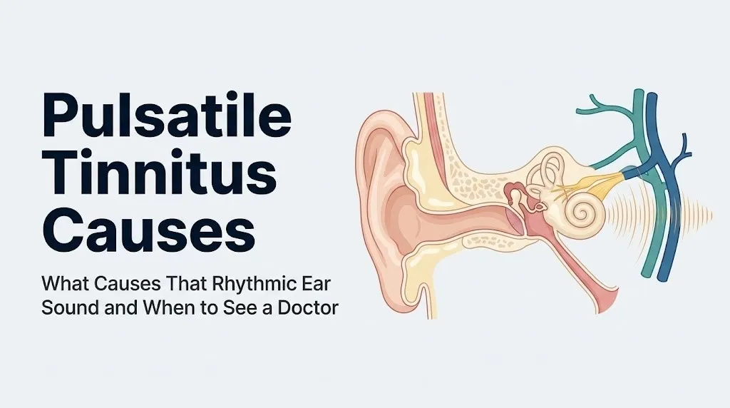
Pulsatile tinnitus is a rhythmic sound in one or both ears that beats in synchrony with the heartbeat, and it reaches the auditory system through a physically real pathway: mechanical pressure waves generated by abnormal vascular or pressure dynamics travel through bone, fluid, or tissue to reach the organ of Corti in the cochlea.
Understanding why you hear a sound that matches your pulse requires understanding the anatomy between your blood vessels and your hearing apparatus. The cochlea is a fluid-filled, snail-shaped structure in the petrous portion of the temporal bone, surrounded by dense bone on most surfaces. Normally, the only sounds that enter the cochlea are those transmitted through the ossicular chain (hammer, anvil, and stirrup) from the tympanic membrane, or through bone conduction from external sources.
A blood vessel generating turbulent or amplified flow does not normally produce sound loud enough to reach the cochlea above ambient noise. Pulsatile tinnitus occurs when one of three abnormal situations arises. First, a vascular structure has become unusually close to or has lost its normal bony separation from the cochlea or middle ear. Second, a structural defect (like a hole in the bone covering the sigmoid sinus or superior semicircular canal) creates an abnormal acoustic window. Third, intracranial pressure elevates to the point where it mechanically loads the cochlear aqueduct (the tiny bony channel connecting the perilymphatic space of the cochlea to the subarachnoid cerebrospinal fluid space), altering the pressure dynamics within the cochlea itself.
In all three scenarios, mechanical energy at heart-rate frequency reaches the basilar membrane of the organ of Corti. Hair cells on the basilar membrane are displaced by these pressure waves, generating neural signals that travel along the cochlear branch of cranial nerve VIII to the primary auditory cortex in Heschl’s gyrus of the superior temporal plane. The brain registers this signal as sound: specifically, a rhythmic whooshing, thumping, or pulsing that beats in time with the heart.
People with sensorineural hearing loss may actually perceive pulsatile tinnitus more clearly at certain frequencies because the damaged hair cells in those frequency ranges have less competing background signal to mask the vascular sound.
Objective vs Subjective Pulsatile Tinnitus: Why the Distinction Matters
Objective pulsatile tinnitus is a sound that can be heard by a clinician using a stethoscope or Doppler probe placed near the patient’s ear, neck, or skull, while subjective pulsatile tinnitus is perceived only by the patient, and this distinction carries direct implications for which diagnostic pathway is most likely to identify the underlying cause.
Objective pulsatile tinnitus, by definition, involves an acoustic event with enough physical energy to be detected externally. The presence of an audible bruit (vascular murmur) heard on auscultation over the mastoid, neck, or eye confirms that turbulent blood flow is occurring near enough to the ear to generate externally detectable sound. Conditions most likely to produce objective pulsatile tinnitus include dural arteriovenous fistulas, carotid cavernous fistulas, glomus tumors, and high-output states from severe anemia or thyrotoxicosis.
Subjective pulsatile tinnitus, where no bruit is audible, does not mean the cause is less serious. Sigmoid sinus wall anomalies, superior semicircular canal dehiscence, and idiopathic intracranial hypertension all produce subjective pulsatile tinnitus because their mechanism involves transmitting vascular sounds that are already present in normal tissue at higher amplitude to the cochlea, rather than generating an abnormally loud new sound source. These causes require specific imaging rather than physical examination to identify.
The American Academy of Otolaryngology-Head and Neck Surgery recommends that any clinician evaluating pulsatile tinnitus begin with careful auscultation over the mastoid, posterior auricular region, neck, and periorbital area, because the presence or absence of an audible bruit substantially narrows the differential diagnosis and guides the sequence of imaging studies ordered.
Women make up the majority of patients referred for pulsatile tinnitus evaluation in tertiary clinical settings, which reflects the higher prevalence of IIH and sigmoid sinus wall anomalies in females. Men with pulsatile tinnitus, while less frequently affected, should be evaluated with particular attention to atherosclerotic vascular causes, which are more prevalent in older males.
Key Takeaway: Whether your doctor can hear the sound too (objective pulsatile tinnitus) or only you can (subjective) is one of the most clinically useful pieces of information for guiding which imaging studies are ordered first.
High Blood Pressure and Atherosclerosis as Causes of Pulsatile Tinnitus
High blood pressure and atherosclerosis cause pulsatile tinnitus by increasing the mechanical energy of blood flow and creating irregularities in vessel wall geometry that generate turbulence, producing pressure fluctuations at heart-rate frequency that propagate through surrounding tissue to the cochlea.
In a healthy artery with smooth walls and normal blood pressure, blood flows in a laminar pattern: orderly parallel streams with no lateral mixing. Laminar flow generates minimal mechanical energy transfer to surrounding tissue. When blood pressure rises, flow velocity increases, and when atherosclerotic plaques roughen the inner surface of vessel walls, the smooth laminar pattern breaks into chaotic, swirling turbulence. Turbulent flow is described mathematically by the Reynold’s number, and once turbulence is established, the vessel wall vibrates at the frequency of blood pulsations, transmitting mechanical energy to surrounding bone and soft tissue.
The vessels most likely to transmit turbulence-related sound to the cochlea are those running through or near the temporal bone: the internal carotid artery as it passes through the carotid canal adjacent to the middle ear, and the branches of the external carotid artery, which supply the dura and skull base structures immediately surrounding the cochlea.
The American Heart Association identifies hypertension as affecting approximately 47% of adults in the United States, making it the single most prevalent systemic condition associated with pulsatile tinnitus. Lowering blood pressure to guideline targets frequently reduces or eliminates pulsatile tinnitus in hypertension-related cases, which is one of the few causes that may respond to medical management without procedural intervention.
People over 60 with poorly controlled hypertension and pulsatile tinnitus should have their blood pressure evaluated by their primary care physician before pursuing advanced vascular imaging, because achieving blood pressure control is both diagnostically informative and therapeutically meaningful.
Carotid Artery Stenosis and Pulsatile Tinnitus
Carotid artery stenosis causes pulsatile tinnitus by generating a high-velocity turbulent jet at the narrowed segment of the carotid artery bifurcation or internal carotid artery, producing a pressure wave that propagates through the skull base and temporal bone to reach the cochlea as a synchronous pulsatile sound.
The mechanism is anatomically specific. The carotid bifurcation (where the common carotid artery divides into the internal and external carotid arteries) sits in the neck at approximately the level of the thyroid cartilage, just 3 to 4 centimeters from the temporal bone and cochlea. When an atherosclerotic plaque narrows the lumen of the carotid artery at or near the bifurcation, blood accelerates through the narrowing and emerges as a turbulent jet on the downstream side. This jet creates pressure oscillations at cardiac cycle frequency that conduct through the soft tissues and skull base directly to the structures of the inner ear.
Think of this like pressing your thumb over the end of a garden hose. The water that was flowing smoothly before suddenly becomes a turbulent, hissing jet. In your carotid artery, that turbulent jet is happening centimeters from your cochlea, which acts as an extraordinarily sensitive pressure-detection device.
A carotid bruit, audible to a clinician using a stethoscope placed over the neck, is a clinical finding that raises the probability of carotid stenosis as the source of pulsatile tinnitus. However, the Mayo Clinic notes that not all carotid bruits indicate stenosis severe enough to cause tinnitus, and not all significant stenoses produce an audible bruit. Duplex carotid Doppler ultrasound is the standard first-line imaging for carotid artery assessment and can quantify the degree of stenosis and flow velocity at the bifurcation.
Tobacco smokers with pulsatile tinnitus warrant expedited carotid evaluation because smoking is the most modifiable independent risk factor for carotid atherosclerosis and accelerates plaque progression.
Dural Arteriovenous Fistula and AVM as Causes of Pulsatile Tinnitus
A dural arteriovenous fistula (dAVF) and an arteriovenous malformation (AVM) cause pulsatile tinnitus by creating an abnormal direct connection between high-pressure arterial blood and low-pressure venous structures adjacent to the cochlea, forcing arterial-pressure pulsatile flow through venous structures that lack the arterial wall architecture to dampen those pressure waves before they reach the inner ear.
In normal circulation, the arterial-to-venous pressure gradient is dissipated across the capillary bed. Capillaries are too small and too numerous to transmit arterial pressure pulsations directly to veins. A dAVF short-circuits this system entirely. Arterial blood flows directly into a dural venous sinus at arterial pressure, producing a high-velocity turbulent stream within the sinus that generates a loud, rhythmically pulsing bruit. When the fistula involves the transverse sinus or sigmoid sinus adjacent to the temporal bone, this turbulent flow is transmitted directly to the cochlea.
The AJNR: American Journal of Neuroradiology has published extensive series establishing dAVF as one of the most reliably identified causes of objective pulsatile tinnitus on advanced imaging, with the pulsatile tinnitus resolving in the majority of patients following endovascular embolization or surgical obliteration of the fistula. The clinical significance of identifying a dAVF extends beyond tinnitus management: high-grade dAVFs (Cognard classification types III-V) carry a risk of intracranial hemorrhage or progressive neurological deficit if untreated.
Digital subtraction angiography (DSA) remains the gold standard for dAVF diagnosis, though MRI with gadolinium and MR angiography can suggest the diagnosis before DSA is performed. Any person with pulsatile tinnitus, a subjective or objective bruit, and negative initial MRI should discuss catheter angiography with their managing neuroradiologist or neurovascular surgeon.
People with a history of head trauma, prior craniotomy, or venous sinus thrombosis have elevated risk for dAVF formation, as these events can trigger abnormal arteriovenous connections during the healing process.
Sigmoid Sinus Wall Anomalies and Pulsatile Tinnitus
Sigmoid sinus wall anomalies (SSWA), including sigmoid sinus diverticulum and sigmoid sinus dehiscence, cause pulsatile tinnitus by eliminating the normal bony separation between the turbulently flowing blood in the sigmoid venous sinus and the adjacent cochlea or middle ear, allowing venous pressure pulsations to be transmitted directly to inner ear structures.
The sigmoid sinus is a large venous channel that runs through the posterior cranial fossa immediately adjacent to the mastoid portion of the temporal bone. Normally, a layer of bone several millimeters thick separates the sigmoid sinus from the mastoid air cells and the structures of the inner ear. In sigmoid sinus dehiscence, this bony wall is absent over a segment of the sinus, leaving only a thin membrane between pulsating venous blood and the air cells or inner ear. In sigmoid sinus diverticulum, an outpouching of the sinus wall extends toward or through the temporal bone, placing turbulent, pulsating venous flow in even closer proximity to the cochlea.
Research published in Otology and Neurotology has established SSWA as one of the most frequently identified structural causes of unilateral pulsatile tinnitus in the era of high-resolution computed tomography (CT) temporal bone imaging, accounting for a substantial proportion of previously “idiopathic” cases when appropriate CT protocols are used. The resolution required to identify SSWA is higher than that of standard brain CT, and many cases are missed when only routine head CT or standard MRI is performed.
Diagnosis requires dedicated high-resolution CT of the temporal bone using thin-slice protocols, which is distinctly different from standard brain CT and must be specifically requested. SSWA is more frequently identified in middle-aged women, and many affected individuals have no other identifiable vascular risk factors.
Treatment approaches, which may include sigmoid sinus wall resurfacing or other otologic procedures, are managed by a board-certified otolaryngologist with subspecialty training in neurotology.
Key Takeaway: Sigmoid sinus wall anomaly is one of the most commonly missed causes of pulsatile tinnitus because it requires dedicated high-resolution CT temporal bone imaging, not standard brain CT or MRI, to identify.
Superior Semicircular Canal Dehiscence and Pulsatile Tinnitus
Superior semicircular canal dehiscence (SSCD) causes pulsatile tinnitus by creating a pathological third mobile window into the inner ear at the superior semicircular canal, allowing vascular pressure pulsations that would normally be reflected away from the cochlea to enter the inner ear fluid spaces and be registered as sound.
The normal inner ear has two mobile membrane windows connecting it to the middle ear: the oval window (where the stapes footplate transmits sound) and the round window (which allows pressure equalization). These two windows account for all normal mechanical input to the cochlea. In SSCD, a portion of the thin bone covering the superior semicircular canal is absent, creating a third window directly into the membranous labyrinth. This abnormal opening does not create a new sound source. Instead, it reduces the acoustic impedance of the inner ear in a way that allows vascular pressure pulsations already present in the surrounding bone and tissue to more efficiently drive fluid motion within the cochlea.
Research from the team of Lloyd Minor at Johns Hopkins, published in Annals of Otology, Rhinology and Laryngology, originally described SSCD and has been extensively replicated and expanded. The audiological signature of SSCD includes a specific low-frequency conductive hearing component on audiogram alongside the pulsatile tinnitus, and the condition can also produce autophony (hearing one’s own voice or footsteps amplified), sound-induced vertigo (Tullio phenomenon), and pressure-induced vertigo.
Diagnosis is made with high-resolution CT of the temporal bone and confirmed with audiological testing including cervical and ocular vestibular evoked myogenic potentials (cVEMP and oVEMP). SSCD is bilateral in approximately 25% of cases, according to research published in Otology and Neurotology.
Women appear to be more frequently affected by SSCD-related pulsatile tinnitus, though the anatomical prevalence of dehiscence may not differ substantially by sex, and the reason for the clinical presentation difference is not yet established.
Idiopathic Intracranial Hypertension as a Cause of Pulsatile Tinnitus
Idiopathic intracranial hypertension (IIH), also called pseudotumor cerebri, causes pulsatile tinnitus by elevating cerebrospinal fluid pressure to the point where it is transmitted through the cochlear aqueduct into the perilymphatic space of the cochlea, mechanically loading the cochlear fluid system in a way that amplifies normally inaudible vascular pressure pulsations into perceptible sound.
IIH is defined by elevated opening pressure on lumbar puncture (greater than 250 mmH2O in adults, or greater than 200 mmH2O in children, according to the Mayo Clinic) in the absence of an identifiable structural cause. The elevated CSF pressure is transmitted throughout the intracranial compartment, including to the cochlear aqueduct and the perilymphatic space. This pressure loading alters the mechanical properties of the cochlea, effectively reducing the acoustic impedance threshold at which vascular pulsations become perceptible as sound.
Pulsatile tinnitus is one of the most consistent early symptoms of IIH, often appearing before or alongside the other classical features: headache (typically positional and worse with Valsalva maneuver), visual changes (transient visual obscurations, blurring, or double vision), and pulsatile tinnitus that characteristically improves when the jugular vein is compressed on the affected side (a clinical test performed by an examining physician).
IIH overwhelmingly affects women of reproductive age with elevated body mass index. The National Institute of Neurological Disorders and Stroke (NINDS/NIH) notes that IIH occurs at a rate approximately 15 times higher in obese women of childbearing age than in the general population. Oral contraceptive use and certain medications including tetracycline antibiotics, vitamin A derivatives (retinoids), and long-term corticosteroid withdrawal are recognized risk factors.
The emergency threshold within IIH is papilledema with progressive visual field loss, specifically inferior nasal step or arcuate field defects on formal visual field testing, because untreated elevated CSF pressure can cause permanent vision loss through optic nerve compression. Any person with pulsatile tinnitus alongside headache and visual disturbance should be evaluated urgently, with fundoscopic examination for papilledema, by a primary care physician, neurologist, or neuro-ophthalmologist the same day.
Cerebral Venous Sinus Thrombosis and Pulsatile Tinnitus
Cerebral venous sinus thrombosis (CVST) causes pulsatile tinnitus by obstructing the normal venous outflow from the intracranial compartment, producing downstream venous hypertension and increased intracranial pressure that loads the perilymphatic spaces of both cochleae, typically producing bilateral pulsatile tinnitus alongside other signs of intracranial pressure elevation.
The intracranial venous drainage system relies on patent venous sinuses (primarily the superior sagittal sinus, transverse sinuses, and sigmoid sinuses) to return blood from the brain to the internal jugular veins. When thrombosis (blood clot formation) obstructs one or more of these sinuses, venous outflow resistance increases, raising intracranial venous pressure and consequently intracranial CSF pressure. This pressure elevation is transmitted to both cochleae simultaneously via the cochlear aqueducts, which explains why CVST-associated pulsatile tinnitus is frequently bilateral, distinguishing it from most structural causes that produce unilateral symptoms.
CVST most frequently affects women, particularly those taking combined oral contraceptives, those in the postpartum period, or those with inherited or acquired thrombophilias (Factor V Leiden mutation, prothrombin gene mutation, antiphospholipid antibody syndrome). The Journal of the American Medical Association has published data establishing oral contraceptive use as the most prevalent modifiable risk factor for CVST in reproductive-age women.
CVST is a potentially life-threatening emergency. Bilateral pulsatile tinnitus with new severe headache, visual changes, nausea, vomiting, or any focal neurological deficit in a woman of reproductive age on oral contraceptives requires immediate emergency department evaluation. The diagnosis is made with MRI of the brain with MR venography, which shows absent flow signal in the affected sinus. Treatment with anticoagulation is typically initiated immediately upon diagnosis.
Key Takeaway: Bilateral pulsatile tinnitus with severe headache in a reproductive-age woman on oral contraceptives is a pattern requiring same-day emergency evaluation because cerebral venous sinus thrombosis, a treatable but potentially fatal condition, fits this presentation exactly.
Glomus Tumors and Vascular Neoplasms Causing Pulsatile Tinnitus
Glomus tumors (also called paragangliomas of the temporal bone) cause pulsatile tinnitus by growing as highly vascular neoplasms within the middle ear cavity (glomus tympanicum) or the jugular fossa (glomus jugulare), where their dense vascular networks generate pulsatile blood flow that is directly transmitted to the middle ear ossicular chain and cochlea.
Glomus tumors arise from neural crest-derived paraganglia normally present in the adventitia of the jugular bulb and along Jacobson’s nerve (the tympanic branch of cranial nerve IX) in the middle ear. The tumor tissue is supplied by branches of the ascending pharyngeal artery and other external carotid artery branches, creating a tumor mass that is extraordinarily well-vascularized. Blood flowing through the tumor’s abnormal vascular architecture generates turbulent pulsatile flow directly within the temporal bone, producing a loud, objective pulsatile tinnitus audible on auscultation over the mastoid and sometimes audible to people in the same quiet room as the patient.
A classic physical examination finding in glomus tympanicum is a reddish-blue pulsating mass visible through the intact tympanic membrane on otoscopic examination, often described as a “rising sun” appearance when the tumor reaches a visible size. This finding, when present, is one of the most specific clinical signs in all of otolaryngology.
The Cleveland Clinic identifies glomus tumors as uncommon but consistently identified causes of pulsatile tinnitus, with glomus jugulare tumors additionally capable of causing lower cranial nerve deficits (hoarseness, dysphagia, and shoulder weakness from involvement of cranial nerves IX, X, XI at the jugular foramen). MRI with gadolinium demonstrates the highly vascular nature of these tumors with characteristic “salt and pepper” signal pattern on T2-weighted sequences.
Women are affected by glomus tumors approximately three to six times more often than men, according to data reviewed in the Laryngoscope. The reason for this sex predilection is not fully established but may involve hormonal influences on paraganglioma cell proliferation.
Systemic Causes of Pulsatile Tinnitus: Anemia, Thyroid Disease, and Pregnancy
Systemic causes of pulsatile tinnitus produce the symptom by increasing cardiac output, reducing blood viscosity, or both, which together cause blood to flow faster and more turbulently through the vessels near the cochlea, generating pressure oscillations that the inner ear registers as a pulsatile sound.
Iron-deficiency anemia is one of the most clinically underappreciated causes of pulsatile tinnitus. When red blood cell count and hemoglobin concentration fall, the blood becomes less viscous. Less viscous blood flows at higher velocity through the same vessel diameter, dramatically increasing the likelihood of turbulent rather than laminar flow (increasing the Reynold’s number above the turbulence threshold). Simultaneously, the heart increases cardiac output to compensate for the reduced oxygen-carrying capacity of each unit of blood, further increasing flow velocity through the great vessels and their branches near the temporal bone.
Hyperthyroidism (thyrotoxicosis) causes pulsatile tinnitus through a similar high-output mechanism. Excess thyroid hormone (T3 and T4, produced by the thyroid gland under excess stimulation) increases heart rate, stroke volume, and overall cardiac output, producing hyperdynamic circulation. Blood moves faster through all vessels, increasing turbulence at any anatomical irregularity near the cochlea. The pulsatile tinnitus of hyperthyroidism typically resolves when thyroid hormone levels are normalized.
Pregnancy elevates plasma volume by approximately 40 to 50% above pre-pregnancy baseline by the third trimester, as noted by the American College of Obstetricians and Gynecologists. This volume expansion increases cardiac output substantially. Combined with physiological anemia of pregnancy (dilutional anemia from plasma expansion exceeding red cell mass expansion) and the hormonal effects of estrogen and progesterone on vascular tone, pregnancy creates a high-output, low-viscosity circulatory state that generates turbulent flow in vessels near the cochlea. Pulsatile tinnitus during pregnancy is worth reporting to an obstetrician-gynecologist because it may also be the presenting symptom of IIH, which has elevated risk during pregnancy, rather than a purely physiological finding.
Unilateral vs Bilateral Pulsatile Tinnitus: What the Pattern Tells Your Doctor
Unilateral pulsatile tinnitus (sound in one ear only) suggests a structural or localized vascular cause on that side, while bilateral pulsatile tinnitus (sound in both ears) suggests a systemic, elevated-pressure, or bilateral venous cause, and this distinction is one of the most clinically informative features of a patient’s history.
The anatomical logic is straightforward. Structural causes, including sigmoid sinus wall anomalies, superior semicircular canal dehiscence, carotid artery stenosis at a bifurcation, and glomus tumors, are localized to one side of the skull. The pulsatile sound they generate reaches the cochlea on that side because the anomaly is ipsilateral to the ear hearing the sound. A unilateral pulsatile tinnitus is therefore a prompt to image both the affected temporal bone and the ipsilateral vascular structures.
Bilateral pulsatile tinnitus has a shorter differential diagnosis. Conditions that elevate intracranial pressure symmetrically (IIH, CVST, severe hypertension) load both cochlear aqueducts simultaneously, producing bilateral symptoms. High-output systemic states (severe anemia, thyrotoxicosis, pregnancy) increase turbulence throughout the vascular system, producing bilateral sound. Bilateral pulsatile tinnitus therefore directs clinical attention toward systemic and intracranial pressure causes rather than localized structural causes.
The NIDCD recommends that the laterality of pulsatile tinnitus be precisely documented in the clinical history, including whether the sound is perceived differently in each ear, whether it is heard equally bilaterally, and whether changing head position or neck position alters the perceived sound, as these features collectively guide the imaging sequence.
People who cannot reliably determine whether their pulsatile tinnitus is unilateral or bilateral should press gently on the tragus of each ear in a quiet room to reduce external sound, then assess whether the internal pulsing is equally present on both sides. This simple self-assessment provides useful clinical information to report at their appointment.
How Doctors Diagnose the Cause of Pulsatile Tinnitus
The diagnostic evaluation for pulsatile tinnitus follows a systematic sequence that begins with clinical history and physical examination, advances to targeted imaging based on clinical findings, and may culminate in catheter-based vascular imaging when non-invasive studies are inconclusive.
To prepare for a productive evaluation with a board-certified otolaryngologist or primary care physician:
- Document the character of the sound precisely: rhythmic (matching heartbeat) or not, pulsing or whooshing, constant or intermittent, position-dependent (worse lying down, worse turning head), affected by Valsalva maneuver or gentle jugular vein compression.
- Establish laterality clearly: which ear, both ears equally, or predominantly one side.
- Identify onset timing: sudden onset vs. gradual, any preceding event (head injury, illness, new medication, pregnancy).
- List all medications currently being taken, including oral contraceptives, tetracyclines, retinoids, blood pressure medications, and NSAIDs, as these can cause or mask relevant conditions.
- Note associated symptoms: headache (especially positional), visual changes (blurring, transient blackouts of vision, double vision), hearing changes, vertigo, ear fullness, facial weakness, or voice changes.
The diagnostic imaging sequence a clinician typically follows:
- MRI of the brain and internal auditory canals with gadolinium: Screens for intracranial pathology, venous sinus abnormality, and acoustic neuroma
- MR angiography and MR venography: Evaluates arterial and venous structures for AVMs, dAVFs, CVST, and venous stenosis
- High-resolution CT of the temporal bone (thin-slice protocol): Required to identify sigmoid sinus wall anomalies and SSCD, which are invisible on MRI
- Carotid Doppler ultrasound: First-line evaluation for carotid artery stenosis
- Digital subtraction angiography (DSA): Gold standard for dAVF when non-invasive imaging is equivocal
- Lumbar puncture with opening pressure measurement: Definitive for IIH when clinical suspicion is high
Key Takeaway: High-resolution CT of the temporal bone is a distinct, specific imaging study that must be explicitly requested to identify sigmoid sinus wall anomalies and SSCD: standard brain CT and routine MRI will miss both of these diagnoses.
Emergency Symptoms: When to Call 911 or Go to the ER
Certain presentations associated with pulsatile tinnitus require immediate emergency evaluation. Do not wait to see if these resolve on their own.
Call 911 or go to the nearest emergency room immediately if you experience:
- Sudden onset of severe headache unlike any previous headache (“thunderclap headache”), which can indicate subarachnoid hemorrhage from a ruptured AVM or aneurysm adjacent to an auditory structure
- Sudden loss of vision in one or both eyes, or sudden onset of double vision, alongside pulsatile tinnitus, which can indicate elevated intracranial pressure from CVST or IIH with optic nerve compromise
- Any facial weakness, arm or leg weakness, slurred speech, or sudden difficulty speaking alongside pulsatile tinnitus, which indicates possible stroke or dural arteriovenous fistula with intracranial hemorrhage
- Sudden severe vertigo with complete loss of hearing in one ear alongside pulsatile tinnitus, which can indicate labyrinthine artery occlusion or superior semicircular canal dehiscence with acute vestibular crisis
- Pulsatile tinnitus in a reproductive-age woman on oral contraceptives with new severe headache and any visual change, indicating possible cerebral venous sinus thrombosis
- New pulsatile tinnitus following head trauma, which can indicate traumatic dural arteriovenous fistula or carotid cavernous fistula requiring emergency angiography
These presentations can indicate ruptured arteriovenous malformation, subarachnoid hemorrhage, cerebral venous sinus thrombosis, or acute intracranial hypertension with optic nerve compromise, all of which require emergency medical assessment, not a scheduled appointment.
Frequently Asked Questions About Pulsatile Tinnitus Causes
Is pulsatile tinnitus serious or can it go away on its own?
Pulsatile tinnitus is more clinically serious than ordinary tinnitus because it usually has an identifiable structural or vascular cause that warrants investigation, and some causes are associated with genuine neurological risk.
Pulsatile tinnitus caused by reversible systemic conditions (anemia, hyperthyroidism, hypertension, pregnancy) may resolve when the underlying condition is treated, but this can only be determined after a physician evaluates the cause.
Any new pulsatile tinnitus should be evaluated by a primary care physician initially and, if persistent beyond 2 to 4 weeks, referred to a board-certified otolaryngologist for further workup.
What is the most common cause of pulsatile tinnitus?
The most frequently identified causes of pulsatile tinnitus in clinical series are hypertension, atherosclerotic vascular disease, and sigmoid sinus wall anomalies, though the relative frequency depends significantly on the population studied and the imaging technology available.
A 2014 study in Otology and Neurotology found that sigmoid sinus wall anomalies were identified in a substantial proportion of previously “unexplained” pulsatile tinnitus cases when dedicated high-resolution CT temporal bone protocols were used.
The cause identified most often in any individual depends on their age, sex, cardiovascular risk factors, and whether advanced temporal bone imaging is part of their workup.
Should I go to the ER for pulsatile tinnitus?
Pulsatile tinnitus alone without other symptoms does not typically require emergency room evaluation, but it does require timely clinical evaluation by a physician within days to a few weeks.
Call 911 or go to the emergency room immediately if pulsatile tinnitus is accompanied by sudden severe headache, any visual change, sudden hearing loss, facial weakness, limb weakness, or slurred speech.
A reproductive-age woman on oral contraceptives who develops new bilateral pulsatile tinnitus with headache should go to the emergency room the same day to rule out cerebral venous sinus thrombosis.
Can high blood pressure cause a whooshing sound in the ear?
Yes, high blood pressure causes pulsatile tinnitus by increasing blood flow velocity to the point where turbulent flow develops in vessels near the cochlea, generating pressure waves that are transmitted to the inner ear as a rhythmic whooshing sound.
The internal carotid artery, which runs through the carotid canal immediately adjacent to the middle ear, is the most clinically relevant vessel through which hypertension-related turbulent flow reaches the cochlea.
Treating hypertension to guideline targets frequently reduces pulsatile tinnitus in people whose primary cause is elevated blood pressure, though achieving this should be confirmed by a primary care physician through blood pressure monitoring and clinical follow-up.
What type of doctor should I see for pulsatile tinnitus?
Start with your primary care physician for an initial evaluation of blood pressure, thyroid function, complete blood count, and basic cardiovascular assessment.
If the cause is not identified through this initial workup, your primary care physician should refer you to a board-certified otolaryngologist (ENT surgeon) with experience in otology, who will direct further imaging and evaluate for structural causes.
A neuroradiologist or neurovascular surgeon will be involved if imaging suggests a dural arteriovenous fistula, AVM, or other vascular malformation requiring catheter angiography or procedural treatment.
What does it mean if my pulsatile tinnitus is only in one ear?
Unilateral pulsatile tinnitus (in one ear only) strongly suggests a localized structural or vascular cause on that side, such as a sigmoid sinus wall anomaly, superior semicircular canal dehiscence, carotid artery stenosis, dural arteriovenous fistula, or glomus tumor.
Bilateral pulsatile tinnitus is more consistent with systemic causes (anemia, thyroid disease, hypertension) or intracranial pressure conditions (IIH, CVST) that affect both inner ears simultaneously.
Tell your physician precisely which ear you hear the sound in, whether it is ever bilateral, and whether changing head position or pressing gently on the neck vessels alters the sound, as this information directly guides which imaging studies are ordered first.
Closing
Pulsatile tinnitus is not something to attribute to stress and ignore for months. It is a symptom with a source, and that source is identifiable in the majority of cases when the right clinical evaluation and imaging are applied. The key is getting to the right physician with the right information.
Start with your primary care physician. Bring notes on which ear you hear it in, whether it is constant or intermittent, whether it changes with position, and any associated symptoms. Ask specifically for blood pressure evaluation, thyroid function testing, and a complete blood count as initial screening, and ask for referral to an otolaryngologist with otology experience if initial testing does not identify a cause.
If you develop severe headache, visual changes, or any neurological symptoms alongside the pulsatile tinnitus, do not wait for an appointment. You now know enough about this symptom’s causes to recognize when you need emergency care, and that knowledge is the most practically useful thing you can take from this article.