Eye Swelling Causes: What’s Making Your Eye Swell Up
Eye swelling causes range from completely benign conditions like allergies and styes to genuine medical emergencies like orbital cellulitis and cavernous sinus thrombosis, and the single most important factor in determining which category you are dealing with is a piece of anatomy most people have never heard of: the orbital septum. Whether the swelling is in front of or behind that fibrous barrier determines whether you need a warm compress or an emergency room.
According to the National Eye Institute (NEI), the periorbital tissues surrounding the eye contain some of the loosest connective tissue in the human body, which is precisely why the eyelids swell so visibly from even minor inflammatory, infectious, or fluid-based triggers. A small amount of extra fluid in the eyelid tissue produces dramatic swelling that looks alarming even when the underlying cause is self-limiting.
This article covers the specific physiological mechanism behind every major eye swelling cause, from the IgE-mediated mast cell release in allergic edema to the TSH receptor antibody mechanism in thyroid eye disease. You’ll learn the clinical patterns that distinguish urgent from non-urgent presentations, which symptoms require a 911 call, and the exact type of physician to contact for each specific clinical picture.
Eye Swelling Causes: The Anatomy That Determines Urgency
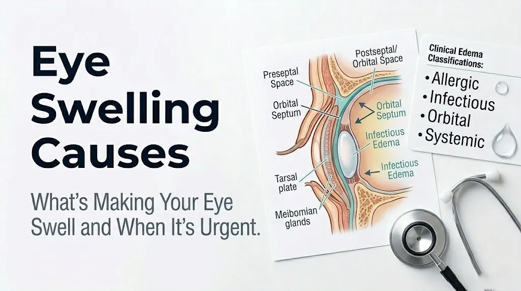
The anatomical structure that determines whether any eye swelling cause is benign or potentially sight-threatening is the orbital septum, a thin but clinically decisive sheet of fibrous connective tissue that runs from the orbital rim (the bony edge of the eye socket) to the tarsal plates of the upper and lower eyelids.
Think of the orbital septum as a locked gate. The eyelid side of the gate is the preseptal space: conditions here are confined to the eyelid skin, the glands along the eyelid margin, and the subcutaneous tissue of the periorbital area. The socket side is the postseptal (orbital) space: conditions here involve the globe (eyeball), the optic nerve, the extraocular muscles, the orbital fat, and the superior and inferior ophthalmic veins that drain toward the cavernous sinus inside the skull. When infection breaches the orbital septum and enters the postseptal space, it has the potential to reach the optic nerve, compress it, and destroy vision, or to track along the ophthalmic veins into the cavernous sinus and produce a life-threatening intracranial infection.
Every cause of eye swelling in this article sits on one side of that gate or the other. Most causes are preseptal and self-limited. A small number are postseptal and demand emergency evaluation.
Anatomical framework for evaluating eye swelling:
| Space | Location | Key Contents | Clinical Significance |
|---|---|---|---|
| Preseptal (periorbital) | Anterior to orbital septum | Eyelid skin, subcutaneous fat, Meibomian glands, orbicularis muscle | Conditions confined here are generally not sight-threatening |
| Postseptal (orbital) | Posterior to orbital septum | Globe, optic nerve, extraocular muscles, orbital fat, ophthalmic veins | Conditions here can threaten vision, eye movement, and intracranial structures |
The tarsal plate (the firm fibrous plate that gives each eyelid its shape, housing the Meibomian glands) and the orbicularis oculi muscle (the sphincter muscle that closes the eyelid) are preseptal structures. The levator palpebrae superioris (the muscle that raises the upper eyelid) has its anterior attachment in the preseptal space but its origin in the postseptal orbital apex, which is why orbital disease affecting the posterior orbit can produce ptosis (drooping of the eyelid).
Children under 5 are at higher risk for preseptal-to-orbital disease progression because their orbital septum is thinner and less developed than in adults, providing a less reliable barrier against spread from eyelid or sinus infection.
Preseptal Versus Orbital Eye Swelling: Why the Distinction Matters
The distinction between preseptal and orbital eye swelling is the most clinically consequential piece of knowledge in evaluating eye swelling causes, because it determines the difference between home management and a hospital admission.
Preseptal cellulitis (also called periorbital cellulitis) involves infection of the eyelid skin and subcutaneous tissue anterior to the orbital septum. The eye itself, the extraocular muscles, and the optic nerve are not involved. The person can move their eye normally in all directions without pain, their vision is normal, and the globe is not displaced forward. Preseptal cellulitis is treated with oral antibiotics under physician supervision in most cases and does not require hospitalization in immunocompetent adults with mild presentations.
Orbital cellulitis (postseptal cellulitis) has breached the orbital septum. The infection is now inside the orbit, around the globe and optic nerve. Clinical features that distinguish orbital from preseptal cellulitis include:
- Proptosis: the eyeball is displaced forward because the infected, edematous orbital fat is pushing it out
- Ophthalmoplegia or restricted eye movement: the extraocular muscles are swollen, inflamed, or compressed
- Pain on eye movement: the inflamed orbital structures produce pain when the extraocular muscles contract
- Diplopia (double vision): misalignment of the eyes from restricted muscle movement
- Decreased visual acuity: optic nerve compression or ischemia from elevated orbital pressure
- Chemosis: edema of the bulbar conjunctiva, appearing as a gelatinous swelling of the white of the eye
According to the American Academy of Ophthalmology (AAO), orbital cellulitis most commonly originates from contiguous spread from the paranasal sinuses, particularly the ethmoid sinuses in children, because the paper-thin bone of the medial orbital wall (the lamina papyracea) that separates the ethmoid sinus from the orbit provides minimal bacterial resistance when infected.
The clinical staging system most frequently used in orbital cellulitis evaluation (Chandler classification, modified) ranks infections from preseptal edema through subperiosteal abscess, orbital abscess, and cavernous sinus thrombosis, each stage requiring escalating urgency of intervention.
Postmenopausal women and older adults may present with blunted fever responses even in severe orbital cellulitis, which can mask the severity. Any older adult with progressive unilateral eye swelling and systemic illness should be evaluated by an emergency medicine physician with low threshold for CT orbit imaging.
Allergies as a Cause of Eye Swelling
Allergic reactions are among the most frequent causes of bilateral eye swelling, producing periorbital edema through a well-characterized IgE-mediated (type I) hypersensitivity response in the eyelid tissue.
When a sensitized person encounters an allergen, airborne particles such as tree pollen, dust mite fecal proteins, pet dander (specifically cat Fel d 1 protein or dog Can f 1 protein), or mold spores land on the conjunctival surface and come into contact with mast cells in the eyelid skin and conjunctiva. Allergen molecules cross-link IgE antibodies bound to FcεRI receptors on the mast cell surface. This cross-linking triggers mast cell degranulation, releasing a cascade of preformed and newly synthesized mediators.
Histamine is released immediately on degranulation. It binds H1 receptors on eyelid arterioles, causing vasodilation and increased capillary permeability. Protein-rich plasma fluid leaks into the loose areolar connective tissue of the eyelid. Because periorbital connective tissue has minimal structural resistance compared to denser facial tissue, even a small amount of leaked fluid produces visible, dramatic swelling within minutes to hours.
According to the American Academy of Allergy Asthma and Immunology (AAAAI), allergic conjunctivitis affects an estimated 40% of the population at some point, with periorbital swelling (allergic periorbital edema) being one of its most visually prominent manifestations. Seasonal allergic conjunctivitis produces bilateral swelling tied to pollen seasons, while perennial allergic conjunctivitis from dust mites or pet allergens may produce year-round morning eyelid puffiness.
Key features that indicate allergic rather than infectious eye swelling:
- Bilateral rather than unilateral swelling
- Associated intense itching (histamine-mediated H1 receptor activation in conjunctival sensory nerves)
- Clear or watery eye discharge rather than purulent (yellow or green) discharge
- Associated nasal symptoms (sneezing, runny nose, congestion)
- Rapid onset tied to allergen exposure, often resolving within hours of removal
People with atopic dermatitis (eczema) are at substantially higher risk for allergic periorbital reactions because their eyelid skin barrier is already disrupted, allowing greater allergen penetration, and their mast cells are more sensitized to a broader range of allergens than those of non-atopic individuals.
Key Takeaway: Allergic eye swelling is almost always bilateral and itchy, with clear discharge and rapid onset tied to allergen exposure; any eye swelling that is unilateral, painful, or accompanied by purulent discharge is not typical of simple allergy and warrants professional evaluation.
Angioedema and Contact Dermatitis Causing Eye Swelling
Angioedema produces periorbital swelling through a mechanism distinct from standard allergic edema, and the distinction matters clinically because one type responds to antihistamines and the other does not.
Standard allergic angioedema is histamine-mediated: IgE-dependent mast cell activation releases histamine causing deep dermal and submucosal vascular leakage. The periorbital area is particularly susceptible because of its distensible loose connective tissue. This type responds to antihistamines and epinephrine when severe.
Hereditary angioedema (HAE) is fundamentally different. It is caused by deficiency or dysfunction of C1 esterase inhibitor (C1-INH), a serine protease inhibitor that normally controls activation of the complement pathway and the plasma kallikrein-kinin system. Without functional C1-INH, bradykinin accumulates in plasma and tissues, binding B2 receptors on endothelial cells, producing vascular permeability and deep tissue swelling. HAE does not respond to antihistamines or corticosteroids because it is bradykinin-mediated, not histamine-mediated. This is a critical diagnostic distinction: a person with recurrent unexplained periorbital swelling that does not respond to antihistamines should be evaluated for HAE by a board-certified allergist and immunologist with C4 complement level and C1-INH level and function testing.
Contact dermatitis of the eyelids produces swelling through a type IV delayed hypersensitivity reaction mediated by sensitized T-lymphocytes rather than IgE. Common eyelid allergens include:
- Nail polish ingredients (tosylamide formaldehyde resin), transferred from fingertips touching the eyelid
- Eye cosmetics (mascara preservatives, eyeliner pigments, eyeshadow fragrances)
- Nickel from metal eyeglass frames resting on the nose pad area
- Topical ophthalmic medications (neomycin, tobramycin, preservatives like benzalkonium chloride)
- Hair dye (paraphenylenediamine, PPD) transferred to the eyelids
The T-cell-mediated response typically appears 24 to 72 hours after exposure, producing red, swollen, eczematous, scaly eyelid skin that itches intensely but does not produce the watery conjunctival discharge of allergic conjunctivitis. Patch testing by a board-certified allergist and immunologist or contact dermatitis-trained dermatologist identifies the specific allergen.
Older adults who begin using new ophthalmic drops (particularly glaucoma medications containing preservatives) are at elevated risk for contact allergic blepharitis, as cumulative lifetime sensitization to preservatives increases with age and repeated topical exposure.
Stye and Hordeolum as Causes of Swollen Eyelid
A stye (hordeolum) causes eyelid swelling through a bacterial infection of one of the oil-secreting or sweat-secreting glands along the eyelid margin, producing a localized abscess with surrounding inflammatory edema.
Two anatomically distinct gland types produce two distinct types of hordeolum. An external hordeolum involves either the glands of Zeis (sebaceous glands associated with eyelash follicles) or the glands of Moll (modified apocrine sweat glands at the eyelid margin). An internal hordeolum involves the Meibomian glands (also called tarsal glands), which are the larger oil-secreting glands embedded within the tarsal plate. External hordeola are visible at the eyelash line; internal hordeola present as swelling deeper within the eyelid, sometimes visible as a red nodule through the conjunctival surface when the eyelid is everted.
The causative organism in the vast majority of hordeola is Staphylococcus aureus. S. aureus colonizes the eyelid margin and eyelash follicles in many people without producing disease. When a gland orifice becomes obstructed (by dried secretion, blepharitis-related scale, or dead skin cells), bacteria multiply in the obstructed duct, release toxins including protein A, coagulase, and alpha-toxin, and trigger localized innate immune activation with neutrophil recruitment, pus formation, and surrounding inflammatory edema.
According to the American Academy of Ophthalmology, most external hordeola drain spontaneously within 1 to 2 weeks with consistent warm compresses applied for 5 to 10 minutes, four times daily, which soften the obstructing material and promote drainage. Internal hordeola often require longer resolution times and may benefit from physician-prescribed topical or oral antibiotic therapy if they are large or associated with significant surrounding cellulitis.
Clinical features of hordeolum by type:
| Feature | External Hordeolum | Internal Hordeolum |
|---|---|---|
| Location | At eyelash line, eyelid margin | Within eyelid substance, tarsal plate |
| Visible pus | At eyelid margin, eyelash base | Visible on conjunctival surface when eyelid everted |
| Tenderness | Localized at one point on lid margin | Diffuse eyelid tenderness, deeper |
| Typical organism | Staphylococcus aureus | Staphylococcus aureus |
| Resolution timeline | 1-2 weeks with warm compress | 2-4 weeks; may require intervention |
People with diabetes mellitus develop hordeola more frequently and with greater severity because of impaired neutrophil chemotaxis reducing abscess containment efficiency. A stye that is enlarging rapidly, associated with spreading periorbital redness beyond the eyelid, or in a person with diabetes should be evaluated by a primary care physician or ophthalmologist within 24 to 48 hours rather than monitored at home.
Chalazion as a Cause of Swollen Eyelid
A chalazion causes eyelid swelling through a lipogranulomatous inflammatory reaction that is mechanistically distinct from the bacterial infection of a hordeolum, even though the two conditions are often confused by patients and sometimes by clinicians.
The Meibomian glands, approximately 25 to 30 per eyelid in the tarsal plate, secrete a lipid-rich mixture called meibum that contributes to the outer lipid layer of the tear film, preventing tear evaporation. When a Meibomian gland duct becomes obstructed (from thickened meibum due to Meibomian gland dysfunction, rosacea, or eyelid inflammation), the lipid secretion cannot drain normally and accumulates within the gland. Eventually, the gland wall ruptures, releasing the lipid material into the surrounding tarsal tissue.
The immune system encounters this extruded lipid as a foreign body. Macrophages attempt to engulf the lipid droplets, transforming into lipid-laden foam cells. CD4+ T-lymphocytes and multinucleated giant cells aggregate around the lipid material in a granulomatous pattern. This organized granulomatous inflammation produces the firm, rubbery, non-tender nodule palpable within the eyelid substance that characterizes a chalazion, with surrounding inflammatory edema making the eyelid appear swollen.
The critical clinical distinction from a hordeolum: a chalazion is typically non-tender (because it is a granulomatous, not acute bacterial, reaction), while a hordeolum is acutely tender and usually warmer. A chalazion may enlarge slowly over weeks, while a hordeolum typically forms within 24 to 48 hours.
According to the Mayo Clinic, chalazia resolve spontaneously in approximately 50% of cases within 2 to 8 weeks with warm compresses and gentle lid massage. Chalazia that persist beyond 4 to 6 weeks, or that are large enough to distort vision by pressing on the cornea (producing astigmatism), require evaluation and possible incision and curettage by a board-certified ophthalmologist.
Adults with rosacea are at substantially higher risk for recurrent chalazia because rosacea produces chronic Meibomian gland inflammation and abnormal meibum viscosity, creating the duct obstruction conditions that initiate chalazion formation repeatedly. Any adult with multiple recurrent chalazia in the same area of the same eyelid should be evaluated by an ophthalmologist to exclude the rare possibility of a sebaceous gland carcinoma, which can mimic a recurrent chalazion.
Key Takeaway: A chalazion is a non-tender, slowly developing lipogranuloma from a blocked Meibomian gland, while a hordeolum is an acutely tender bacterial abscess at the eyelid margin; they require different management and should not be self-treated identically, with any chalazion unresolved at 6 weeks and any hordeolum worsening beyond 2 days warranting ophthalmologist evaluation.
Blepharitis as a Cause of Chronic Eyelid Swelling
Blepharitis is chronic inflammation of the eyelid margins that produces recurrent or persistent eyelid swelling, redness, crusting, and irritation through a combination of bacterial colonization, Meibomian gland dysfunction, and the skin inflammatory response.
Two primary types of blepharitis produce eyelid swelling through distinct mechanisms. Anterior blepharitis primarily affects the anterior eyelid margin where the eyelashes emerge. It is most often associated with Staphylococcus aureus colonization of the eyelash follicles, producing exotoxins that trigger chronic low-grade inflammatory responses in the eyelid skin, with collarette scales around the eyelash bases and associated eyelid margin swelling. Seborrheic anterior blepharitis is associated with dysfunction of the sebaceous glands throughout the face and scalp, producing greasy scaling similar to seborrheic dermatitis.
Posterior blepharitis involves the Meibomian glands and eyelid margin posterior to the lash line. Meibomian gland dysfunction alters the composition of meibum, making it thicker, more viscous, and prone to duct obstruction. The abnormal meibum provides an ideal substrate for the lipase-producing bacteria Demodex folliculorum and Demodex brevis (microscopic mites that live in eyelash follicles and Meibomian gland orifices) to generate free fatty acids from meibum lipids. These free fatty acids are directly irritating to the eyelid margin tissue, producing inflammation, swelling, and the sensation of chronic grittiness.
According to research published in the journal Ophthalmology, Demodex infestation of the eyelid is found in virtually all adults over age 60 and in a substantial proportion of adults over 40 who present with blepharitis symptoms. The clinical implication is that chronic eyelid swelling and irritation in older adults should specifically prompt evaluation for Demodex blepharitis, which responds to specific eyelid hygiene regimens rather than standard antibiotic drops.
Blepharitis is a chronic condition that does not resolve permanently without ongoing management. Recurrent episodes of eyelid margin swelling, morning crusting, and intermittent stye formation in the same eyelid are the typical clinical pattern. Adults with rosacea or seborrheic dermatitis affecting the scalp and face have substantially higher rates of concurrent blepharitis because the same sebaceous gland dysfunction affects eyelid glands as it does other facial skin glands.
Conjunctivitis and Eye Infection Causing Eye Swelling
Conjunctivitis (inflammation of the conjunctiva, the transparent mucous membrane covering the white of the eye and lining the inner eyelid surfaces) causes periorbital swelling through the vascular response in the richly vascular conjunctival tissue, with edema extending into the surrounding eyelid.
Bacterial conjunctivitis, most frequently caused by Staphylococcus aureus, Streptococcus pneumoniae, or Haemophilus influenzae (particularly in children), produces a purulent discharge, conjunctival injection (redness), and chemosis (conjunctival edema, appearing as a gelatinous ballooning of the conjunctiva). The eyelid swelling in bacterial conjunctivitis is typically mild and secondary to the conjunctival inflammation. The discharge is characteristically thick, yellow or green, and causes the eyelids to stick together overnight, which is why many parents report their child’s eyes are “glued shut” in the morning with bacterial conjunctivitis.
Viral conjunctivitis, most commonly caused by adenovirus, produces a watery rather than purulent discharge, intense conjunctival redness, occasional chemosis, and mild eyelid swelling. Adenoviral conjunctivitis can produce a particularly dramatic form called epidemic keratoconjunctivitis (EKC), which causes intense periorbital swelling, photophobia, and preauricular lymph node enlargement. EKC is highly contagious through direct contact and on shared surfaces, and hand hygiene is the primary measure to prevent spread.
According to the American Academy of Pediatrics (AAP), bacterial conjunctivitis in neonates (under 28 days of age) requires urgent evaluation because gonococcal conjunctivitis from Neisseria gonorrhoeae can rapidly penetrate the cornea and cause blindness within hours if untreated. Neonatal conjunctivitis with any degree of eyelid swelling warrants same-day pediatric evaluation regardless of apparent severity.
Herpes simplex virus (HSV) keratoconjunctivitis produces periorbital swelling alongside vesicular lesions on the eyelid skin and should be evaluated by an ophthalmologist the same day, because HSV can infect the cornea (herpetic keratitis), producing dendritic ulcers visible on slit-lamp examination that threaten vision. Immunocompromised individuals, including those on corticosteroids, those with HIV, and organ transplant recipients, are at risk for more severe and rapidly progressing HSV ocular disease.
Periorbital and Orbital Cellulitis as Causes of Eye Swelling
Periorbital cellulitis (preseptal cellulitis) and orbital cellulitis are two distinct conditions that both produce dramatic eyelid swelling and redness, but their anatomical location relative to the orbital septum determines their clinical danger level entirely.
Periorbital cellulitis is a bacterial infection confined to the eyelid skin and preseptal soft tissue. The most common sources are: a skin break or insect bite on the eyelid providing a direct entry point; spread from a nearby hordeolum or dacryocystitis (infection of the lacrimal sac); or bacteremic seeding from an upper respiratory tract infection. The causative organisms are typically Staphylococcus aureus (including MRSA) and Streptococcal species. The eyelid is red, warm, swollen, and tender, but the globe moves freely in all directions, visual acuity is normal, and the eyeball is not displaced forward.
Orbital cellulitis is a postseptal infection with dramatically higher consequences. According to the American Academy of Pediatrics, the most common source of orbital cellulitis in children is ethmoid sinusitis spreading through the lamina papyracea, the paper-thin medial orbital wall bone. Adults more often develop orbital cellulitis from dental infections, trauma, or complicated sinusitis. The offending organisms frequently include anaerobes from sinus flora alongside aerobes, making empirical antibiotic coverage broader than for preseptal cellulitis.
Clinical signs distinguishing periorbital from orbital cellulitis:
- Periorbital: eyelid swelling, redness, tenderness; normal globe position; full painless eye movement; normal vision; may or may not have fever
- Orbital: same eyelid changes PLUS proptosis, restricted and painful eye movement, diplopia, decreased visual acuity, chemosis, and usually high fever
The feared complication of untreated orbital cellulitis is cavernous sinus thrombosis (CST). Bacteria spread from the orbital infection through the superior and inferior ophthalmic veins, which drain directly into the cavernous sinus without valves (valveless venous drainage allows bidirectional flow). Once the cavernous sinus is infected and thrombosed, the clinical picture escalates to bilateral eye involvement, high fever, headache, meningismus, and altered consciousness. Mortality from CST remains approximately 20 to 30% even with modern antibiotic therapy, according to cohort data published in the British Journal of Ophthalmology.
Any child with unilateral eyelid swelling and fever should be evaluated in a pediatric emergency department the same day. Distinguishing preseptal from orbital cellulitis in young children who cannot cooperate with eye movement assessment requires a CT scan of the orbit with contrast, ordered by the treating emergency medicine physician.
Key Takeaway: Orbital cellulitis is diagnosed on the opposite side of the orbital septum from periorbital cellulitis; the key distinguishing clinical signs are proptosis, painful or restricted eye movement, and decreased visual acuity, and any of these features in a swollen eye require emergency evaluation, not a next-day appointment.
Herpes Zoster Ophthalmicus Causing Eye Swelling
Herpes zoster ophthalmicus (HZO) causes periorbital swelling and eyelid edema through reactivation of the varicella-zoster virus (VZV) within the trigeminal ganglion, specifically along the ophthalmic branch (V1) of the trigeminal nerve, which supplies sensation to the forehead, upper eyelid, and tip of the nose.
VZV establishes latency in sensory nerve ganglia after primary varicella (chickenpox) infection. When cellular immunity declines, which occurs with advancing age, immunosuppression from HIV, transplant medications, or chemotherapy, or occasionally from psychological stress, VZV reactivates, travels down the sensory axons of the V1 branch, and erupts at the skin surface as a painful unilateral vesicular rash in the dermatomal distribution of V1: the forehead, upper eyelid, and periorbital skin on one side only.
The eyelid swelling in HZO is produced by neurogenic inflammation: VZV infection of sensory nerve fibers triggers release of neuropeptides including substance P and calcitonin gene-related peptide (CGRP) from the nerve terminals in the eyelid skin. These neuropeptides cause arteriolar dilation and increased vascular permeability, producing edema. The VZV-infected epidermis simultaneously mounts an immune response involving CD4+ and CD8+ T-lymphocytes, contributing to the inflammatory swelling.
Hutchinson’s sign, a vesicular rash involving the tip or side of the nose (the nasociliary nerve territory), is a clinically reliable indicator that the nasociliary branch of V1 is involved, predicting a substantially higher risk of ocular complications including herpetic keratitis (corneal infection), uveitis (anterior chamber inflammation), and episcleritis. According to the Mayo Clinic, approximately 50% of HZO cases develop ocular complications, and the presence of Hutchinson’s sign is associated with even higher rates of corneal involvement.
Any person with a unilateral vesicular rash on the forehead or upper eyelid should be evaluated by an ophthalmologist or emergency medicine physician the same day, not in a week, because antiviral therapy (physician-prescribed oral or intravenous antivirals) initiated within 72 hours of rash onset substantially reduces the duration and severity of ocular complications, according to clinical trial data cited by the American Academy of Ophthalmology.
Adults over age 60 and immunocompromised individuals of any age are at the highest risk for HZO because of their reduced VZV-specific T-cell immunity. The Shingrix vaccine (recombinant zoster vaccine), recommended by the Centers for Disease Control and Prevention (CDC) for adults 50 and older, reduces the risk of herpes zoster and its ocular complications.
Thyroid Eye Disease and Graves’ Ophthalmopathy Causing Eye Swelling
Thyroid eye disease (TED), also called Graves’ ophthalmopathy, is an autoimmune orbital condition that causes bilateral eye swelling, proptosis, and eyelid retraction through a distinct mechanism involving TSH receptor antibodies (TRAb) stimulating orbital fibroblasts to produce excess glycosaminoglycans.
The mechanism begins with the autoimmune process of Graves’ disease, in which the immune system produces thyroid-stimulating immunoglobulins (TSI) that bind and activate the TSH receptor on thyroid follicular cells, causing uncontrolled thyroid hormone production. The same TSH receptor is expressed on orbital fibroblast cells in the connective tissue and fat of the orbital space. When TSI and TRAb bind these orbital fibroblast TSH receptors, they trigger a cascade of pathological changes: fibroblasts differentiate into adipocytes (fat cells), increasing orbital fat volume; they produce massive amounts of hyaluronan (a glycosaminoglycan that binds water avidly); and they recruit T-lymphocytes and mast cells, establishing an inflammatory environment.
The result of expanded orbital fat volume and glycosaminoglycan-bound water accumulation is increased orbital pressure. The globe is displaced forward (proptosis or exophthalmos) because the expanding orbital contents push it outward. The eyelids appear swollen and retracted because the expanded orbital contents pull on the eyelid retractors. The levator palpebrae superioris in the upper eyelid and the inferior rectus muscle are particularly prone to inflammation and fibrosis, causing upper eyelid retraction (giving the characteristic wide-eyed stare of thyroid eye disease) and restricted upward gaze.
According to a 2022 clinical review published in JAMA Ophthalmology, thyroid eye disease affects approximately 25 to 50% of people with Graves’ hyperthyroidism to some degree, with severe, sight-threatening disease occurring in approximately 3 to 5% of TED patients. The disease is approximately 5 to 6 times more common in females than in males, with peak onset in the 4th to 6th decades of life.
Key clinical features of thyroid eye disease:
- Bilateral (though often asymmetric) eyelid and periorbital swelling
- Proptosis (forward displacement of the globe), measurable with exophthalmometry
- Upper eyelid retraction, producing a wide, staring appearance
- Restricted upward or lateral gaze from inferior rectus or lateral rectus fibrosis
- Double vision (diplopia) from extraocular muscle involvement
- Dry eye and corneal exposure from incomplete eyelid closure over the proptotic globe
- Compression of the optic nerve at the orbital apex in severe cases, producing vision loss
Thyroid eye disease requires co-management by an endocrinologist for thyroid disease control and a board-certified ophthalmologist with oculoplastic surgery training for orbital and eyelid management. Smokers have a substantially worse prognosis with thyroid eye disease because tobacco smoking independently activates orbital fibroblasts and worsens glycosaminoglycan accumulation, according to evidence cited by the Thyroid Eye Disease Charitable Trust.
Systemic Conditions Causing Eye Swelling: Kidney, Heart, and Liver Disease
Eye swelling from systemic conditions is typically bilateral, non-inflammatory, and most prominent in the morning, reflecting the overnight accumulation of fluid in the most distensible periorbital tissue while the person lies flat.
Nephrotic syndrome produces periorbital edema through a mechanism centered on massive urinary protein loss. When the glomerular filtration barrier in the kidney is damaged (from conditions including minimal change disease, focal segmental glomerulosclerosis, or diabetic nephropathy), protein, primarily albumin, leaks into the urine rather than being retained in the bloodstream. Serum albumin falls below 2.5 grams per deciliter, reducing plasma oncotic pressure substantially. Without sufficient oncotic force pulling fluid back into capillaries, fluid accumulates in the loose periorbital connective tissue, in the legs, and in the abdomen. The periorbital edema of nephrotic syndrome is characteristically worse in the morning and improves as gravity redistributes fluid toward the lower extremities during the day.
According to the National Institute of Diabetes and Digestive and Kidney Diseases (NIDDK), periorbital edema is one of the classic presenting signs of nephrotic syndrome, particularly in children, in whom minimal change disease producing nephrotic-range proteinuria is most common. Any child presenting with significant facial and periorbital puffiness should have a urinalysis ordered by their pediatrician to check for proteinuria.
Heart failure produces periorbital swelling through elevated systemic venous pressure increasing capillary hydrostatic pressure throughout the body, including the periorbital tissue, with sodium and water retention from RAAS and ADH activation amplifying total body fluid volume. The periorbital puffiness from heart failure is typically less dramatic than from nephrotic syndrome and is accompanied by dependent leg edema and dyspnea.
Liver disease and hypoalbuminemia from cirrhosis cause periorbital edema through the same mechanism as nephrotic syndrome: reduced albumin production by damaged hepatocytes reduces plasma oncotic pressure.
Older adults taking corticosteroids chronically (for rheumatoid arthritis, asthma, or inflammatory bowel disease) develop facial puffiness including periorbital swelling from mineralocorticoid receptor-mediated sodium and water retention, a pattern called Cushingoid facies that appears within weeks of starting high-dose steroid therapy.
Key Takeaway: Bilateral morning periorbital puffiness that is non-itchy, non-painful, and not associated with redness or discharge can indicate nephrotic syndrome (especially in children), heart failure, or liver disease, and warrants a urinalysis, serum albumin, and basic metabolic panel ordered by a primary care physician before assuming it is simply allergies or fatigue.
Trauma and Injury as Causes of Eye Swelling
Trauma is the most straightforward cause of unilateral eye swelling, producing periorbital edema through direct tissue disruption and the rapid local inflammatory response that follows any mechanical injury to the eyelid and periorbital soft tissue.
A direct blunt impact to the periorbital area ruptures small blood vessels in the loose subcutaneous tissue of the eyelid, producing a periorbital hematoma (commonly called a “black eye”). Blood dissects into the distensible periorbital space rapidly after the impact, causing swelling that appears within minutes and deepens in color from red to purple to blue-black over the first 24 to 48 hours as hemoglobin in the extravasated blood undergoes oxidation. The swelling from a periorbital hematoma resolves over 10 to 14 days as macrophages clear the hemoglobin breakdown products and the tissue returns to normal.
Not all trauma-related eye swelling is benign. A blowout fracture of the orbital floor (the thin bone forming the floor of the orbit, separating it from the maxillary sinus) can occur from blunt impact. When the orbital floor shatters, orbital fat and occasionally the inferior rectus muscle herniate into the maxillary sinus below. Orbital swelling accompanies the fracture, but the key findings suggesting blowout fracture rather than simple contusion are: restricted upward gaze (inferior rectus entrapment), diplopia, numbness of the ipsilateral cheek or upper lip (infraorbital nerve injury), and enophthalmos (sunken appearance of the globe once swelling subsides).
According to the American Academy of Ophthalmology, any trauma-related eye swelling accompanied by visible deformity, inability to move the eye normally, significant vision change, or blood in the anterior chamber of the eye (hyphema) requires emergency evaluation by an ophthalmologist or emergency medicine physician the same day.
Contact lens wearers who develop unilateral eye swelling should be aware that contact lens-related microbial keratitis (bacterial corneal infection) can produce periorbital reactive edema alongside corneal pain and photophobia. Any contact lens wearer with eye pain, discharge, photophobia, or vision changes alongside eyelid swelling should remove the lens immediately, not re-insert it, and be evaluated by an ophthalmologist the same day, because contact lens-associated corneal infections can progress rapidly if the lens remains in place.
Eye Swelling Causes in Children and Older Adults
The causes of eye swelling differ in clinical priority and presentation pattern between children and older adults because of age-specific physiological and anatomical differences that affect both the types of conditions that develop and how they present.
In children, the three highest-priority causes to identify are orbital cellulitis (covered in its own section), periorbital cellulitis from skin breaks or upper respiratory sources, and nephrotic syndrome producing morning facial puffiness. Children are particularly susceptible to orbital cellulitis from ethmoid sinusitis because their ethmoid sinuses develop and pneumatize between ages 2 and 12, creating a period of maximal proximity between the ethmoid air cells and the thin lamina papyracea of the medial orbital wall. The AAP recommends that any child under 5 with eyelid swelling and fever be evaluated in a pediatric emergency department the same day, with CT orbit imaging if proptosis, restricted eye movement, or high fever is present.
Neonates (under 28 days) with eyelid swelling and discharge require same-day evaluation without exception, because gonococcal ophthalmia neonatorum from Neisseria gonorrhoeae acquired during vaginal delivery can penetrate the corneal stroma within 24 hours and cause perforation and blindness. Chlamydial ophthalmia neonatorum is more common but less immediately destructive.
Juvenile idiopathic arthritis (JIA), particularly the oligoarticular subtype, is associated with anterior uveitis (inflammation of the iris and ciliary body) that can produce mild periorbital erythema and swelling alongside white eye photophobia. The uveitis is frequently asymptomatic, which is why the AAP recommends routine slit-lamp ophthalmology surveillance in children with JIA, regardless of ocular symptoms.
In older adults, the highest priority concerns for new eye swelling are thyroid eye disease (particularly in women aged 50 to 70), giant cell arteritis (temporal arteritis) producing periorbital swelling and ischemic optic neuropathy in adults over 50, and the increased prevalence of Meibomian gland dysfunction driving recurrent chalazia and chronic blepharitis. Giant cell arteritis should be considered in any older adult with new headache, jaw claudication, scalp tenderness, and any visual change alongside periorbital or temporal soft tissue swelling, as it can cause sudden permanent vision loss through anterior ischemic optic neuropathy.
According to the American Academy of Ophthalmology, adults over 50 with new unilateral visual symptoms alongside periorbital swelling and headache should have an erythrocyte sedimentation rate (ESR) and C-reactive protein (CRP) measured urgently, with same-day rheumatology or ophthalmology referral if results suggest giant cell arteritis.
What Causes One Swollen Eyelid Versus Both Eyes Swollen
Whether one eye or both eyes are swollen is one of the most practically useful clinical distinguishing features in evaluating eye swelling causes.
Unilateral eye swelling (one eye swollen) strongly suggests a local cause confined to that eye or periorbital area: a stye, a chalazion, unilateral blepharitis, preseptal or orbital cellulitis, herpes zoster ophthalmicus (always unilateral because it follows a single nerve’s dermatomal territory), trauma, insect bite, contact lens complication, or a unilateral lacrimal sac infection (dacryocystitis).
Bilateral eye swelling shifts the diagnostic probability toward systemic or diffuse causes: allergic reaction (airborne allergens affect both eyes simultaneously), angioedema (affects periorbital tissue bilaterally), systemic edema from nephrotic syndrome or heart failure (affects both sides equally), thyroid eye disease (bilateral though often asymmetric), or crying (bilateral from lacrimal drainage overload and rubbing). Bilateral bacterial conjunctivitis can occur but typically starts unilaterally before spreading to the second eye within days.
Comparison of unilateral versus bilateral eye swelling by most likely cause:
| Pattern | Most Likely Cause Categories | Key Associated Features |
|---|---|---|
| Unilateral, painful, warm, with fever | Orbital or periorbital cellulitis, herpes zoster | Fever, skin redness, eyelid tenderness |
| Unilateral, painless nodule | Chalazion, stye | Firm nodule palpable in eyelid |
| Unilateral, vesicular rash forehead | Herpes zoster ophthalmicus | Unilateral dermatomal rash, pain |
| Bilateral, itchy, watery discharge | Allergic conjunctivitis, angioedema | Seasonal pattern, known allergen |
| Bilateral, worse in morning, non-itchy | Nephrotic syndrome, heart failure, systemic edema | Associated systemic symptoms |
| Bilateral, asymmetric proptosis | Thyroid eye disease | Wide-eyed stare, restricted upgaze |
| Bilateral, after direct blow or fall | Trauma (bilateral blowout fracture rare) | History of impact, pain with movement |
The one exception to watch for: thyroid eye disease frequently appears bilateral but markedly asymmetric, with one eye appearing much more proptotic and swollen than the other. This asymmetry can be misread as a unilateral condition until the more subtle changes in the other eye are recognized on careful examination by an ophthalmologist.
Pregnant women frequently develop bilateral morning periorbital puffiness in the second and third trimesters from the combination of increased plasma volume, reduced oncotic pressure from blood volume dilution, and sodium and water retention from elevated progesterone and estrogen. This is physiological and does not require investigation unless accompanied by other symptoms of preeclampsia (headache, visual changes, elevated blood pressure).
Emergency Symptoms: When to Call 911 or Go to the ER
Certain eye swelling patterns associated with their underlying causes require immediate emergency evaluation. Do not wait to see if these resolve on their own.
Call 911 or go to the nearest emergency room immediately if you experience:
- Eye swelling with proptosis (the eye appears to be pushed forward or bulging out), restricted or painful eye movement, or diplopia (double vision): these indicate orbital cellulitis or cavernous sinus thrombosis, both of which threaten vision and life
- Eye swelling with fever above 38.5 degrees C (101.3 degrees F) and systemic illness in any child, particularly under age 5: this presentation requires same-day CT imaging to distinguish preseptal from orbital cellulitis, which cannot be reliably done without imaging in young children
- Rapidly progressive bilateral eye and facial swelling with throat tightening, difficulty swallowing, or difficulty breathing: this is anaphylaxis or severe angioedema with airway compromise, requiring immediate epinephrine and emergency airway management
- Eye swelling with sudden vision loss, severe eye pain, or visible deformity of the eye or surrounding bone following trauma: indicates potential globe rupture, hyphema, or orbital blowout fracture requiring emergency ophthalmology
- Unilateral eye swelling with high fever, headache, altered consciousness, or neck stiffness: indicates possible cavernous sinus thrombosis or meningitis extending from orbital infection
- A neonate under 28 days old with any eyelid swelling and discharge: requires same-day pediatric evaluation to exclude gonococcal ophthalmia neonatorum
- Eye swelling with severe headache, jaw pain when chewing, and scalp tenderness in an adult over 50: indicates possible giant cell arteritis with risk of sudden permanent vision loss within hours if untreated
These presentations can indicate orbital cellulitis, cavernous sinus thrombosis, anaphylaxis, globe rupture, or giant cell arteritis-related ischemic optic neuropathy, and they require emergency medical assessment with imaging, laboratory evaluation, and specialist involvement, not an urgent care appointment the following morning.
When Eye Swelling Is an Emergency and How It Is Diagnosed
Most eye swelling is not a medical emergency. The framework for identifying the cases that are relies on recognizing specific clinical patterns rather than the severity of swelling alone.
The diagnostic approach a physician or ophthalmologist uses for eye swelling of uncertain cause follows a structured sequence based on clinical history, examination, and targeted testing.
How eye swelling is evaluated clinically:
- History: onset (sudden versus gradual), duration, unilateral versus bilateral, pain (localized versus with eye movement), fever, vision changes, discharge character (clear/watery versus purulent/mucoid), recent trauma, insect bite, allergen exposure, contact lens use, recent upper respiratory infection, prior similar episodes, current medications, thyroid disease history, systemic symptoms
- External examination: pattern and extent of swelling, skin changes (vesicles, erythema, warmth), presence of palpable nodule in eyelid, pupil reactivity and symmetry, globe position (proptosis assessment), eyelid position and movement
- Ocular examination: visual acuity testing (Snellen chart or equivalent), extraocular movement in all directions (restricted movement indicates orbital involvement), assessment for diplopia, intraocular pressure measurement, slit-lamp examination of anterior segment (conjunctiva, cornea, anterior chamber), funduscopic examination for optic nerve assessment
- Imaging: CT scan of the orbit with contrast is the first-line imaging for suspected orbital cellulitis, subperiosteal abscess, or orbital mass; MRI orbit with gadolinium provides superior soft tissue detail for evaluating thyroid eye disease, orbital pseudotumor, and optic nerve involvement
- Blood tests based on clinical suspicion: CBC with differential for infection, blood cultures for suspected orbital cellulitis, ESR and CRP for inflammatory conditions and giant cell arteritis, thyroid panel (TSH, free T4, TSI) for suspected thyroid eye disease, serum albumin and urinalysis for systemic edema evaluation
- Specialist referral: board-certified ophthalmologist for any visual change, proptosis, restricted eye movement, or corneal involvement; pediatric emergency medicine physician for febrile children with eyelid swelling; board-certified allergist and immunologist for recurrent unexplained angioedema; endocrinologist for thyroid eye disease; rheumatologist or ophthalmologist for suspected giant cell arteritis
According to the American Academy of Ophthalmology, the single most reliable bedside clinical test for distinguishing preseptal from orbital cellulitis is the assessment of extraocular motility (ability to move the eye in all directions) and the presence or absence of proptosis, which is why any clinician evaluating eyelid swelling must actively test eye movement rather than observe the swollen eyelid in isolation.
When presenting to a physician for eye swelling evaluation, bring the following:
- A photograph of the eye at its worst (swelling often partially improves between home observation and the appointment)
- A list of all eye drops, contact lenses, and eye cosmetics currently in use
- A description of any preceding upper respiratory illness, dental procedure, or facial trauma in the prior 2 to 3 weeks
- Any systemic symptoms: fever, headache, weight changes, palpitations, or bowel changes
Frequently Asked Questions About Eye Swelling Causes
What is the most common cause of eye swelling?
Allergic reactions are the most common cause of bilateral eye swelling in the general population, producing periorbital edema through IgE-mediated mast cell degranulation releasing histamine into the loose eyelid connective tissue.
For unilateral eye swelling, styes (hordeola) and chalazia are the most frequent causes, both involving glands in the eyelid margin.
Periorbital cellulitis from skin infection or a nearby hordeolum is the most common cause requiring antibiotic treatment, while simple allergic swelling typically resolves when the allergen is removed.
How do I know if my swollen eye is an emergency?
A swollen eye is an emergency if it is accompanied by proptosis (the eye appears pushed forward), pain or restriction when moving the eye, double vision, or decreased visual acuity, because these features indicate orbital cellulitis or cavernous sinus thrombosis requiring immediate evaluation.
Any child under 5 with a swollen eye and fever should be evaluated in a pediatric emergency department the same day without waiting to assess at home.
Rapid facial and eye swelling with throat tightening or difficulty breathing is anaphylaxis and requires calling 911 immediately.
What causes only one eye to swell?
Unilateral eye swelling almost always points toward a local cause confined to that side: a stye, chalazion, preseptal or orbital cellulitis, herpes zoster ophthalmicus (which follows a single nerve’s territory and is always unilateral), trauma, or an insect bite.
Allergic reactions from airborne allergens typically cause bilateral swelling; unilateral eye allergy swelling usually follows direct contact of an allergen with that specific eye (a finger that touched an allergen touching only one eye).
Any acute unilateral swelling with fever or restricted eye movement warrants same-day evaluation by a primary care physician or emergency medicine physician.
Why do my eyes swell in the morning?
Bilateral morning eye puffiness that improves during the day is most often produced by one of three mechanisms: allergic swelling from overnight exposure to dust mite allergens in bedding, fluid accumulation during horizontal sleep in the loose periorbital tissue from systemic fluid retention, or inflammatory arthritis-related morning fluid pooling in facial and periorbital tissue.
In children, significant morning periorbital puffiness that improves during the day and is non-itchy may indicate nephrotic syndrome with proteinuria and should prompt a urine test.
Persistent morning puffiness unrelated to allergen exposure and not improving warrants evaluation by a primary care physician with urinalysis, serum albumin, and thyroid function testing to exclude systemic causes.
Can a swollen eye be a sign of something serious?
Eye swelling can indicate serious conditions including orbital cellulitis (which can cause blindness and spread to the brain), cavernous sinus thrombosis (which has a mortality rate of approximately 20-30%), thyroid eye disease with optic nerve compression, and giant cell arteritis with ischemic optic neuropathy.
Bilateral eye swelling in a child can be an early presenting sign of nephrotic syndrome, and in an adult over 50 with headache and jaw pain, it may indicate giant cell arteritis.
The clinical features that convert eye swelling from likely benign to potentially serious are proptosis, restricted eye movement, vision change, high fever, or a vesicular rash on the forehead and nose.
When should I see an eye doctor for a swollen eyelid?
See a board-certified ophthalmologist the same day if eye swelling is accompanied by any vision change, proptosis, restricted or painful eye movement, significant eye pain, or a vesicular rash on the eyelid or forehead.
A stye or chalazion can be monitored at home with warm compresses for up to 2 weeks (stye) or 4 to 6 weeks (chalazion) before requiring ophthalmologist evaluation if not improving.
Recurrent chalazia in the same eyelid location, eyelid swelling in any contact lens wearer with eye pain or discharge, and any eyelid swelling in an immunocompromised person all warrant ophthalmologist evaluation without waiting for home treatment to fail.
Acting on What Your Eye Is Telling You
The path from noticing eye swelling to knowing what to do next depends on two clinical observations you can make yourself: is it one eye or both, and is there any pain, restricted movement, or vision change. Bilateral itchy swelling with clear discharge in a known allergy season is almost certainly allergic. Unilateral swelling with a painful firm bump at the eyelid margin is almost certainly a stye or chalazion.
The presentations that change that calculus are the ones involving the orbit itself. Proptosis, restricted eye movement, diplopia, vision change alongside eye swelling, or fever with eyelid swelling in a child: these require emergency evaluation, not a warm compress and a wait.
For anything in between, the right next step is a primary care physician appointment with your full medication list, eye drop list, and a description of exactly when the swelling started and what accompanied it. You now have the clinical framework to describe your situation precisely and ask the right questions when you get there.